




No CrossRef data available.
Article contents
Identification of the first pyrimidine nucleobase transporter in Leishmania: similarities with the Trypanosoma brucei U1 transporter and antileishmanial activity of uracil analogues
Published online by Cambridge University Press: 27 October 2004
Abstract
While purine transport has been widely studied in protozoa, almost nothing is known about their capacity to salvage pyrimidines. Here, we report a Leishmania major transporter with high affinity for uracil (Km=0·32±0·07 μM) which we designated LmU1. This transporter displayed a high degree of specificity, as it had virtually no affinity for cytosine, thymine or purine nucleobases, nor did it transport pyrimidine nucleosides. Highest affinity was for 5-fluorouracil. The results show that the permeant binding site of LmU1 interacts strongly with the keto groups of uracil, as shown by a low affinity for 2-thio- and 4-thiouracil. LmU1 appears to further bind uracil through a weak hydrogen bond with N(1)H of the pyrimidine ring in addition to a stronger H-bond with N(3)H. Substrate binding and selectivity were strikingly similar to that of the U1 transporter in the related kinetoplastid Trypanosoma brucei. Uracil analogues likely to be transported by LmU1 were also screened for antileishmanial activity, with 5-fluorouracil displaying strong activity against promastigotes and intracellular amastigotes. Overall, the results show that, like purine nucleobase transport, pyrimidine nucleobase transport function is very similar in L. major and T. brucei insect forms.
Keywords
Information
- Type
- Research Article
- Information
- Copyright
- 2005 Cambridge University Press
INTRODUCTION
The limitations of the current treatment for leishmaniasis, because of drug resistance and severe side-effects of existing therapeutic agents, and the urgent need for new therapeutic approaches are well documented (Croft & Yardley, 2000; Croft, 2001; Sundar, 2001). The identification of metabolic differences between a parasite and its mammalian host provides fertile ground for therapeutic exploitation of parasitic diseases.
Existing antiprotozoal agents often derive selectivity from selective accumulation by the parasite rather than the host cells (De Koning, 2001). Exploitation of this phenomenon for the delivery of novel therapeutic agents requires the identification of parasite-specific high-affinity transporters for nutrients with a low concentration in the host environment. Preferably, these transporters will be energy dependent and actively accumulate the antimetabolite within the parasite, to levels exceeding the extracellular concentration. The kinetoplastid transporters for purines and pyrimidines appear to meet these criteria very well, being high affinity/high capacity transporters of low-abundance substrates (De Koning & Diallinas, 2000; Carter, Landfear & Ullman, 2001; Al-Salabi, Wallace & De Koning, 2003). In addition, several have been shown to be proton symporters (De Koning & Jarvis, 1997a,b; De Koning, Watson & Jarvis, 1998; Stein et al. 2003) and nucleoside and nucleobase analogues have an excellent record as therapeutics in infectious diseases and malignancies (Isono, 1991; Kolb, 1997; De Koning & Diallinas, 2000). One of only a handful of clinically used antileishmanials, allopurinol (Das et al. 2001; Abrishami et al. 2002; Momeni, Reiszadae & Aminjavaheri, 2002), is a nucleobase analogue and efficiently taken up by a high-affinity purine nucleobase transporter (Al-Salabi et al. 2003; Al Salabi & De Koning, unpublished obsevations).
We have recently shown that the L. major and T. brucei purine nucleobase transporters are very similar and will mediate the selective accumulation of the same antimetabolites (Wallace, Candlish & De Koning, 2002; Al Salabi et al. 2003), potentially facilitating the commercial development of novel drugs. Here, we investigate whether Leishmania species are also able to salvage pyrimidine nucleobases and whether the similarities between the nucleobase transporters of the two pathogens extend to the pyrimidine transporters. Apart from a Giardia thymidine transporter (Davey, Ey & Mayrhofer, 1991), the uracil-specific pyrimidine transporter TbU1 in T. b. brucei procyclics (De Koning & Jarvis, 1998) is the only pyrimidine-specific protozoan transporter reported to date, and it is currently unclear whether such transporters would belong to the Equilibrative Nucleoside Transporter (ENT) gene to which the recently cloned protozoan purine nucleobase transporters TbNBT1, TbNT8.1 and LmNT3 belong (Burchmore et al. 2003; Henriques et al. 2003; Sanchez et al. 2004). The identification of the uracil transport activity here shows that a pyrimidine-specific nucleobase transporter is encoded in the Leishmania genome and that the architecture of the transporter-binding pocket is completely different from those of the previously characterized purine nucleobase transporters in T. b. brucei (Wallace et al. 2002) or L. major (Al Salabi et al. 2003), possibly indicating it may not belong to the same gene family. The identification and detailed characterization of novel pyrimidine transporters in situ is thus a necessary prelude to the cloning of a novel transporter family and may aid the identification of similar genes in mammalian species. We also investigate the potential for the accumulation of antileishmanial compounds through pyrimidine transporters.
MATERIALS AND METHODS
Pyrimidine analogues and radio-isotope labelled compounds
[3H] uracil (2·07 TBq/mmol) was purchased from Moravek (Brea, CA, USA). Natural pyrimidines and pyrimidine analogues were obtained from Sigma, Aldrich, Acros or ICN as indicated in Table 1.
Table 1. Michaelis-Menten constants (Km) or inhibition constants (Ki) of various pyrimidine and purine analogues for the Leishmania major and Trypanosoma brucei brucei U1 uracil transporters (Averages of 3–6 independent experiments and standard errors are given. NI, no inhibition at the indicated concentration in μM. ND, not determined. For structures see Fig. 2. The source of each compound is indicated. SIG, Sigma; ALD, Aldrich; ACR, Acros. [dagger]Km value. §Value from De Koning & Jarvis, 1998.)

Leishmania culture
L. major promastigotes were cultured at 26 °C in DMEM containing 10% fetal calf serum (Iovannisci & Ullman, 1983). Parasites from the late log phase were centrifuged, washed twice with the assay buffer (33 mM HEPES, 98 mM NaCl, 4·6 mM KCl, 0·55 mM CaCl2, 0·07 mM MgSO4, 5·8 mM NaH2PO4, 0·3 mM MgCl2, 23 mM NaHCO3 and 14 mM glucose, pH 7·3) and resuspended at 108 cells/ml (Al-Salabi et al. 2003).
Uptake assays
Uptake of radio-isotope labelled uracil in L. major was determined by mixing 100 μl of the parasite suspension (~107 cells), 100 μl of assay buffer containing [3H] uracil (final concentration of 20 nM) and test inhibitor using a rapid oil-stop protocol exactly as previously described (Al-Salabi et al. 2003). Zero uptake values were considered equal to [3H] uracil transport in the presence of 10 mM unlabelled uracil performed at 0 °C.
In vitro assays for antileishmanial activity of nucleobases
Cytotoxicity of uracil analogues, pentamidine, allopurinol and aminopurinol was determined using the fluorescent dye Alamar Blue (Trek Diagnostic Systems, East Grinstead, UK), essentially as described for T. b. brucei (Räz et al. 1997) and L. major promastigotes (Mikus & Steverding, 2000), with small changes. Series of doubling dilutions of test compound were made in a final volume of 100 μl of HOMEM medium (Gibco) in 96-well plates and 100 μl of L. major promastigote culture, diluted to 2×10⁵ promastigotes/ml, was added to each well followed by incubation at 25 °C for 24 h. Then 20 μl of Alamar Blue solution was added and the plates incubated for an additional 48 h. Fluorescence was determined in a Perkin-Elmer LS55B fluorimeter (λexc=480 nm, λem=530 nm).
Infection of macrophages and flow cytometry
Macrophages of the J774 line (ATCC, USA) were plated in 96-well flat bottom microculture plates at 106/ml of Medium 199. Lesion-derived L. major promastigotes were used in infection studies, at a ratio of 10 parasites per cell. The microculture plates were incubated at 37 °C in an atmosphere of 5% CO2 for 5 h. After removal of non-adherent parasites, macrophages were washed 3 times and incubated in medium without or with various concentrations of 5-fluorouracil for a further 72 h. Washed cells were analysed by FACS. The analysis was performed on 12000 events and numeric data were processed with Cellquest software. Amastigote-containing macrophages were detected with a polyclonal mouse antibody raised against amastigotes as previously described (Papageorgiou & Soteriadou, 2002). Briefly, cells were incubated with antibodies in permeabilization buffer (0·1% sodium azide, 1% BSA, 1% FCS, 0·1% saponin in PBS pH 7·4) and then with FITC-conjugated anti-mouse immunoglobulins (Sigma, 1[ratio ]1000), washed twice and then run on a FacSort analytical cytometer (Becton-Dickinson).
Data analysis
All experiments were carried out in triplicate. Data were fitted to the appropriate equations with the use of GraphPad PRISM to obtain Km and IC50 values. IC50 values for compounds inhibiting the uptake of radio-isotope labelled permeant were determined from full dose-response curves with a minimum of 8 points spread over the relevant range. In all cases the Hill-coefficients were close to −1. The Cheng-Prusoff equation was used to give the Ki values for the analogues:

in which L is the permeant concentration. It should be noted that the Ki value is an inhibition constant and implies binding to the transporter but does not indicate that the ligand is being transported across the membrane. Gibbs free energy ΔG0 was calculated from:

in which R is the gas constant and T is the absolute temperature. Errors given in tables and shown as bars in graphs are standard errors.
RESULTS
Kinetic parameters of uracil transport in Leishmania major promastigotes
Uptake of [3H] uracil at 110 nM by L. major promastigotes was rapid and linear for at least 40 s, with a rate of 0·010±0·0004 pmol (107 cells)−1 sec−1 (linear regression; r2=0·98). In the presence of 10 mM unlabelled uracil, uptake was not detectable during this interval. In all subsequent experiments a [3H] uracil concentration of 20 nM and an incubation time of 20 sec were used. Uracil transport at 22 °C was saturable and conformed to Michaelis-Menten kinetics, with a Km value of 0·32±0·07 μM and a Vmax value of 0·68±0·15 pmol (107 cells)−1 sec−1 (n=3) (Fig. 1A). Average Hill-coefficients of the uracil saturation plots were approximately −1, consistent with the presence of a single, high-affinity transporter, which was tentatively designated LmU1.

Fig. 1. (A) Inhibition of 20 nM [3H] uracil by increasing concentrations of unlabelled uracil (IC50=0·13 μM for this experiment). Inset: Michaelis-Menten plot, calculated Km and Vmax values for this experiment: 0·097±0·009 μM and 0·55±0·02 pmol (107 cells)−1 s−1. (B) Substrate specificity of LmU1. Uptake of 20 nM [3H] uracil by Leishmania major promastigotes was measured in the presence of various inhibitors and expressed as a percentage of control, defined as [3H] uracil uptake in the absence of inhibitor; 5-fluorouracil ([dtrif ]), 2-thiouracil ([squf ]) and 4-thiouracil (□). IC50 values were calculated assuming maximum inhibition by analogue, equalling inhibition by 1 mM uracil.
Substrate specificity for LmU1
To further investigate [3H] uracil uptake by L. major promastigotes, the initial rate of uptake of 20 nM [3H] uracil was measured in the presence of varying concentrations of test compounds. LmU1 displayed very low affinity for the other natural pyrimidine nucleobases, thymine and cytosine, with >700-fold lower Ki values than for uracil, or the purine nucleobases adenine, hypoxanthine and guanine (Table 1). The results were all consistent with transport by a single type of transporter.
Substrate recognition motifs of the Leishmania major uracil transporter
By calculating the Gibbs free energy involved in the interaction between the transporter and substrates, a model for permeant binding can be constructed (De Koning & Jarvis, 1999; Wallace et al. 2002; Al-Salabi et al. 2003; De Koning et al. 2003).
Transport of [3H] uracil by LmU1 was assessed in the presence of high concentrations of nucleobases and nucleosides. Of the analogues tested (Table 1; structures see Fig. 2), 5-fluorouracil displayed the highest affinity for both carriers (Fig. 1B), while 5-chlorouracil, 5-carboxyuracil, 5-methyluracil (thymine) and 5,6-dimethyluracil were ineffective inhibitors (Table 1), suggesting size limitations on position 5 substitutions. Substitutions at position 6 (6-methyluracil, 6-azauracil) similarly led to profound loss of LmU1 binding affinity.

Fig. 2. Preferred tautomeric isomers of some tested pyrimidine analogues. Numbering of the pyrimidine ring is indicated. Sources: Elguero et al. (1976), Beak et al. (1980), Katritsky et al. (1991), Brown (1994).
Both 2-thio- and 4-thiouracil displayed a reduced affinity for LmU1, compared to uracil (Fig. 1B). This indicates that LmU1 forms hydrogen bonds with the keto groups of uracil, as the predominant form of the thiouracils is as thione (=S) rather than a thiol (-SH) (Brown, 1994) and the thione group is much less capable of forming an H-bond than the keto group (Donohue, 1969). If the thione group does engage in any H-bonding with the LmU1 binding pocket, this would lead to an underestimation of the ΔG0 for the keto groups. However, the energy of any S2 or S4 H-bond would be expected to be an order of magnitude less than from the corresponding keto group (Pitha & Scheit, 1975). The presence of an amino group at position 2 (isocytosine), caused a complete loss of affinity for the LmU1 transporter (Table 1), indicating again that an H-bond acceptor rather than an H-bond donor is required at this position. The estimated bond energies for the two keto groups are very similar, based on similar δ(ΔG0) of 2-thio and 4-thiouracil versus uracil (10·9 and 11·5 kJ/mol, respectively).
One additional interaction between uracil and LmU1 appears to involve N(1)H. This clearly follows from the Δ(ΔG0) between 4(3H)pyrimidone, lacking both 2-keto and N(1)H, and uracil of 14·9 kJ/mol. This value could be attributed to the loss of 10·9 kJ/mol from O(2) plus 4 kJ/mol from N(1)H. The fact that isocytosine did not inhibit uracil transport at the tested concentrations would also be consistent with a role for N(1)H in uracil binding, since the dominant tautomeric form of isocytosine is N(3)H (see Fig. 2).
The final clue for the binding model of uracil comes from the observed δ(ΔG0) for 2-pyrimidone, 16·6 kJ/mol. This can be attributed only partly to the loss of the H-bond at position 4 (11·5 kJ/mol) and the rest (5·1 kJ/mol) must be the result of loss of an H-bond to either N(1)H or N(3)H, as only one of these is protonated in this analogue (Fig. 2), which is planar and aromatic. If only N(1)H was involved in hydrogen bonding with the transporter, no additional loss of interaction energy would be observed as N1 and N3 are equivalent in 2-pyrimidone (2-fold symmetry along the axis through positions 2 and 5) and the orientation in the binding pocket will be optimized for maximum energy gain. It follows that both nitrogen residues are involved in hydrogen bonding while protonated and that N(3)H contributes more than N(1)H. Since the total ΔG0 for 2-pyrimidone is 20·5 kJ/mol, this then appears to consist of 10·9 kJ/mol from 2-keto and 9·6 kJ/mol from N(3)H.
The analogue 5,6-dihydrouracil, lacking a conjugated π-system, displayed a 170-fold lower affinity than uracil for LmU1. This appears to indicate that part of the high interaction energy between uracil and LmU1 derives from stacking of π-orbitals of the pyrimidine ring with the respective orbitals of aromatic residues in the LmU1 binding pocket. However, the loss of interaction energy may also derive from steric effects of the additional hydrogen atoms at positions 5 and 6, and a loss of planarity, by 0·136 Å (Chem3D Ultra, CambridgeSoft). The steric interference of lost aromaticity and subsequent planarity is even more obvious with the compound glutarimide, which lacks a conjugated π-system, is non-planar by 0·20 Å and had no effect on uracil transport at the tested concentrations (Table 1), despite the availability of N(3)H and both keto groups for H-bonding.
The fact that most changes to the pyrimidine ring will affect size, bond angles, bond length, planarity, charge distribution in the π-orbitals etc. (Brown, 1994), makes really quantitative modelling of the transporter-permeant interactions impracticable using currently available techniques. The model presented in Fig. 3 must therefore be regarded as a best possible estimate. The sum of the individual bond energy estimates is 36·5 kJ/mol, within 2% of the estimated ΔG0 for uracil binding (37·1 kJ/mol) based on its Km value. Table 1 lists the effects of the uracil analogues on LmU1 alongside their effects on the corresponding T. b. brucei transporter TbU1. The low affinity for the thiouracils, 2- and 4-pyrimidone and glutarimide allow very similar conclusions about H-bonds between substrate and binding pocket. Also, the same size exclusion is evident at positions 5 and 6, as well as the apparent necessity for planarity.

Fig. 3. Model of the interaction between uracil and the LmU1 transporter. Shaded areas depict areas of interaction with amino acid residues in the transporter-binding pocket, most probably H-bonds. Apparent ΔG0 for the interactions is given in kJ/mol.
Antileishmanial activity of nucleobases
Most pyrimidine analogues listed in Table 1 had little or no effect on the viability of L. major promastigotes as determined by the Alamar Blue fluorescent assay (Mikus & Steverding, 2000; Fumarolo, Spinelli & Brandonisio, 2004). ED50 values for 4-pyrimidone, 6-azauracil, 5,6-dihydrouracil, 2-thiouracil and 4-thiouracil were all well over 100 μM. Allopurinol and its amino analogue, 4-aminopyrazolo[3,4-d]-pyrimidine (4-APP, aminopurinol), did display antileishmanial activity (Fig. 4), with IC50 values of 330±80 and 24±9 μM, respectively). Pentamidine was used as a further positive control and displayed an in vitro ED50 of 1·6±0·5 μM. Fluorescent assays were validated by manual cell counts, which yielded virtually identical estimates of ED50. Of the pyrimidines tested, only 5-fluorouracil displayed strong antileishmanial activity, consistently performing better than either allopurinol or aminopurinol against promastigotes, with an ED50 of 5·1±1·6 μM (Fig. 4).

Fig. 4. Promastigotes of Leishmania major (2×10⁴ cells), in 96-well plates, were incubated with test substance for 48 h before addition of the Alamar Blue reagent. Further incubation in the presence of the reagent was for 48 h after which fluorescence was determined and cells counted (inset). ED50 values for pentamidine ([squf ]) determined by fluorescence or cell counts were identical. Allopurinol: [utrif ]; Aminopurinol: [dtrif ]; 5-Fluorouracil: diamond.
Because of its relatively strong effect on promastigotes, the antileishmanial action of 5-FU was further assessed on L. major-infected macrophages and analysed by FACS. Fig. 5 clearly displays two well-defined populations corresponding to non-amastigote-containing cells and infected ones. The flow cytometric assay could not separate cells containing a single parasite from cells with more than one amastigote, but the results clearly indicated that 10 μM 5-FU drastically reduces the parasite burden of macrophages. As little as 10 nM 5-FU appears to induce a minor reduction in parasite burden.

Fig. 5. FACS histogram acquired with macrophages infected with Leishmania major V39. The macrophages were infected for 5 h, washed and incubated in medium without or with 10 μM or 10 nM 5-fluorouracil for a further 72 h. The cultures were washed again and processed for FACS analysis as described in the Materials and Methods section.
DISCUSSION
Nucleoside transporters of the kinetoplastids, T. b. brucei and Leishmania species generally exhibit a broad specificity (Vasudevan et al. 1998; De Koning & Jarvis, 1999; Ghosh & Mukherjee, 2000; Matovu et al. 2003). Purine nucleobase transporters of these organisms are similarly reported to mediate transport of a wide range of natural purine bases and analogues, sometimes including nucleosides (De Koning & Jarvis, 1997a,b; Al-Salabi et al. 2003; Burchmore et al. 2003). In sharp contrast, the T. brucei TbU1 pyrimidine nucleobase transporter appears to exclusively recognize uracil. To investigate whether this was a common feature in kinetoplastids, we examined uracil transport in L. major promastigotes. We have established the presence of a single, high-affinity uracil-specific transporter, designated LmU1.
Based on the data from inhibition plots, the binding energies for the interactions of various compounds with the transporter were determined and a model was proposed for the interaction between uracil and LmU1 permeant site. The most striking feature of the kinetoplastid uracil transporters proved to be the combination of submicromolar affinity combined with almost total selectivity. We have been unable to find any modification of the uracil molecule that did not result in a loss of affinity by at least two orders of magnitude. The sole exception was the substitution of hydrogen for fluorine at position 5, the smallest possible increase in atomic radius, but even in this case significant loss of affinity occurred. Their selectivity lies, therefore, in large part in the restrictions on the size and planarity of the permeant. Selectivity is further enhanced by the requirement for a H-bond acceptor at position 4 and a H-bond donor at position 3, both absent in cytosine. The high affinity is the result of using each available H-bond donor or acceptor on the uracil molecule in binding with the transporter.
The current structure/activity analysis also demonstrates that LmU1 and TbU1 bind their substrate in a very similar way and that the 3-dimensional architecture of these transporters could be very similar, probably engaging in hydrogen bonds to the same positions of the pyrimidine ring, similar size exclusion and a need for planarity. It was recently reported that the L. major and T. brucei purine nucleobase transporters LmNBT1 and TbH2 also bind their substrates in a virtually identical way (Al-Salabi et al. 2003). In addition, the substrate specificity of the much-studied hexose transporters of Leishmania promastigotes and T. brucei procyclics are also virtually identical, with similar Km values and capacity to transport fructose, 2-deoxyglucose and mannose (Tetaud et al. 1997; Barrett et al. 1998). It thus appears that transporters of Leishmania and Trypanosoma species are very similar with respect to specificity and the mechanisms of substrate recognition. The NBT1 nucleobase transporter of L. major was recently described as a functional homologue of the T. b. brucei H2 transporter (Al-Salabi et al. 2003) and this seems an appropriate description for the uracil transporters, and probably the glucose transporters as well.
The uracil transporters of the respective species have not been cloned, so it is impossible at this point to verify whether the functional homology is the result of high levels of sequence similarity. However, amino acid sequence identity between the T. b. brucei HT2 and the L. major GT2 hexose transporters is only 40·7%, whereas a single amino acid substitution could alter substrate specificity or function. Very recently, broad specificity purine nucleobase transporters with very similar Km values and substrate specificities have been cloned from L. major (Sanchez et al. 2004) and T. b. brucei (Burchmore et al. 2003; Henriques et al. 2003) and found to be 50% identical at amino acid level.
We speculate that function, and particularly the architecture of the binding pocket, is far more conserved than primary sequence. A comparative genomic study of equilibrative nucleoside transporters (ENT) recently provided strong evidence for this hypothesis (Sankar et al. 2002). Efforts to identify the uracil transporter genes, which, our results suggest, may not belong to the ENT family, are currently proceeding and it is anticipated that this will substantially increase our understanding of the evolution of transporter function. The detailed characterization of the uracil transporter described in this paper contributes towards this goal. The cloning of the first pyrimidine-specific transporter is one of the last main challenges in the nucleotide metabolism of protozoa. Our observation that 5-FU has strong anti-leishmanial activity further highlights the importance of pyrimidine transporters in protozoa and, being the only high-affinity substrate besides uracil for LmU1, suggests a correlation between efficient salvage and therapeutic effect.
This work was supported by The Wellcome Trust and the Greek General Secretariat of Research and Technology.
References
REFERENCES
ABRISHAMI, M.,
SOHEILIAN, M.,
FARAHI, A. &
DOWLATI, Y. (
2002).
Successful treatment of ocular leishmaniasis.
European Journal of Dermatology 12,
88–89.Google Scholar
AL-SALABI, M. I.,
WALLACE, L. J. M. &
DE KONING, H. P. (
2003).
A Leishmania major nucleobase transporter is a functional homolog of the Trypanosoma brucei H2 transporter.
Molecular Pharmacology 63,
814–820.CrossRefGoogle Scholar
BARRETT, M. P.,
TETAUD, E.,
SEYFANG, A.,
BRINGAUD, F. &
BALTZ, T. (
1998).
Trypanosome glucose transporters.
Molecular and Biochemical Parasitology 91,
195–205.CrossRefGoogle Scholar
BEAK, P.,
COVINGTON, J. B.,
SMITH, S. G.,
WHITE, J. M. &
ZEIGLER, J. M. (
1980).
Displacement of protomeric equilibria by self-association: Hydroxypyridine – pyridone and mercaptopyridine – thiopyridone.
Journal of Organic Chemistry 45,
1354–1362.CrossRefGoogle Scholar
BROWN, D. J. (
1994).
The Chemistry of Heterocyclic Compounds. Vol. 52: The Pyrimidines.
John Wiley and Sons,
New York.CrossRef
BURCHMORE, R. J. S.,
WALLACE, L. J. M.,
CANDLISH, D.,
AL-SALABI, M. I.,
BEAL, P. R.,
BARRETT, M. P.,
BALDWIN, S. A. &
DE KONING, H. P. (
2003).
Cloning, heterologous expression and in situ characterization of the first high affinity nucleobase transporter from a protozoan.
Journal of Biological Chemistry 278,
23502–23507.CrossRefGoogle Scholar
CARTER, N. S.,
LANDFEAR, S. M. &
ULLMAN, B. (
2001).
Nucleoside transporters in parasitic protozoa.
Trends in Parasitology 17,
142–145.CrossRefGoogle Scholar
CROFT, S. L. &
YARDLEY, V. (
2000).
Chemotherapy of leishmaniasis.
Current Pharmacological Design 8,
319–342.Google Scholar
CROFT, S. L. (
2001).
Monitoring drug resistance in leishmaniasis.
Tropical Medicine and International Health 6,
899–905.CrossRefGoogle Scholar
DAS, V. N.,
RANJAN, A.,
SINHA, A. N.,
VERMA, N.,
LAL, C. S.,
GUPTA, A. K.,
SIDDIQUI, N. A. &
KAR, S. K. (
2001).
A randomized clinical trial of low dosage combination of pentamidine and allopurinol in the treatment of antimony unresponsive cases of visceral leishmaniasis.
Journal of the Association of Physicians India 49,
609–613.Google Scholar
DAVEY, R. A.,
EY, P. L. &
MAYRHOFER, G. (
1991).
Characteristics of thymidine transport in Giardia intestinalis trophozoites.
Molecular and Biochemical Parasitology 48,
163–172.CrossRefGoogle Scholar
DE KONING, H. P. (
2001).
Transporters in African trypanosomes: role in drug action and resistance.
International Journal for Parasitology 31,
512–522.CrossRefGoogle Scholar
DE KONING, H. P.,
AL-SALABI, M. I.,
COHEN, A.,
COOMBS, G. H. &
WASTLING, J. M. (
2003).
Identification and characterisation of high affinity purine nucleoside and nucleobase transporters in Toxoplasma gondii.
International Journal for Parasitology 33,
821–831.CrossRefGoogle Scholar
DE KONING, H. P. &
DIALLINAS, G. (
2000).
Nucleobase transporters (review).
Molecular Membrane Biology 17,
75–94.Google Scholar
DE KONING, H. P. &
JARVIS, S. M. (
1997 a).
Hypoxanthine uptake through a purine-selective nucleobase transporter in Trypanosoma brucei brucei procyclic cells is driven by protonmotive force.
European Journal of Biochemistry 247,
1102–1110.Google Scholar
DE KONING, H. P. &
JARVIS, S. M. (
1997 b).
Purine nucleobase transport in bloodstream forms of Trypanosoma brucei is mediated by two novel transporters.
Molecular and Biochemical Parasitology 89,
245–258.Google Scholar
DE KONING, H. P. &
JARVIS, S. M. (
1998).
A highly selective, high-affinity transporter for uracil in Trypanosoma brucei brucei: evidence for proton-dependent transport.
Biochemistry and Cell Biology 76,
853–858.CrossRefGoogle Scholar
DE KONING, H. P. &
JARVIS, S. M. (
1999).
Adenosine transporters in bloodstream forms of Trypanosoma brucei brucei: substrate recognition motifs and affinity for trypanocidal drugs.
Molecular Pharmacology 56,
1162–1170.CrossRefGoogle Scholar
DE KONING, H. P.,
WATSON, C. J. &
JARVIS, S. M. (
1998).
Characterisation of a nucleoside/proton symporter in procyclic Trypanosoma brucei brucei.
Journal of Biological Chemistry 273,
9486–9494.CrossRefGoogle Scholar
DONOHUE, J. (
1969).
On N-H…S hydrogen bonds.
Journal of Molecular Biology 45,
231–235.CrossRefGoogle Scholar
ELGUERO, J.,
MARZIN, C.,
KATRITZKY, A. R. &
LINDA, P. (
1976).
The tautomerism of heterocycles. In
Advances in Heterocyclic Chemistry, Supplement 1.
Academic Press,
New York.
FUMAROLA, L.,
SPINELLI, R. &
BRANDONISIO, O. (
2004).
In vitro assays for evaluation of drug activity against Leishmania spp.
Research in Microbiology 155,
224–230.CrossRefGoogle Scholar
GHOSH, M. &
MUKHERJEE, T. (
2000).
Stage-specific development of a novel adenosine transporter in Leishmania donovani amastigotes.
Molecular and Biochemical Parasitology 108,
93–99.CrossRefGoogle Scholar
HENRIQUES, C.,
SANCHEZ, M. A.,
TRYON, R. &
LANDFEAR, S. M. (
2003).
Molecular and functional characterization of the first nucleobase transporter gene from African trypanosomes.
Molecular and Biochemical Parasitology 130,
101–110.CrossRefGoogle Scholar
ISONO, K. (
1991).
Current progress on nucleoside antibiotics.
Pharmacology and Therapeutics 52,
269–286.CrossRefGoogle Scholar
IOVANNISCI, D. M. &
ULLMAN, B. (
1983).
High efficiency plating method for Leishmania promastigotes in semi-defined or completely-defined medium.
Journal of Parasitology 69,
633–636.CrossRefGoogle Scholar
KATRITZKY, A. R.,
KARELSON, M. &
HARRIS, P. A. (
1991).
Prototropic tautomerism of heteroaromatic compounds.
Heterocycles 32,
329–369.CrossRefGoogle Scholar
KOLB, V. M. (
1997).
Novel and unusual nucleosides as drugs.
Progress in Drug Research 48,
195–232.CrossRefGoogle Scholar
MATOVU, E.,
STEWART, M.,
GEISER, F.,
BRUN, R.,
MÄSER, P.,
WALLACE, L. J. M.,
BURCHMORE, R. J.,
ENYARU, J. C. K.,
BARRETT, M. P.,
KAMINSKY, R.,
SEEBECK, T. &
DE KONING, H. P. (
2003).
The mechanisms of arsenical and diamidine uptake and resistance in Trypanosoma brucei.
Eukaryotic Cell 2,
1003–1008.CrossRefGoogle Scholar
MIKUS, J. &
STEVERDING, D. (
2000).
A simple colorimetric method to screen drug cytotoxicity against Leishmania using the dye Alamar Blue.
Parasitology International 48,
265–269.CrossRefGoogle Scholar
MOMENI, A. Z.,
REISZADAE, M. R. &
AMINJAVAHERI, M. (
2002).
Treatment of cutaneous leishmaniasis with a combination of allopurinol and low-dose meglumine antimoniate.
International Journal of Dermatology 41,
441–443.CrossRefGoogle Scholar
PAPAGEORGIOU, F. T. &
SOTERIADOU, K. P. (
2002).
Expression of a novel Leishmania gene encoding a histone H1-like protein in Leishmania major modulates parasite infectivity in vitro.
Infection and Immunity 70,
6976–6986.CrossRefGoogle Scholar
PITHA, J. &
SCHEIT, K. H. (
1975).
Hydrogen bonding abilities of 2,4-dithiouridine derivatives.
Biochemistry 14,
554–557.CrossRefGoogle Scholar
RÄZ, B.,
ITEN, M.,
GRETHER-BÜHLER, Y.,
KAMINSKY, R. &
BRUN, R. (
1997).
The Alamar Blue assay to determine drug sensitivity of African trypanosomes (T. b. rhodesiense and T. b. gambiense) in vitro.
Acta Tropica 68,
139–147.Google Scholar
SANCHEZ, M. A.,
TRYON, R.,
PIERCE, S.,
VASUDEVAN, G. &
LANDFEAR, S. M. (
2004).
Functional expression of a purine nucleobase transporter gene from Leishmania major.
Molecular Membrane Biology 21,
11–18.CrossRefGoogle Scholar
SANKAR, N.,
MACHADO, J.,
ABDULLA, P.,
HILLIKER, A. J. &
COE, I. R. (
2002).
Comparative genomic analysis of equilibrative nucleoside transporters suggests conserved protein structure despite limited sequence identity.
Nucleic Acids Research 30,
4339–4350.CrossRefGoogle Scholar
STEIN, A.,
VASEDUVAN, G.,
CARTER, N. S.,
ULLMAN, B.,
LANDFEAR, S. M. &
KAVANAUGH, M. P. (
2003).
Equilibrative Nucleoside Transporter family members from Leishmania donovani are electrogenic proton symporters.
Journal of Biological Chemistry 278,
35127–35134.CrossRefGoogle Scholar
SUNDAR, S. (
2001).
Drug resistance in Indian visceral leishmaniasis.
Tropical Medicine and International Health 6,
849–854.CrossRefGoogle Scholar
TETAUD, E.,
BARRETT, M. P.,
BRINGAUD, F. &
BALTZ, T. (
1997).
Kinetoplast glucose transporters.
Biochemical Journal 325,
569–580.CrossRefGoogle Scholar
VASUDEVAN, G.,
CARTER, N. S.,
DREW, M. E.,
BEVERLEY, S. M.,
SANCHEZ, M. A.,
SEYFANG, A.,
ULLMMAN, B. &
LANDFEAR, S. M. (
1998).
Cloning of Leishmania nucleoside transporter genes by rescue of a transport-deficient mutant.
Proceedings of the National Academy of Sciences, USA 95,
9873–9878.CrossRefGoogle Scholar
WALLACE, L. J. M.,
CANDLISH, D. &
DE KONING, H. P. (
2002).
Different substrate recognition motifs of human and trypanosome nucleobase transporters: selective uptake of purine antimetabolites.
Journal of Biological Chemistry 277,
26149–26156.CrossRefGoogle Scholar
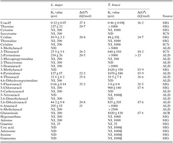
Table 1. Michaelis-Menten constants (Km) or inhibition constants (Ki) of various pyrimidine and purine analogues for the Leishmania major and Trypanosoma brucei brucei U1 uracil transporters

Fig. 1. (A) Inhibition of 20 nM [3H] uracil by increasing concentrations of unlabelled uracil (IC50=0·13 μM for this experiment). Inset: Michaelis-Menten plot, calculated Km and Vmax values for this experiment: 0·097±0·009 μM and 0·55±0·02 pmol (107 cells)−1 s−1. (B) Substrate specificity of LmU1. Uptake of 20 nM [3H] uracil by Leishmania major promastigotes was measured in the presence of various inhibitors and expressed as a percentage of control, defined as [3H] uracil uptake in the absence of inhibitor; 5-fluorouracil ([dtrif ]), 2-thiouracil ([squf ]) and 4-thiouracil (□). IC50 values were calculated assuming maximum inhibition by analogue, equalling inhibition by 1 mM uracil.
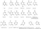
Fig. 2. Preferred tautomeric isomers of some tested pyrimidine analogues. Numbering of the pyrimidine ring is indicated. Sources: Elguero et al. (1976), Beak et al. (1980), Katritsky et al. (1991), Brown (1994).
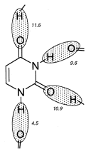
Fig. 3. Model of the interaction between uracil and the LmU1 transporter. Shaded areas depict areas of interaction with amino acid residues in the transporter-binding pocket, most probably H-bonds. Apparent ΔG0 for the interactions is given in kJ/mol.

Fig. 4. Promastigotes of Leishmania major (2×10⁴ cells), in 96-well plates, were incubated with test substance for 48 h before addition of the Alamar Blue reagent. Further incubation in the presence of the reagent was for 48 h after which fluorescence was determined and cells counted (inset). ED50 values for pentamidine ([squf ]) determined by fluorescence or cell counts were identical. Allopurinol: [utrif ]; Aminopurinol: [dtrif ]; 5-Fluorouracil: diamond.
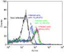
Fig. 5. FACS histogram acquired with macrophages infected with Leishmania major V39. The macrophages were infected for 5 h, washed and incubated in medium without or with 10 μM or 10 nM 5-fluorouracil for a further 72 h. The cultures were washed again and processed for FACS analysis as described in the Materials and Methods section.
 You have
Access
- 37
- Cited by