Introduction
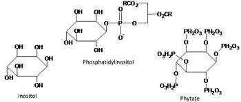
Inositol is a cyclohexitol sugar alcohol that exists as nine possible stereoisomers, of which myo-inositol is the most predominant form having biological activity (McDowell, Reference MCDOWELL2000). In this review, myo-inositol will be named inositol, unless otherwise specified. In nature, inositol is found mainly in its free form, as inositol phospholipids and as phytic acid ( Figure 1 ). Average human daily intake of inositol in the United States is around 1 g, primarily from the consumption of meat, poultry, fish and dairy products (Kohlmeier, Reference KOHLMEIER2003). In animals, inositol largely exists in its free form as well as phosphatidylinositol as part of the polar head group. In contrast, plant sources contain higher quantities of inositol polyphosphates, most commonly in the form of phytic acid or as its salt, phytate (inositol hexaphosphate; IP6). These serve as important phosphate reserves in the seed of cereals, and therefore phytate can be found in a number of diets for both humans and animals, with levels ranging from 5-25 g/kg (Cowieson et al., Reference COWIESON, WILCOCK and BEDFORD2011). The nutritional impact of phytate in animal diets, particularly its anti-nutritional effect on phosphorus (P) retention and mineral metabolism, has been given considerable attention and is well reviewed (Bedford and Schulze, Reference BEDFORD and SCHULZE1998; Bedford, Reference BEDFORD2000).
Chemical structures of free inositol, and inositol as part of phytate and phosphatidylinositol.

Much of our current knowledge has come from studies that have focused on improvements in animal performance as a result of supplementing diets with phytase for the liberation of phosphate groups from phytate. The more recent concept of superdosing phytases (Cowieson et al., Reference COWIESON, WILCOCK and BEDFORD2011) has demonstrated improvements in weight gain and performance compared to the standard enzyme dose. It has been proposed that these higher levels of phytase enable an almost complete dephosphorylation of phytate, and, as a consequence, increase inositol concentration in the intestinal tract (Walk et al., Reference WALK, SANTOS and BEDFORD2014). This leads to the question as to whether this free inositol plays a role in the improved growth performance reported for phytase super dosed animals, and by which mechanisms it might be acting.
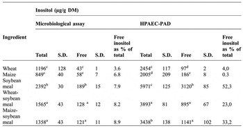
To date, there is limited evidence relating to the effects of free inositol on the metabolism and performance of monogastrics. Duliñski et al. (Reference DULIÑSKI, STARZYNSKA-JANISZEWSKA, STODOLAK and ZYLA2011) reported the content of free and total inositol levels in common feed ingredients ( Table 1 ) using two different detection methods. Although there are differences in the values produced using these methods, it would appear that the contribution of free inositol to the total inositol content in feed is minor. Therefore, animal response to inositol can be investigated through addition of inositol itself or phytase into the diet. Some of the first studies to investigate inositol as a required nutrient were performed using rodents. For example, inositol deficiency in mice has been shown to result in alopecia, retarded growth and even death, with re-supplementation alleviating these effects (Woolley, Reference WOOLLEY1940; Reference WOOLLEY1941). Tissue culture work has also revealed inositol as an essential growth factor for human cells (Eagle et al., Reference EAGLE, OYAMA, LEVY and FREEMAN1956). Together, these studies suggest that inositol is required for normal growth. The importance of inositol for growth in chickens was first reported over 70 years by Hegsted et al. (Reference HEGSTED, BRIGGS, MILLS, ELVEHJEM and HART1941). The addition of inositol to the chick ration demonstrated growth responses ranging from 18 to 52 g in 4 week-old birds. Since then, there has been minimal work investigating this suggested growth-response from inositol. However, more recent studies have begun to reveal potential benefits of supplementing poultry diets with inositol through improvements in body weight gain (BWG) and feed conversion ratio (FCR), particularly when diets are mineral-deficient, namely in calcium (Ca2+) and phosphorus (P).
The recent work of Cowieson et al. (Reference COWIESON, PTAK, MACKOWIAK, SASSEK, PRUSZYNSKA-OSZMALEK, ZYLA, SWIATKIEWICZ, KACZMAREK and JÓZEFIAK2013) has demonstrated the potential of inositol addition into broiler diets to improve growth performance, notably in the grower and finisher phases. Inclusion of 0.15% inositol increased BWG of broilers fed diets low in Ca2+ and digestible P (dP) as well as significantly reducing FCR over the trial period. However, birds fed diets high in Ca2+ and dP had a reduced BWG with inositol supplementation. During the starter phase (d 1-10), inositol supplementation resulted in a reduced BWG and higher FCR in birds fed diets either deficient or adequate both Ca2+ and dP. The reasoning for this effect may be due to the less well developed digestive tract and microflora population of the younger bird. This effect was partially alleviated, however, in the presence of a phytase, suggesting positive interaction between inositol and phytase. Moreover, Zyla et al. (Reference ZYLA, MIKA, STODOLAK, WIKIERA, KORELESKI and SWIATKIEWICZ2004) reported an improvement in BWG in broilers fed P-deficient diets supplemented with 0.1% inositol from d 1-21, and Pirgozliev et al. (Reference PIRGOZLIEV, ALLYMEHR, SARWAR, ACAMOVIC and BEDFORD2007) reported an improvement in BWG with inositol supplementation at 0.25% to a low P diet in broilers from d 7-17.
A simple trial where 0 or 3 kg of inositol was fed factorially with 0 or 1500 FTU of phytase in a corn-soy diet to broilers over a 42 d period (Bedford, unpublished) revealed a significant interaction as a result of the inositol only returning a performance benefit in the absence of the phytase. Performance in the presence of either diets supplemented with the phytase was identical, suggested a part, but not all of the response to phytase superdosing is due to inositol provision. Our estimate from this trial was that inositol was responsible for 30-35% of the response accorded to superdosing.
Free and total inositol content of feed as determined by microbiological assay and high-performance anion-exchange chromatography-pulsed amperometric detection (HPAEC-PAD). Values taken from Duliñski et al. (Reference DULIÑSKI, STARZYNSKA-JANISZEWSKA, STODOLAK and ZYLA2011).

Means within columns followed by the same letter are not significantly different (P = 0.05)
n=3 for microbiological assay, n=4 for HPAEC-PAD.
Although these trials suggest a beneficial effect of inositol through improvements in BWG, the reasons for this growth improvement are less well understood. Current knowledge indicates that inositol has a number of important biological functions within the body, with a number of tissues capable of synthesising and catabolising this polyol. This review will focus on inositol uptake, metabolism and biological function within the monogastic animal, to ascertain the mechanisms by which inositol may be implicated in the reported growth response.
Digestion and absorption
With negligible amounts of free inositol in animal feed, the majority is consumed as part of the phytate molecule. It is generally well accepted that the inositol and phosphate in phytate is poorly available for digestion by monogastric animals. Contrary to previous conceptions, this is not the result of poor phytase activity in these animals; in fact, poultry have been shown to possess efficient phytase activity in the small intestinal mucosa (Maenz and Classen, Reference MAENZ and CLASSEN1998). However, low phytate solubility in this section of the tract appears to be implicated. Lower IP esters are more soluble (Schlemmer et al., Reference SCHLEMMER, JANY, BERK, SCHULZ and RECHKEMMER2001), and these lower esters also form much weaker complexes with other nutrients such as minerals and proteins, thereby posing less of an anti-nutritional impact. Phytases catalyses the step-wise release of phosphate groups from phytate, releasing intermediary products including penta- (IP5), tetra- (IP4), tri- (IP3), di- (IP2) and mono- (IP1) phosphate esters of inositol. Exogenous phytase supplementation initiates the hydrolysis of phytate in the acidic conditions of the animal's stomach or gizzard, thereby releasing these lower esters into the intestinal tract. The animal's own alkaline phosphatase then completes the process through hydrolysis of IP1, resulting in the release of free inositol.
Free inositol and lipid-bound inositol are absorbed with high efficiency from the small intestine (Kohlmeier, Reference KOHLMEIER2003). In vitro incubation of hamster small intestine segments revealed that uptake and accumulation of inositol occurred against a concentration gradient (Caspary and Crane, Reference CASPARY and CRANE1970), thereby signifying the active transport of inositol into cells. Free inositol uptake is mediated by the sodium-dependant inositol transporter (SMIT1 or SLC5A3). Although the uptake of glucose and inositol appears to be through different transport systems, the inhibition of inositol uptake in the presence of glucose has been noted (Lerner and Smagula, Reference LERNER and SMAGULA1979). Addition of 16.6 mM glucose, a concentration often used in nutrient transport studies to simulate the intestinal milieu, suppressed the transport of inositol at concentrations ranging from 1-20 mM. This may therefore suggest that in semi-synthetic diets where glucose is added, there could be an effect on inositol uptake and hence an unexpected influence on the results by suppressing performance or fat metabolism. Moreover, in starch rich diets where the starch is rapidly digested, for example as a result of extensive processing and subsequent gelatinisation, then there may be more competition in the proximal part of the small intestine than in diets where more slowly digested starch sources are used. If glucose truly competes with inositol for absorption then the rate of generation of glucose from maltose at the brush border may have significant implications also. Assuming that inositol might be of functional importance to the enterocyte itself, then such a divergence in supply between proximal and distal small intestine may have performance, health or metabolism effects. Hydrolysis of phosphatidylinositol by pancreatic phospholipases can also occur in the small intestine (Kohlmeier, Reference KOHLMEIER2003), generating free fatty acids and lysophosphatidylinositol. However, further hydrolysis of this lysophospholipid, through removal of another fatty acid and phosphate group, is required before free inositol can be released. An acyltransferase in enterocytes can also convert lysophosphatidylinositol back to phosphatidylinositol, which is released as a component of lipoprotein (Berdanier, Reference BERDANIER1997).
In the enterocyte, it is possible that inositol will be specifically phosphorylated and used as mediator of several signalling pathways, as described later in this review. Moreover, it is likely that the majority of absorbed inositol is circulated in the blood and distributed to various tissues throughout the body. Early studies detected inositol polyphosphates, including phytate, in the blood of humans (Grases et al., Reference GRASES, SIMONET, VUCENIK, PRIETO, COSTA-BAUZA, MARCH and SHAMSUDDIN2001). The presence of IP6 in the erythrocytes of chickens was first reported by Rapoport (Reference RAPOPORT1940), with levels ranging from 61.2-68.1 mg/100ml (Rapoport and Guest, Reference RAPOPORT and GUEST1941). Oshima et al. (Reference OSHIMA, TAYLOR and WILLIAMS1964) showed increasing levels of IP6 in the blood of chickens during the first three weeks post-hatch. However, more recent evidence suggests that this is not the case, with IP6 being undetectable in the blood (Wilson et al., Reference WILSON, BULLEY, PISANI, IRVINE and SAIARDI2015). This discrepancy could be due to the differences in detection methods used. Typically, the majority of inositol in the blood is present in its free form, at approximately 29 µmol/l in humans, 50 µmol/l in rats (Burton et al., Reference BURTON, RAY, BRADFORD, ORR, NICKERSON and WELLS1976) and 280 µmol/l (2-5 mg/100ml) in chickens (Oshima et al., Reference OSHIMA, TAYLOR and WILLIAMS1964). Fewer quantities are transported as phosphatidylinositol, as a component of lipoproteins.
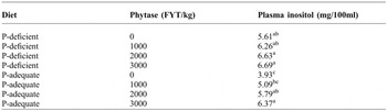
Compared to previous inositol detection methods using enzyme-linked assays (MacGregor and Matschinsky, Reference MACGREGOR and MATSCHINSKY1984; Ashizawa et al., Reference ASHIZAWA, YOSHIDA and AOTSUKA2000) or gas chromatographic methods (Niwa et al., Reference NIWA, YAMAMOTO, MAEDA, YAMADA, OHKI and MORI1983; Lee and Chung, Reference LEE and CHUNG2006), Frieler et al. (Reference FRIELER, MITTENESS, GOLOVKO, GIENGER and ROSENBERGER2009) reported a high-performance liquid chromatographic method for accurately measuring inositol in blood and tissue samples. In this method, samples underwent a pre-column derivatisation reaction using benzoyl chloride before separation of the benzoylated derivatives using reversed-phase HPLC. Mass spectrometry was subsequently used to quantify inositol levels via absorbance detection at 231 nm. Modification of this method, using a LC-MS/MS technique (Kindt et al., Reference KINDT, SHUM, BADURA, SNYDER, BRANT, FOUNTAIN and SZEKELY-KLEPSER2004; Leung et al., Reference LEUNG, MILLS, BURREN, COPP and GREENE2011), allows separation of inositol from other molecules of similar weight, such as glucose or other inositol isomers. Using this method, Cowieson et al. (Reference COWIESON, AURELI, GUGGENBUHL and FRU-NJI2015) was able to show an increase in plasma inositol concentration in broiler chickens fed a phytase supplemented nutritionally adequate diet (including P) and a Ca2+, P-deficient diet ( Table 2 ). This increase was shown to be significant for birds fed the PC diet supplemented with higher doses of phytate. These results suggest a greater dephosphorylation of phytate by dietary phytase, thereby increasing free inositol release and absorption. Detection of plasma inositol concentrations, using methods such as this, can therefore be a useful tool for comparing different dietary treatments and the supply of inositol to the animal. Once in circulation, inositol enters the brain, kidney and other tissues via the SMIT1 transporter (Berry et al., Reference BERRY, MALLEE, KWON, RIM, MULLA, MUENKE and SPINNER1995).
Effect of diet and different levels of phytase on plasma inositol concentrations. Data taken from Cowieson et al. (Reference COWIESON, AURELI, GUGGENBUHL and FRU-NJI2015).

a,b,c Means within columns followed by the same letter are not significantly different (P = 0.05).
Metabolism and function
Although the potential mechanisms by which inositol might stimulate a growth response have not yet been fully elucidated, there is evidence indicating its importance as a precursor in a number of biological functions. These include cell signalling, regulation of cell osmolality and growth, as well as membrane structure. Induced inositol deficiencies as well as disease states have provided valuable information on some of the essential functions of inositol in the body. The metabolic fate of inositol appears to involve its catabolism into CO2, application in gluconeogenesis and synthesis of phospholipids.
inositol synthesis
All cells appear to contain inositol, although some tissues appear to maintain higher levels than others ( Table 3 ). Sources of cytosolic inositol come from de novo synthesis, the recycling of inositol-containing compounds such as phosphoinositides, and uptake from extracellular fluid. The inositol requirements for humans and other animals are generally met by endogenous synthesis. Tissues that contain higher levels of inositol, such as the brain, liver, kidney and testis (Eisenberg and Bolden, Reference EISENBERG and BOLDEN1963; Hauser and Finelli, Reference HAUSER and FINELLI1963) have been shown to synthesis relatively large quantities from glucose ( Figure 2 ). For example, the kidney has been shown to synthesise inositol at an approximate rate of 4 g/d in the dinephric human (Clements and Diethelm, Reference CLEMENTS and DIETHELM1979). In a rate-limiting step, inositol-1-phosphate synthase (EC5.5.1.4) catalyses the conversion of glucose-6-phsphate into inositol-1-phosphate. Subsequent removal of the phosphate group by inositol-1-monophosphatase (EC3.1.3.25) results in the circulatory release of free inositol.
inositol catabolism
The kidney is the major site for inositol catabolism (Howard and Anderson, Reference HOWARD and ANDERSON1967). Inositol can be oxidised to CO2 or participate in gluconeogenesis via the glucuronic acid and pentose phosphate pathways (Charalampous and Lyras, Reference CHARALAMPOUS and LYRAS1957; Moscatelli and Larner, Reference MOSCATELLI and LARNER1959; Hankes et al., Reference HANKES, POLITZER, TOUSTER and ANDERSON1970). The catabolism process ( Figure 2 ) is initiated by a specific inositol oxygenase that catalyses the conversion of inositol into D-glucuronic acid. The latter is then transported to the liver where it is metabolised into D-xylulose 5-phosphate, and subsequently enters the pentose phosphate pathway. This process is essential for regulation of inositol homeostasis, with impaired kidney function being concomitant to an increased blood inositol levels (Holub, Reference HOLUB1986). Studies have shown minimal amounts of inositol released in the urine of rats and humans (Lewin et al., Reference LEWIN, YANNAI, SULIMOVICI and KRAICER1976; Clements and Diethelm, Reference CLEMENTS and DIETHELM1979).
Free inositol distribution in tissues of various species.

1 Dawson and Freinkel (Reference DAWSON and FREINKEL1961)- rats fed diets containing 0.065% free and 0.114% total myo-inositol; 2 Battaglia et al. (Reference BATTAGLIA, MESCHIA, BLECHNER and BARRON1961)- diet unknown; 3 Chu and Geyer (Reference CHU and GEYER1983)- animals fed diets containing 0.3% total myo-inositol.
Pathways of inositol synthesis and catabolism. Adapted from Kohlmeier (Reference KOHLMEIER2003).

inositol-containing phospholipid
One of inositol's most well-known functions is its involvement as part of the phospholipid component of cell membranes and lipoproteins, as an essential precursor for the synthesis of phosphatidylinositol. Typically, inositol-containing phospholipids are minor yet wide-spread constituents of the eukaryotic cell membrane, with phosphatidylinositol making up the majority. This inositol-containing phospholipid tends to have a high proportion of stearic acid in the sn-1 positions and arachidonic acid in the sn-2 position of the glycerol backbone (Holub, Reference HOLUB1986). Phosphatidylinositol is synthesised in the endoplasmic reticulum and transported to the cell membrane via vesicles or cytosolic transfer proteins. The primary localisation of phosphatidylinositol- PtdIns(4)P is shown to be in the Golgi, PtdIns(3)P in the endosome and PtdIns(4,5)P2 in the plasma membrane of Chinese hamster ovary cells (Di Paolo and De Camilli, Reference DI PAOLO and DE CAMILLI2006). The fact that the inositol head group can be phosphorylated at various positions allows for the production of specific phosphoinositides that play different functional roles in the cell's signalling pathway. Cellular phosphoinositides are maintained and interconverted by multiple kinases and phosphatases in response to specific stimuli.
signalling pathways
Hokin and Hokin (Reference HOKIN and HOKIN1955) were the first to distinguish a unique role of phosphoinositides from that of other membrane phospholipids. Incubation of pancreatic and brain cortex slices with acetylcholine resulted in the majority of 32P being incorporated into the phosphoinositide phospholipid fraction. The rapid turnover of phosphoinositides under various stimulants suggested a likely connection to signal transduction across the cell membrane (Michell, Reference MICHELL1975). This functional role is now generally well accepted. In response to stimulation, phosphoinositide-specific phospholipase C (PLC) hydrolysis of PtdIns(4,5)P2 results in the release of two second messengers ( Figure 3 ); 1,2-diacylglycerol (DAG) that activates protein kinase C (PKC), and Ins (1,4,5)P3 that simulates the release of Ca2+ from internal stores (Berridge and Irvine, Reference BERRIDGE and IRVINE1984). Inositol lipids have also been found to perform other cellular functions, including the anchoring of cell surface proteins and insulin signal transduction. Signalling pathways involving phosphoinositides have been reported to mediate cell growth, differentiation, apoptosis, vesicle trafficking, insulin action, cytoskeletal organisation and motility (Toker, Reference TOKER2002). Moreover, the presence of phosphoinositides in the nucleus of cells and the discovery that the nucleus has its own phosphatidylinositol metabolism independent to the rest of the cell, have given much attention to their function in this organelle. Several reviews have been written covering this topic (Martelli et al., Reference MARTELLI, CAPITANI and NERI1999; Reference MARTELLI, BORTUL, TABELLINI, ALUIGI, PERUZZI, BAREGGI, NARDUCCI and COCCO2001; (Irvine, Reference IRVINE2003). Evidence now suggests a role of phosphoinositides in various nuclear processes, including rRNA synthesis, mRNA-splicing, chromatin structure, all of which can alter cell physiology and function.
Signal induced hydrolysis of PtdIns(4,5)P2 by phosphoinositide-specific phospholipase C (PLC), generating the second messengers Ins(1,4,5)P3 and 1,2-diacylglycerol (DAG). Ins(1,4,5)P3 leads to Ca2+ release from intracellular stores, while DAG activates PKC, resulting in a cascade of protein phosphorylation events.

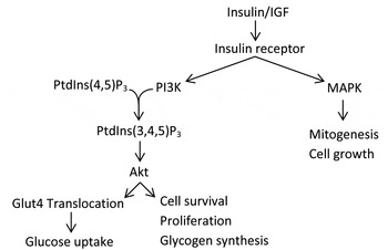
It is well established that the binding of insulin or insulin-like growth factor (IGF) to the insulin receptor leads to the recruitment of phosphoinositide 3-kinase (PI3K) and mitogen-activated protein kinase (MAPK) ( Figure 4 ). Stimulation of the IP3K signalling pathway generates PtdIns(3,4,5)P3, leading to the activation of the Akt (protein kinase B) cascade, which regulates cellular functions such as cell survival and metabolism (White, Reference WHITE2003). Activation of the MAPK pathway supports cascades that regulate mitogenesis and cell growth (Guo, Reference GUO2014). Insulin signalling in muscles and adipocytes also stimulates translocation of the glucose transporter, GLUT4, to the plasma membrane, thereby enhancing glucose uptake into these cells (Leto and Saltiel, Reference LETO and SALTIEL2012).
Insulin signalling through the PI3K/Akt and MAPK pathways.

In subjects with insulin resistance disorders such as polycystic ovary syndrome, gestational diabetes mellitus or metabolic syndrome, the inositol content of certain tissues is reduced, suggesting a potential link between inositol and insulin sensitivity (Croze and Soulage, Reference CROZE and SOULAGE2013). The majority of studies investigating the beneficial effects of inositol supplementation on insulin sensitivity in both animal and human subjects have primarily used the isoform D-chiro-inositol (Ortmeyer et al., Reference ORTMEYER, HUANG, ZHANG, HANSEN and LARNER1993; Reference ORTMEYER, LARNER and HANSEN1995; Nestler et al., Reference NESTLER, JAKUBOWICZ, REAMER, GUNN and ALLAN1999; Larner, Reference LARNER2002; Yao et al., Reference YAO, SHAN, BIAN, CHEN, WANG and REN2008). Since insulin sensitive tissues, such as adipose, skeletal muscle and liver, are capable of converting inositol into D-chiro-inositol (Pak et al., Reference PAK, HONG, KIM, PICCARIELLO, FARESE and LARNER1998; Larner, Reference LARNER2002), this suggested that administration of inositol may also regulate insulin sensitivity. The lowering of plasma and urine glucose concentrations in diabetic monkeys and humans has been demonstrated with inositol administration, without the concomitant increase in insulin concentrations (Ortmeyer, Reference ORTMEYER1996; Corrado et al., Reference CORRADO, D'ANNA, DI VIESTE, GIORDANO, PINTAUDI, SANTAMARIA and DI BENEDETTO2011). It is possible that an insulin-mimicking effect of inositol phosphoglycans (IPGs) is generating this response, with the increased availability of inositol in cells (Croze and Soulage, Reference CROZE and SOULAGE2013). However, the mechanisms of this mimicking effect still need further investigation.
inositol in development
The importance of inositol during rapid postnatal development can be argued due to the relatively high concentrations found in milk, at levels of 1840 µmol/l in humans (Pereira et al., Reference PEREIRA, BAKER, EGLER, CORCORAN and CHIAVACCI1990) and 1600 µmol/l in rats (Burton and Wells, Reference BURTON and WELLS1974). There does not currently appear to be any information on the content of inositol in sow's milk. In the chick, however, all pre-hatch nutrient supply comes from the albumin and yolk of the egg. In 1949, unpublished data from Celmer reported the occurrence of inositol in the phospholipid component of egg yolks (Celmer and Carter, Reference CELMER and CARTER1952). Inositol has been shown to account for approximately 13% of the lipid phosphate in egg yolk (Rhodes and Lea, Reference RHODES and LEA1957). Minimal quantities of lipid are found in egg albumin (0.02% by weight), with approximately 14% of this lipid being comprised of phospholipid (Sato et al., Reference SATO, WATANABE and TAKAHASHI1973). This would therefore suggest that the developing chick has a supply of inositol. However, due to the independence of the chicken post-hatch, with all subsequent nutrients coming directly from feed, inositol from dietary phytate as well as de novo synthesis will be the primary supply of inositol to the growing chick.
bone formation
To better understand what role inositol might play in specific tissues through development and in adulthood, mice devoid of the inositol transport SMIT1 (SMIT1-/-) have been generated. SMIT1-/- mice show significantly lower inositol concentrations in a number of tissues, defective nervous systems, respiratory failure and die soon after birth (Chau et al., Reference CHAU, LEE, LAW, CHUNG and CHUNG2005). However, neonatal mortality could be prevented through prenatal supplementation of inositol to maternal drinking water. SMIT1-/- mice have been used to investigate the function of inositol and its transporter in skeletal development and osteoblastogenesis. Dai et al. (Reference DAI, CHUNG, MIAO, LAU, CHAN and KUNG2011) described inositol rescued SMIT1-/- adult mice with shorter limbs, reduced bone mineral density and abnormal bone architecture. This work revealed down-regulation of several transcription factors in SMIT1-/- mice that are essential for osteogenesis, including Runx2, osterix and NFATc1, resulting in poor bone formation in adults. Post-weaning inositol supplementation partially alleviated the abnormal bone defects in SMIT1-/- adult mice, while increasing bone strength in SMIT1+/+ mice. These results suggest an important role of inositol in bone formation, osteogenesis and bone mineral density.
peripheral nerve development
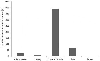
Abnormal development of peripheral nerves, including the brachial plexus, facial, vagus and intercostal nerves, and phrenic nerve in SMIT1-/- mice have also been shown (Chau et al., Reference CHAU, LEE, LAW, CHUNG and CHUNG2005). The phrenic nerve is essential for the control of breathing through innervation of the diaphragm, and thus its underdevelopment is mostly likely the cause of respiratory failure as seen in SMIT1-/- mice. Prenatal inositol supplementation was able to prevent these defects, allowing the SMIT1-/- mice to survive and enter adulthood. However, these adults were shown to have reduced levels of inositol in the sciatic nerve, kidney, skeletal muscle, liver and brain, at approximately 10, 22, 24, 38 and 39% of that measured in their SMIT1+/+ siblings, respectively. Post-weaning supplementation of inositol to SMIT1-/- mice increased levels in these tissues by around 25, 8, 391, 69 and 4%, respectively ( Figure 5 ). This indicates that other transporters, such as SMIT2 and HMIT (H+/myo-inositol transporter) (Uldry et al., Reference ULDRY, IBBERSON, HORISBERGER, CHATTON, RIEDERER and THORENS2001; Coady et al., Reference COADY, WALLENDORFF, GAGNON and LAPOINTE2002), may be facilitating inositol uptake into tissues when plasma concentrations are high. The fact that there is a relatively large increase in skeletal muscle inositol content with supplementation may suggest that this organ has an essential requirement for inositol, yet has a lower capacity to synthesise it de novo.
Relative increases in inositol content of various tissues from SMIT -/- mice with post-weaning supplementation of inositol. Data adapted from Chau et al. (Reference CHAU, LEE, LAW, CHUNG and CHUNG2005).

inositol in the brain
It is now well-established that inositol phospholipids serve as precursors for intracellular second messengers in both neural and non-neuronal tissues (Hawthorne and Pickard, Reference HAWTHORNE and PICKARD1979). Relatively high concentrations of inositol in the brains of numerous species suggest a possible functional role of inositol in the nervous system. Although the brain is capable of synthesising inositol de novo, it is also able to cross the blood-brain-barrier (Spector, Reference SPECTOR1988). SMIT1, the inositol transporter, is dispersed widely throughout the central nervous system (CNS). High expression of SMIT1 mRNA and protein in the brain of rats has been shown to decline postnatally (Guo et al., Reference GUO, SHIMADA, TAJIRI, YAMAUCHI, YAMASHITA, OKADA and TOHYAMA1997); however, expression in the choroid plexus remained high. This might imply that inositol plays an essential role in CNS development. This is contrary to kidney, whereby an increase in SMIT1 mRNA expression following birth is observed (Guo et al., Reference GUO, SHIMADA, TAJIRI, YAMAUCHI, YAMASHITA, OKADA and TOHYAMA1997). High expression of a H+/ inositol transporter, HMIT in the brain of rats has also been shown (Uldry et al., Reference ULDRY, IBBERSON, HORISBERGER, CHATTON, RIEDERER and THORENS2001), with low levels of expression in adipose and kidney tissue. However, more recent studies have argued that HMIT may not be involved in inositol uptake into neuronal cells, as its location appears to be primarily connected with the endoplasmic reticulum and Golgi complex rather than the plasma membrane (Di Daniel et al., Reference DI DANIEL, MOK, MEAD, MUTINELLI, ZAMBELLO, CABERLOTTO, PELL, LANGMEAD, SHAH, DUDDY, KEW and MAYCOX2009).
skeletal muscle
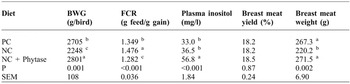
Considering that the involvement of inositol in the growth of poultry has already been proposed (Zyla et al., Reference ZYLA, MIKA, STODOLAK, WIKIERA, KORELESKI and SWIATKIEWICZ2004; Walk et al., Reference WALK, SANTOS and BEDFORD2014; Cowieson et al., Reference COWIESON, AURELI, GUGGENBUHL and FRU-NJI2015), relatively few studies have reported the effect of inositol on gene expression and muscle growth in correlation to performance. Recent work by Schmeisser et al. (Reference SCHMEISSER, SÉON, AURELI, FRIEDEL, GUGGENBUHL, DUVAL, COWIESON and FRU-NJI2016) has begun to shed some light on this area. Supplementing broiler diets with moderate P and Ca2+ deficiency with 1000 U/kg 6-phytase improved BWG and lowered FCR over the five week trial period, compared to both positive and negative control diets ( Table 4 ). Plasma concentrations of inositol were also reported to increase with phytase supplementation, compared to the control diets. This would imply that the phytase is liberating inositol from dietary phytate in the gut where it is absorbed and enters circulation. This increase in circulating inositol would be expected to result in increased uptake into various tissues, including the muscle.
Effect of phytase supplementation on performance, plasma inositol concentration, breast meat yield and weight. Data taken from Schmeisser et al. (Reference SCHMEISSER, SÉON, AURELI, FRIEDEL, GUGGENBUHL, DUVAL, COWIESON and FRU-NJI2016).

PC, positive control diet; NC, negative control diet; + Phytase, NC supplemented with phytase at 1000 U/kg feed.
Means within the same column lacking common superscripts differ (P < 0.05).
When considering muscle growth, phytase supplementation did not significantly affect breast meat yield (%) compared to the control diets. However, an increase in breast meat weight was observed for phytase supplemented birds compared those fed the negative control diet. Microarray analysis with validation by qPCR revealed a number of differentially expressed genes in the breast muscle of birds fed phytases. These genes are known to be involved in two main pathways; the calmodulin/calcineurin and IGF/Akt/mTOR pathways. As previously discussed, inositol has been suggested to mimic the insulin signalling pathway, and thus the increased concentration of inositol in the blood, as the result of phytase action, may have caused this response. The Akt/mTOR pathway in muscle is responsible for protein synthesis and this may be a possible explanation for the increased growth response seen in these birds. In addition, Dang et al. (Reference DANG, MUKAI, YOSHIDA and ASHIDA2010) reported lower concentrations of glucose and insulin in plasma with oral administration of 1 g/kg BW inositol to mice prior to injection of glucose at 2 g/kg BW. This was the result of increased translocation of GLUT4 in skeletal muscle, and thus enhanced glucose uptake into muscle cells. It is possible therefore, that inositol-induced glucose uptake in muscle increases the energy supply to this tissue to support growth. However, it has been noted that chickens do not possess GLUT4 (Seki et al., Reference SEKI, SATO, KONO, ABE and AKIBA2003; Tokushima et al., Reference TOKUSHIMA, TAKAHASHI, SATO and AKIBA2005), and therefore this response may be restricted to mammals.
fat deposition
Phosphatidylinositol also the stimulates the synthesis and secretion of β-lipoprotein, a low-density lipoprotein (LDL), from the liver (Hasan et al., Reference HASAN, KOTAKI and YAGI1970; Reference HASAN, NAKAGAWA, NISHIGAKI and YAGI1971). Hayashi et al. (Reference HAYASHI, MAEDA and TOMITA1974) reported an increase in triglyceride accumulation in the liver of rats fed an inositol deficient diet along with highly saturated fats. However, when highly unsaturated fats were fed with an inositol deficient diet, this effect was not evident. In inositol-deficient gerbils, the secretion rate of LDL was reduced, this being one possible explanation for the accumulation of hepatic lipids (Hoover et al., Reference HOOVER, NICOLOSI, COREY, EL LOZY and HAYES1978). Another explanation was given by Hayashi et al. (Reference HAYASHI, MAEDA and TOMITA1974), who suggested that an increase in liver TAG in inositol-deficient rats was primarily due to the increased rate of fatty acid mobilisation from the adipose tissue to the liver. They found a near three-fold increase in the transfer of radioactively labelled [14C] palmitate from the epididymal fat pads to the liver in inositol-deficient rats. In contrast to rats, diet-induced inositol deficiency in gerbils leads to intestinal lipodystrophy (Hegsted et al., Reference HEGSTED, HAYES, GALLAGHER and HANFORD1973), which is characterised by the accumulation of fat in the mucosal cells of the small intestine. It has been suggested that a deficiency in inositol affects chylomicron assembly and secretion, thereby impairing the lymphatic transport of dietary fat (Chu and Hegsted, Reference CHU and HEGSTED1980). Addition of inositol resulted in an increase in plasma chylomicron and lipid concentrations, followed by disappearance of the accumulated lipids from the intestine (Chu and Geyer, Reference CHU and GEYER1981). The results of these studies suggest that inositol plays an important role as a transporter of dietary fats. Croze et al. (Reference CROZE, VELLA, PILLON, SOULA, HADJI, GUICHARDANT and SOULAGE2013) found that inositol-treated mice showed a significant reduction in adipose accretion associated with reduced adipocyte volume rather than number. The mechanisms by which inositol is causing this reduction in fat mass is not yet known; however, one explanation could be reduced fatty acid synthase activity in adipose tissue (Croze et al., Reference CROZE, GELOEN and SOULAGE2015). It is possible therefore, that inositol could be beneficial for the generation of a leaner animal through the regulation of fat mass. Since inositol appears to encourage fat uptake from the gut and transport from the liver without accretion of fat mass, it is possible that fat is being used as an energy source. Liu et al. (Reference LIU, RU, WANG and XU2010) showed that the addition of phytase into broiler diets can alter blood serum and liver lipid profiles, as well as the mRNA expression of leptin, thereby contributing to lipid metabolism and deposition in the bird. This involvement in fat metabolism may also explain why a higher level of dietary fat is associated with a greater phytase response (Rosen, Reference ROSEN2002). While research in this area is somewhat limited, current data suggest an interesting link between inositol and energy utilisation.
Conclusions
Increasing the content of free inositol in the diet through phytase superdosing or inositol addition has shown an improvement in growth performance in animals. The link between inositol and this growth response, however, remains unclear. It is possible that an increase in inositol concentration in the blood, following intestinal absorption, increases uptake into various tissues. Within cells, the main function of inositol appears to be as a messenger in a number of signalling pathways that regulate various cellular processes, including cell survival, growth and metabolism. Inositol also plays a crucial role in fat transport and deposition, as well as for normal development, with deficient inositol levels severely disrupting these processes. Together these findings suggest that, in some circumstances, inositol could be considered an essential nutrient. In contrast, there seems to be limited information on the response of animals to increased inositol levels and how this might affect inositol-regulated processes. Further investigation is required for a more detailed understanding of tissue specific responses to elevated inositol levels. With this work, the potential mechanisms of inositol in promoting the reported animal growth response may be elucidated.