

Trilobites of the Family Olenidae occur in immense numbers in the Furongian Alum Shales of Scandinavia, and are by far the most dominant fossils. The middle Cambrian through Tremadocian Alum Shales are some 100 metres thick at their maximum, in Skåne, Sweden, the traditional middle Cambrian portion being c.30 m, and the upper Cambrian (Furongian) c.50 m at most. Though the middle Cambrian bears a highly diverse trilobite fauna, that of the upper Cambrian is much more restricted, and most of these are olenids. The environment the olenid inhabited here has been interpreted as being relatively deficient in oxygen (e.g., Anderson et al. Reference Anderson, Dahlman, Gee and Snäll1985; Dworatsek Reference Dworatzek1987; Schovsbo Reference Schovsbo2000, Reference Schovsbo2001). However, even though olenids seem to have adapted to a low-oxygen environment, Balseiro et al. (Reference Balserio, Waisfeld and Buatois2011) showed that some olenids, especially those which lived later in time, are not restricted to dysoxic facies. In the Lower Ordovician of the Argentine Cordillera Oriental, olenids dominated both oxygenated and dysoxic environments. The olenids are usually very well preserved in the Alum Shale; both in three dimensions in carbonate concretions, and flattened in shales. Moreover, juvenile forms commonly occur along with the adults. Many genera and species have been described, and their order of succession is well established; as, in general terms, are their evolutionary relationships. In Norway (Henningsmoen Reference Henningsmoen1957), Sweden (Westergård Reference Westergård1922), and Denmark (Bornholm) (Poulsen Reference Poulsen1923), the Cambrian successions in the Alum Shales permit research in many interlinked fields of palaeobiology, sedimentology, evolution and environment; and, arguably, this region of Scandinavia is one of the few places anywhere in the world where such research can be effected. The ultimate objective of this work would be a synthesis of current knowledge of an extinct environment and the evolution and adaptations of the extinct marine invertebrates that lived within it.
Many studies of the sedimentology, geochemistry and stratigraphical ordering of the Scandinavian Furongian have been undertaken (e.g., Schovsbo Reference Schovsbo2000, Reference Schovsbo2001; Terfelt et al. Reference Terfelt, Eriksson, Ahlberg and Babcock2008, Reference Terfelt, Ahlberg and Eriksson2010), and tie-points using agnostoids have been established for various levels in the sequence (e.g., Ahlberg & Terfelt Reference Ahlberg and Terfelt2012). The most recent stratigraphical paper resurrects the concept of superzones for the Scandinavian Furongian (Nielsen et al. Reference Nielsen, Weidner, Terfelt and Høyberget2014). In addition, since well-preserved juveniles are often found with the adults, several studies of the ontogeny of Scandinavian olenids have been undertaken in recent years (Clarkson & Taylor, Reference Clarkson and Taylor1995a, b; Clarkson et al. Reference Clarkson, Taylor and Ahlberg1997, Reference Clarkson, Ahlgren and Taylor2003; Reference Clarkson, Ahlgren and Taylor2004; Månsson Reference Månsson1998; Clarkson & Ahlberg Reference Clarkson and Ahlberg2002; Bird & Clarkson Reference Bird and Clarkson2003; Månsson & Clarkson Reference Månsson and Clarkson2012). Ontogenies of other Cambrian olenids outside Scandinavia have been described from, for example, Argentina (Tortello & Clarkson Reference Tortello and Clarkson2003, Reference Tortello and Clarkson2008), Canada (Lee & Chatterton Reference Lee and Chatterton2007) and Korea (Lee & Choi Reference Lee and Choi1999). The present paper is a contribution to this databank. Its main purpose is palaeobiological; the study of comparative ontogeny within a close-knit group has a bearing not only on taxonomy but also upon questions of heterochrony and adaptation, as part of the overall interlinked synthesis.
1. Material and stratigraphy
The material upon which this study is based was collected from Bornholm by Dr Arne Torshøj Nielsen some thirty years ago, and was stored in the collections of the Geological Museum, Copenhagen, Denmark. The material was collected at the site that Poulsen (Reference Poulsen1923) named Locality 6, located along the Læsaa Rivulet on southern Bornholm. For a detailed map, see Poulsen (Reference Poulsen1923, fig. 1). Poulsen Reference Poulsen1923 (pp 14, 74) described the section as the most accessible one at Læsaa. The section was about 60 metres long (the northern end of the profile is now almost inaccessible) and about 10 m high. The inclination of the section is such that the older zones are found at the northern end of the section. A number of anthraconite concretions of different sizes may be observed. The section contains six zones. These zones are here reinterpreted according to the most recent zonal schemes (Terfelt et al. Reference Terfelt, Eriksson, Ahlberg and Babcock2008, Nielsen et al. Reference Nielsen, Weidner, Terfelt and Høyberget2014). Zone 8 (Parabolina spinulosa Zone) is followed by Zone 12 (layers with Eurycare latum and Leptoplastus ovatus), then Zone 13 (L. angustatus and L. stenotus), Zone 16 (Ctenopyge flagellifera) and Zone 19 (shale with great numbers of Ctenopyge (M.) tumida and Sphaerophthalmus alatus). The thickness of Zone 19 is about 2.50 metres. These layers contain four big concretions of aragonite, the greatest of which has axes of about 2.50 m and 0.50 m. The highest zone exposed is that containing Peltura scarabaeoides (Zone 22 of Ctenopyge linnarssoni). There are undoubtedly hiatuses within the sequence, which require to be investigated further.
The material described here belongs to Sphaerophthalmus alatus and Ctenopyge (Mesoctenopyge) tumida, the former being the more abundant. There are rare sclerites, belonging to other species but, despite an extensive search, we found no agnostoids. All the specimens of S. alatus and C. (M.) tumida are disarticulated, with uncountable numbers of isolated and usually broken sclerites lying on the bedding planes. This is the usual way in which Sphaerophthalmus is found, with great numbers of disarticulated exuviae, and complete or nearly complete specimens have been found only rarely. Since there are no complete individuals in the Bornholm material, and most of the larger sclerites are broken so as to be too fragmentary to use for studies of ontogeny, it is usually only the smaller sclerites that are well preserved. Accordingly, we have limited our studies to the early ontogenetic stages. Even here, specimens that can be used for this purpose are not common, and the fragmentary nature of the material necessitated an extended search for intact sclerites. This has imposed two particular limitations on what could be established. The first is that it was not possible to produce any kind of morphometric analysis, such as bivariate plots of length and width; there was simply not enough appropriate material. Secondly, many of the reconstructions are inevitably composite, based upon several specimens; though we trust that thus has not diminished their accuracy. The quality of preservation is quite variable, being excellent at some levels, where, for example, long thin spines may be preserved, but less so at others. Eyes are found to be well preserved at some horizons, and lacking in others. It has recently been shown (Schoenemann & Clarkson Reference Schoenemann and Clarkson2015), on the basis of their optical systems, that although S. alatus and C. (M.) tumida are found together, the two trilobite species lived at different depths, and it seems appropriate to describe their ontogenies comparatively in the same paper. S. alatus is believed to have occupied a vagrant benthic habitat, whereas C. (M.) tumida was probably a pelagic swimmer (Schoenemann & Clarkson Reference Schoenemann and Clarkson2015).
1.2. Is Sphaerophthalmus distinct from Ctenopyge?
One major problem that has affected the taxonomic ordering of olenid trilobites is that at least some genera were highly labile; subject to morphological and developmental change through even short time periods (Kaufmann Reference Kaufmann1933, Clarkson et al. Reference Clarkson, Taylor and Ahlberg1997, Lauridsen & Nielsen Reference Lauridsen and Nielsen2005, Ahlberg et al. Reference Ahlberg, Månsson, Clarkson and Taylor2006). Although some olenids seem to follow a constrained ontogenetic pathway, others do not (Månsson & Clarkson Reference Månsson and Clarkson2012) and, because of the limitations of the available material, it is not yet known to which category those of the Sphaerophthalmus/Ctenopyge complex pertain. It is also quite likely that there was a fair degree of regional variation in some geographically widespread olenids. Such issues as these have clouded the production of a sound taxonomy and this has particularly been the case with the leptoplastines of the Sphaerophthalmus/Ctenopyge complex. Where to draw the boundary between these two genera has taxed generations of olenid specialists, as commented upon by Henningsmoen (Reference Henningsmoen1957) and Clarkson et al. (Reference Clarkson, Ahlgren and Taylor2003).
A recent important study by Høyberget & Bruton (Reference Høyberget and Bruton2012) addressed the issue of the confused taxonomy of the Sphaerophthalmus/Ctenopyge complex, based on material from a classic section at Slemmestad, near Oslo, Norway. This work presented a revision of both Sphaerophthalmus and Ctenopyge, and a new genus Triangulopyge was proposed. A detailed stratigraphical log was also given, recording the vertical ranges of all the olenids from the S. postcurrens to the C. linnarssoni Zones. This new work has gone a long way towards resolving a difficult and complex question. One major issue affecting our present work is the proposal, based on fresh and critical material, that what Clarkson et al. (Reference Clarkson, Ahlgren and Taylor2003) described as Ctenopyge (Eoctenopyge) angusta Westergård Reference Westergård1922 was considered by Høyberget & Bruton (Reference Høyberget and Bruton2012) to be Sphaerophthalmus alatus. If this is indeed the case, then at first sight this might seem to make our present work redundant, since the ontogeny of S. alatus has already been described in full. We agree with Høyberget & Bruton (Reference Høyberget and Bruton2012) that C. (Eoctenopyge) is a junior synonym of the genus Sphaerophthalmus, and that C. (Eoctenopyge) angusta described by Clarkson et al. (Reference Clarkson, Ahlgren and Taylor2003) should be regarded as properly belonging to S. alatus. However, we recognise S. alatus as a highly variable species, and most likely geographically clinal; the Bornholm morphs are not identical to those from Västergötland, south-central Sweden, upon which the study of Clarkson et al. (Reference Clarkson, Ahlgren and Taylor2003) were based. Intermediate morphs of S. alatus and S. angustatus have been reported from the Oslo area in Norway by Høyberget & Bruton (Reference Høyberget and Bruton2012). The Bornholm material described herein differs in several respects from that from Norway and Sweden, and in this paper we present some further observations on various aspects of the early ontogeny of S. alatus, based upon specimens from Bornholm.
The remarkable variability found amongst the olenids of the Sphaerophthalmus/Ctenopyge complex, and the problem of intergrading character states and geographical variation, does not make it easy to determine what is rightly a Sphaerophthalmus and what is a Ctenopyge. The study of comparative ontogenies so far has not really contributed to the resolution of this question. It may well be that any future distinction would be best based on relative spinosity of the thorax, rather than on the more traditional characters.
We also describe the early stages of the ontogeny of C. (M.) tumida, including the pygidia, which have not been recorded before. In Clarkson et al. (Reference Clarkson, Ahlgren and Taylor2004), the earlier stages were not described; thus, the present work augments the description of this species.
2. Systematic palaeontology
Illustrated, described and cited specimens are deposited at the Department of Geology, Lund University, Sweden (LO), and at the Geological Museum, University of Oslo, Norway (PMO). Due to the fragmentary nature of the material, the ontogenetic development of the cranidium, librigena, hypostome and pygidium is described separately.
Subclass Libristomata Fortey, Reference Fortey1990
Order Olenida Adrain, Reference Adrain and Zhang2011
Family Olenidae Burmeister, Reference Burmeister1843
Subfamily Leptoplastinae Angelin, Reference Angelin1854
Genus Sphaerophthalmus Angelin, Reference Angelin1854
Type species. Subsequently designated by Linnarsson (Reference Linnarsson1880) Trilobites alatus, Boeck, Reference Boeck and Keihau1838.
Sphaerophthalmus alatus (Boeck, Reference Boeck and Keihau1838) Figs 1–6
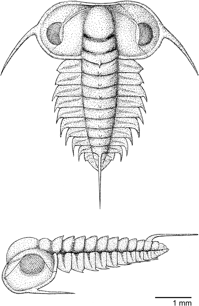
Sphaerophthalmus alatus (Boeck, Reference Boeck and Keihau1838), reconstruction of a late meraspid with nine segments, equivalent to Group 4, in dorsal and lateral views. Based on material from Bornholm.

For complete synonymy lists see Henningsmoen (Reference Henningsmoen1957), Żylińska (Reference Żylińska2001) and Høyberget & Bruton (Reference Høyberget and Bruton2012).
Type data. Størmer (Reference Størmer1940, p. 145) selected a lectotype (PMO 56371) from Gamleby, Oslo, Norway, from Boeck's material. The lectotype has been figured by Henningsmoen (Reference Henningsmoen1957, pl. 22, figs 23–24) and by Høyberget & Bruton (Reference Høyberget and Bruton2012, fig. 5A, D).
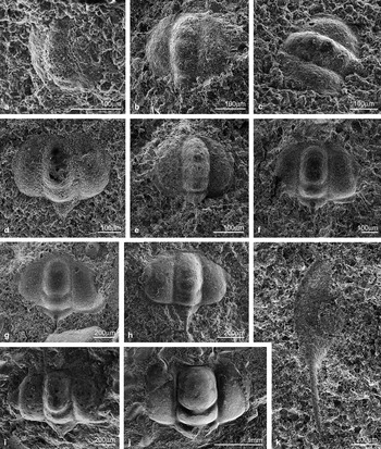
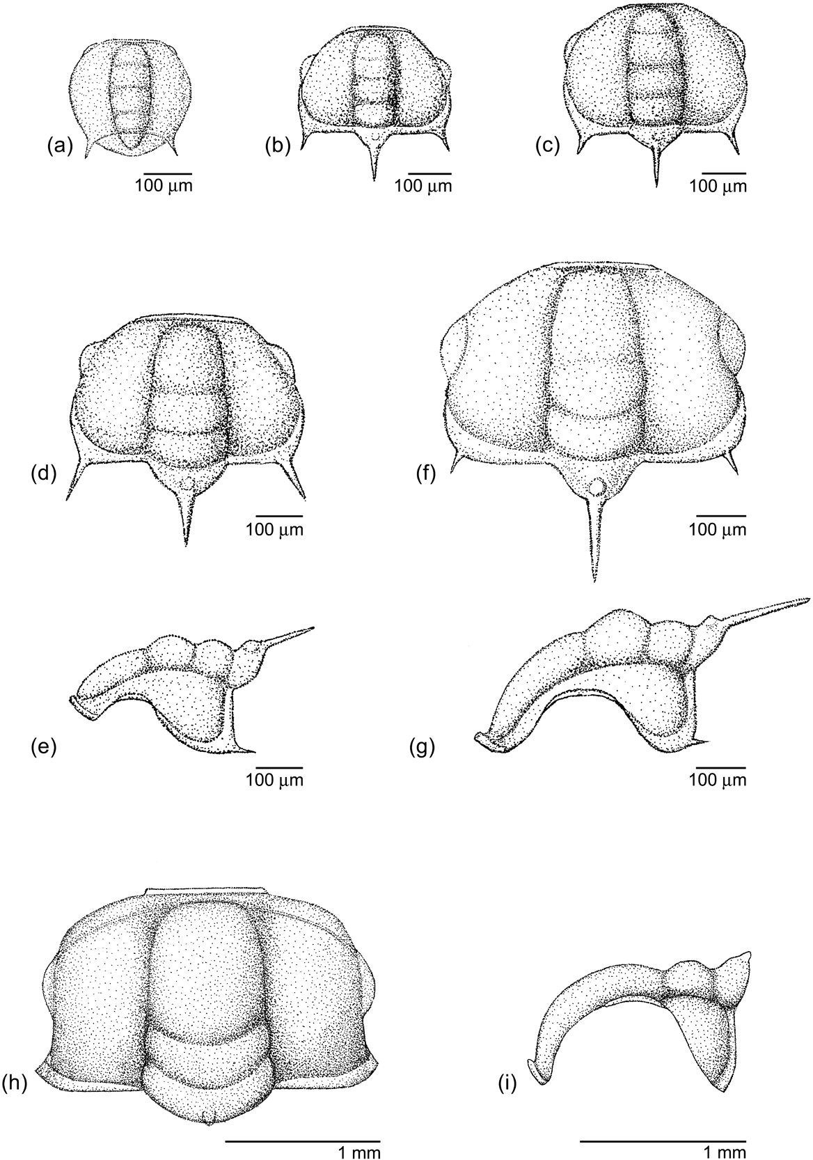
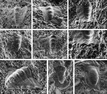
Protaspid stage. (Figs 2a, 3a). The smallest specimens represented in our material are two protaspides, representing an earlier ontogenetic stage than that, for example, figured by Clarkson et al. (Reference Clarkson, Ahlgren and Taylor2003). Each of these is almost circular, being 280 μm long and 280 μm wide, with a well-marked central axis having four axial rings, and a distinct furrow separating the cranidium from the protopygidium. There is a pair of short, sharp intergenal spines. The palbebral lobes are faintly developed.
Development of the meraspid cranidium. (Figs 2b–j, 3b–i). Because the material is fragmentary, we have allocated it into Groups 1, 2, etc., following Whitworth (Reference Whitworth1970), Clarkson & Taylor (Reference Clarkson and Taylor1995b) and Månsson (Reference Månsson1998), rather than attempting to assign detached sclerites to any particular meraspid degree. These groups are mainly based on size and morphological features. These groups do not correspond to moults or instars.
Group 1. (Figs 2b–c, 3b). The two smallest meraspides are 320 μm long (exclusive of spines) and 320 μm wide. The external contour is trapezoidal, with an almost straight anterior border, and the cranidium is widest posteriorly. The axis remains fairly narrow, extending as far as the very narrow anterior border; there are four lobes and the palpebral lobes lie opposite L3. A prominent posterior border has developed, and the occipital ring bears an anterior node with a short, though stout occipital spine. The two intergenal spines are still present. These specimens are likely to represent a degree 1 meraspis.
Sphaerophthalmus alatus (Boeck, Reference Boeck and Keihau1838). (a) Protaspis, LO 12297t. (b–c) Group 1 meraspid cranidia: (b) LO 12298t; (c) LO 12299t. (d–e) Group 2 meraspid cranidia: (d) LO 12300t; (e) LO 12301t. (f–i) Group 3 meraspid cranidia: (f) LO 12302t; (g) LO 12303t; (h) LO 12304t; (i) LO 12305t. (j) Group 4 cranidium of adult proportions, LO 12306t. (k) Librigena, likely belonging to a relatively late meraspid, LO 12307t.

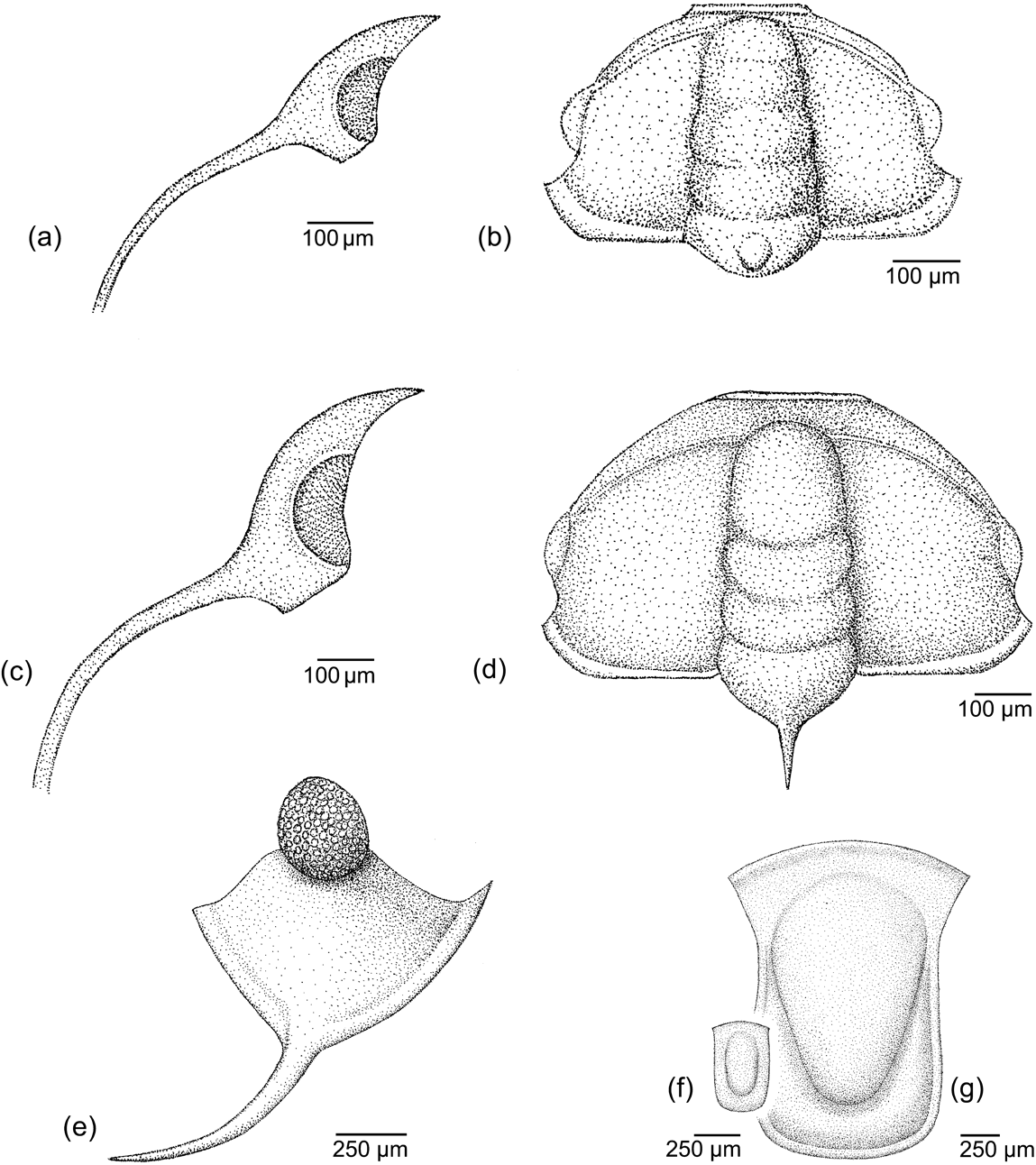
Reconstructions of protaspid and cranidia of Sphaerophthalmus alatus (Boeck, Reference Boeck and Keihau1838): (a) protaspis, dorsal view; (b) Group 1 meraspid cranidium; (c) Group 2 meraspid cranidium; (d–e) Group 3 meraspid, in dorsal and lateral views; (f–g) Group 3 meraspid, in dorsal and lateral views; (h–i) Group 4 cranidium, late meraspid/early holaspid in dorsal and lateral views.

Group 2. (Figs 2d–e, 3c). The next developmental stage is 350 μm long (excluding spines), and 415 μm wide. It retains the morphology of the preceding stage, though is somewhat narrower posteriorly, so that the paired intergenal spines appear to be a little closer together. This cranidium is slightly more inflated than that of the previous stage.
Group 3. (Figs 2f–i, 3d–g). The smallest cranidium within this group is represented by a single imperfectly preserved specimen 370 μm long and 500 μm wide (excluding the spines), and of similar shape to that of the previous degree. There are only three distinct axial lobes; L4 is now effaced. The occipital and intergenal spines are still very distinct, as is the occipital node. The occipital spine is inclined upwards at about 30 degrees. In profile, the cranidium appears moderately convex. A larger specimen still belonging to Group 3 is 400 μm long (excluding spines) and 610 μm wide. Whereas it is generally similar in form to the preceding stage, the S2 furrow is much fainter, and the intergenal spines have become much shorter, though the occipital spine and node remain prominent. There is a distinct increase in convexity and the anterior slope of the glabella is greater, the highest point being at L2. Figure 2i illustrates an oblique anterior view of the largest cranidium in Group 3, which shows an increasing convexity and an upwardly inclined short broken off occipital spine.
Group 4. (Figs 2j, 3h–i). In the latest stage represented in our material, the cranidium is virtually of adult proportions and convexity. The example illustrated is approximately 1.3 mm long and 2.0 mm wide. It probably represents a late meraspis, or an early holaspis. The glabella has broadened so that, as in the adult, it now occupies a third of the total width. There is no trace of the occipital node, and the occipital spine has shrunk to form a small, posteriorly-directed projection. There are no traces of intergenal spines, and the posterior branch of the facial suture now swings slightly outwards as it cuts the posterior border. The palpebral lobes are somewhat longer, lying in front of S1. Of the glabellar furrows, only S0 and S1 remain, and these have become deep and broad, as in the adult. Narrow, curving ocular ridges are now well developed. The convexity of the cranidium is very strong, the anterior part of the glabella sloping almost vertically, and there is hardly any indication of a preglabellar field.
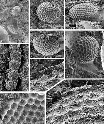
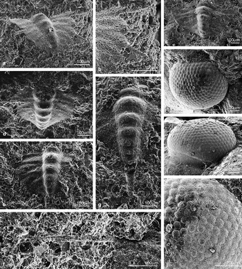
Librigena and eye. (Figs 2k, 4a–i). A small detached librigena, belonging to S. alatus, is illustrated in Figure 2k. It is 0.8 mm long, from the anterior point to the base of the genal spine, and 1.5 mm long to the tip of the spine, as preserved. Exclusive of the eye, the maximum width is about 0.3 mm. The spine is narrow and juts out slightly from the margin of the librigena. The eye, set slightly more than half way along the length of the librigena (excluding the spines), is approximately 250 μm long and is nearly circular; it is crushed down, though still in place. Some traces of the lenses can be discerned. The size would fit a relatively late meraspid. Some eyes of this species were described and figured by Clarkson (Reference Clarkson1973, p. 754, pl. 95, figs 1–2, text-fig. 7a–d). His description was based on material from Andrarum, Skåne and Gamlebyn, Oslo. At some horizons, very well preserved compound eyes of S. alatus are to be found in some abundance. Whereas in some cases the external surfaces are preserved (Fig. 4i), it is more commonly the internal moulds that are seen (Fig. 4a–e, g–h), the lenses having been retained by the external matrix and, thus, wholly or partially stripped off during preparation (Fig. 4f). These illustrations show clearly (Fig. 4a–e, g–i) the original curvature of the visual surfaces, the shapes of the lenses and variation of lens size across the eye. Figure 4b–d shows the same eye in lateral, dorsal and posterior views; this eye is c 400 µm long and 300 µm in height. The lenses average 20 µm in diameter, though there is some variation in lens size; the somewhat larger lenses being in the upper part of the eye in the specimen illustrated in Figure 4e, though this is by no means a constant feature. The contours of the upper and lower surfaces of the thin lenses are visible in the broken surfaces in Figure 4g–i, They are very similar to those illustrated by Lindström (Reference Lindström1901) and Clarkson (Reference Clarkson1979). A discussion of the optics and the eyes of S. alatus is given by Schoenemann & Clarkson (Reference Schoenemann and Clarkson2015).
Sphaerophthalmus alatus (Boeck, Reference Boeck and Keihau1838): (a) internal mould of eye attached to librigena, anterior view, LO 12308t; (b–d) internal mould of eye in lateral, posterior and dorsal views, LO 12309t; (e) well-preserved eye attached to librigena, LO 12310t; (f) string of lenses, partially detached, LO 12311t; (g) vertical broken section through lenses, showing bioconvex form, LO 12312t; (h) internal mould, showing proximal surface of lenses, LO 12313t; (i) internal mould, oblique view showing vertical section of lenses (top) and proximal surfaces of lenses, LO 12314t.

Development of the hypostome. (Figs 5h–i, 6e–f). A juvenile hypostome (Figs 5h, 6e) is 400 µm long and 300 µm wide. It has a relatively broad brim, lacking the U-shaped platform of the adult, and is widest at about three-quarters of the total length from front to rear. The adult hypostome greatly resembles the material found in Västergötland, figured by Clarkson et al. (Reference Clarkson, Ahlgren and Taylor2003, pl. 6, figs 1–5, 8) in having a very similar external contour, pointed anterior wings and a raised middle body tapering posteriorly. The middle body is situated on a U-shaped raised platform, more distinct than that found in the material from Västergötland, outside which lies the brim proper, curled posteriorly, and with a few subdued terrace lines concentric with the margin. The brim is widest at about two-thirds of the distance from front to rear of the hypostome. That figured here (Figs 5i, 6f) belongs to a small holaspis; it is 750 µm long, with a maximum width of 530 µm.
Sphaerophthalmus alatus (Boeck, Reference Boeck and Keihau1838). (a) Group 1 meraspid pygidium, LO 12315t. (b–d) Group 2 meraspid pygidia: (b) LO 12316t; (c) slightly broken and obscured posteriorly, though the posterior downward curling is clear, LO 12317t; (d) LO 12318t. (e–f) pygidium, Group 3: (e) LO 12319t; (f) enlargement of posterior spines of (e). (g) Group 4 meraspid pygidium, showing triangular form, short, upturned marginal spines, and broken spine bases on axial rings, LO 12320t. (h) Meraspid hypostome, LO 12321t. (i) Young holaspid hypostome, LO 12322t.

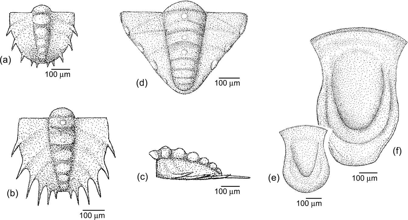
Reconstructions of pygidia and hypostomes of Sphaerophthlamus alatus (Boeck, Reference Boeck and Keihau1838): (a) Group 2 meraspid pygidium; (b–c) Group 3 meraspid pygidium, dorsal and lateral views; (d) Group 4 meraspid pygidium; (e) meraspid hypostome; (f) young holaspid hypostome.

Development of the meraspid pygidium. (Figs 5a–g, 6a–d). We have assigned the pygidia to four groups, as used above in the development of the cranidium, and though some assumptions have been made, we believe that the numbered groups of cranidia and pygidia correspond.
Group 1. (Fig. 5a). Pygidia, being very delicate, are rare in the Bornholm material, and there are no perfect specimens representing the early developmental stages. The smallest, slightly distorted pygidium is 300 μm wide, which would put it in the range of meraspid degree 0–1, by comparison with the specimens figured by Clarkson et al. (Reference Clarkson, Ahlgren and Taylor2003). It is similar in form and dimensions to these, being slightly longer than wide, and apparently flattened by compaction.
Group 2. (Figs 5b–d, 6a). Pygidia in this size range are 320 μm long and 350 μm wide; they are shield-shaped, with a narrow tapering axis, having 5–6 axial rings and faint impressions of the pleural furrows anteriorly. The posterior part remains somewhat downwardly coiled. There are five pairs of short marginal spines.
Group 3. (Figs 5e–f, 6b–c). The pygidium remains shield-shaped; it is 425 μm long and 400 μm wide (excluding spines). There are six axial rings and six pairs of marginal spines. The anterior axial rings are more differentiated and strap-shaped, with a small axial node, and the marginal spines are appreciably longer. Whether or not there is a posterior spine at this stage remains unknown; the reconstruction is made without one.
Group 4. (Figs 5g, Reference Balserio, Waisfeld and Buatois6d). The largest of the pygidia is small and triangular, being 450 μm long and 600 μm in width, and with a faint posterior downward coiling. Although the external contour is like that of the adult, the axis conserves the juvenile state, with six rings, the first being raised and strap-like, the rest decreasing in elevation posteriorly. A pronounced cicatrice on the terminal piece may well indicate the base of a broken pygidial spine (cf. Clarkson et al. Reference Clarkson, Ahlgren and Taylor2004, pl. 8, fig. 5, text-fig. 2). Four pairs of short, stubby marginal spines are present, lining the pleural fields and inclined slightly upwards, and the lateral furrows are faintly impressed.
Remarks. The morphology of the cephalon in S. alatus from Västergötland is virtually identical with that of the reconstruction of S. alatus given by Høyberget & Bruton (Reference Høyberget and Bruton2012), based on the Oslo Fjord specimens; it differs from that of Clarkson et al. (Reference Clarkson, Ahlgren and Taylor2003) only in minor details: a better developed S2 glabellar furrow, a wider axis, genal spines which extend further from the body, and a very long caudal spine. The main difference between the two morphs is in the length of the genal spines. The genal spines of S. alatus found in Västergötland extend horizontally, far under the body, whereas those of S. alatus, as illustrated by Westergård (Reference Westergård1922) and Henningsmoen (Reference Henningsmoen1957), and as found in all the Bornholm material, are relatively short. A holaspid librigena referred to S. alatus from Västergötland, however, with an unusually short spine, was figured by Clarkson et al. (Reference Clarkson, Ahlgren and Taylor2003, pl. 8, fig. 8). Although there are evident similarities between the stages of development of S. alatus from Bornholm and Västergötland, there are some differences; and in particular that the former retains intergenal and occipital spines until relatively late in meraspid ontogeny, while in the latter, such spines are absent from the beginning. Likewise, the marginal spines on the pygidium are appreciably longer in S. alatus from Bornholm during development than they are in the material from Västergötland, and the small triangular pygidium does not closely resemble that of the latter species at any stage of development.
Moreover, throughout ontogeny, each of the occipital rings of the Bornholm S. alatus material invariably bears an occipital spine, whereas only a node is present in the specimens from Västergötland. A reconstruction of S. alatus, based on the Bornholm material, is given in Figure 1.
Lee & Chatterton (Reference Lee and Chatterton2007) stated that olenid protaspides show morphological differences depending on oxygenation conditions. The protaspides of S. alatus are small and round, with spindle shaped axis and a smaller protopygidium, and are lacking anterior and mid fixigenal spine pairs. It fits very well into a description of an olenid protaspid found in a low oxygen environment.
Genus Ctenopyge Linnarsson, Reference Linnarsson1880
Type species. Olenus (Sphaerophthalmus) pecten Salter, Reference Salter1864, from the Merioneth Series of Herefordshire and Worcestershire, England, UK, designated by Vogdes (Reference Vogdes1890). Subgenus Ctenopyge (Mesoctenopyge) Henningsmoen, Reference Henningsmoen1957
Type species. Ctenopyge spectabilis Brøgger, Reference Brögger1882, from the Furongian of Norway. Ctenopyge (Mesoctenopyge) tumida Westergård, Reference Westergård1922 Figs 7–11
Reference Linnarsson1880 Ctenopyge? sp. Indet. [partim] Linnarsson, p. 156, pl. 6, fig. 15.
Reference Westergård1922 Ctenopyge tumida n. sp. [partim] Westergård, p. 155, pl. 11, figs 15–18.
Reference Poulsen1923 Ctenopyge tumida Westergård [partim] Westergård; Poulsen, p. 39, pl. 1, fig. 14.
Reference Westergård1947 Ctenopyge tumida Westergård; Westergård, p. 24.
Reference Henningsmoen1957 Ctenopyge (Mesoctenopyge) tumida Westergård Reference Westergård1922; Henningsmoen, pp 198–199, pl. 5, pl. 20, fig. 16.
Reference Taylor and Rushton1971 Ctenopyge (Mesoctenopyge) tumida Westergård; Rushton, in Taylor & Rushton, p. 32–33, fig. 8a.
Reference Schrank1973 Ctenopyge (Mesoctenopyge) tumida Westergård; Schrank, p. 825, pl. 7, figs 13–20, 22, 23.
Reference Cope and Rushton1992 Ctenopyge (Mesoctenopyge) tumida Westergård; Cope & Rushton, p. 547, fig. 5 m–o.
Reference Żylińska2001 Ctenopyge (Mesoctenopyge) tumida Westergård, 1922; Żylińska, pp 341–342, pl. 1, figs 11–12.
2004 Ctenopyge (Mesoctenopyge) tumida Westergård; Clarkson et al., pp 129ff, figs 17, 18A–D, 19, 20A–D
Type data. As the lectotype, Henningsmoen (Reference Henningsmoen1957) selected a cranidium figured by Westergård (Reference Westergård1922, pl. 11, fig. 16) from Andrarum, Skåne, Sweden.
Description. This species was fully described by Westergård (Reference Westergård1922) and Henningsmoen (Reference Henningsmoen1957). A further description was given by Clarkson et al. (Reference Clarkson, Ahlgren and Taylor2004), based upon excellent material from Västergötland. In all the specimens described so far, however, the pygidium is lacking, and those described below are the only ones so far known.
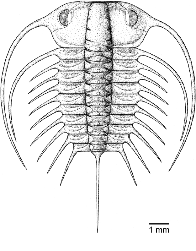
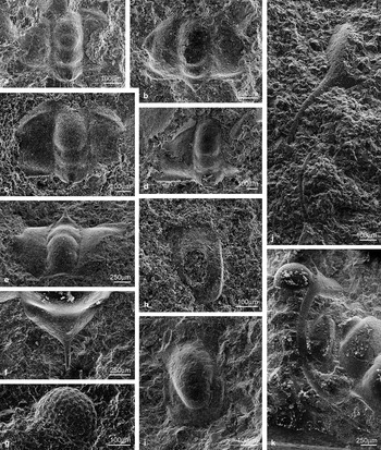
Development of the cranidium. (Figs 8a–f, 9b, d). A few meraspid stages from Västergötland were figured by Clarkson et al. (Reference Clarkson, Ahlgren and Taylor2004). The smallest is an hexagonally shaped cranidium with a single attached thoracic segment, of length 0.43 mm and width 0.5 mm. A larger specimen with five thoracic segments (interpreted as a degree 5 meraspis) has a cranidium of length 0.6 mm and width 0.8 mm. A further individual is a young holaspis with a cranidial length of 0.8 mm and a width of 1.25 mm. The cranidia figured here can be compared with those described from Västergötland. Figures 8a–b and 9b illustrate a cranidium of length 400 μm and width 700 μm. They may belong to a degree 3, or possibly a degree 4 meraspid, compared to that described by Clarkson et al. (Reference Clarkson, Ahlgren and Taylor2004). A larger sized cranidium (Figs 8c, d, 9d), interpreted as a late meraspid stage, appears to be of length 600 μm and width 800 μm, but since both are frayed at the lateral edges, the width is somewhat uncertain. These two specimens resemble those previously figured by Clarkson et al. (Reference Clarkson, Ahlgren and Taylor2004), but the distinctive, very short, deeply sunk glabellar furrows of the typical C (M.) tumida are much less developed. In the smaller specimen, furrows S1, S2 and S3 are continuous over the glabella, though slightly effaced centrally. In the larger specimen, there is a short occipital spine, a short preglabellar field, S0 is continuous, S1 more deeply depressed laterally, and S2 continuous but faint. A late meraspid/early holaspid is shown in antero-lateral view in Reference Boeck and KeihauFigure 8e, showing a short, thin, upturned occipital spine, and tiny tubercles have developed on the occipital ring. Holaspid specimens (Fig. 8f) from Bornholm usually retain the distinct occipital spine, but this seems to be a variable feature. The material from Västergötland, upon which the reconstruction of Clarkson et al. (Reference Clarkson, Ahlgren and Taylor2004, fig. 19; Fig. 7), was based, lacks an occipital spine, though it does have a node.
Ctenopyge (Mesoctenopyge) tumida Westergård, Reference Westergård1922. Reconstruction in dorsal view (from Clarkson et al. Reference Clarkson, Ahlgren and Taylor2004).

Ctenopyge (Mesoctenopyge) tumida Westergård, Reference Westergård1922: (a) cranidium degree 3–4, LO 12323t; (b) cranidium degree 3–4, LO 12324t; (c) late meraspid, cranidium LO 12325t; (d) partial cranidium of a late meraspid, LO 12326t; (e) cranidium of late meraspid or early holaspid, oblique anterior view, LO 12327t; (f) occipital ring and spine of an early holaspid showing tiny tubercles, LO 12328t; (g) juvenile eye, LO 12329t; (h) meraspid hypostome, LO 12330t; (i) young holaspid hypostome, LO 12331t; (j) two librigena, showing distinct curving spines, eyes broken off, LO 12332t; (k) curving genal spine of a late meraspid/early holaspid, LO 12333t.

Librigena and eye. (Figs 8g, j, k, 9a, c, e, 10h–j). Two small librigenae (Figs 8j, 9a, c) show long curving spines which, clearly, were already well developed during the meraspid stage. The eyes are broken off, and the reconstruction presented is tentative. These obviously belong to meraspid stages, but which stage remains uncertain. Figure 9e shows a librigena in lateral view. Here, the lenses, though imperfectly preserved, are large and separate (Fig. 8g), as is common in juvenile holochroal eyes (e.g., in Olenus; Clarkson & Taylor Reference Clarkson and Taylor1995b). In the adults, the eyes, as figured previously (Clarkson et al. Reference Clarkson, Ahlgren and Taylor2004) and also in the present paper, show many lenses of approximately equal dimensions, set on a panoramic visual surface. A feature not previously recorded is a wide, though featureless socle, upon which the visual surface is set (Fig. 10i). Enlarged lenses are shown in Figure 10j. Schoenemann & Clarkson (Reference Schoenemann and Clarkson2015) described the structure and optics of the visual system of C. (M.) tumida.
Reconstructions of librigenae, cranidia and hypostomes of Ctenopyge (Mesoctenopyge) tumida Westergård, Reference Westergård1922: (a, c) librigenae, probably representing similar developmental stages to the cranidia figured here; dimensions of eyes uncertain; (b) meraspid cranidium degree 3 or 4; (d) later meraspid cranidium; (e) librigena in lateral view; (f) juvenile hypostome; (g) adult hypostome.

Development of the hypostome. (Figs 8h–i, 9f–g). A juvenile hypostome (Figs 8h, 9f), 0.55 mm long and 0.32 mm wide, has a slightly curved anterior margin, small anterior wings, and is virtually parallel-sided laterally. The middle body is also almost parallel-sided. A small holaspid hypostome (Figs 8g, 9i) is 2.0 mm long and 1.2 mm wide. The anterior margin is curved and short anterior wings are developed. The middle body is large, inflated and tapering posteriorly to a blunt termination. There is no U-shaped band surrounding the middle body, as in S. alatus. Posteriorly from the anterior wings, the hypostome narrows to a point opposite the widest part of the middle body, thereafter becoming subparallel-sided, expanding slightly rearwards so that it is widest close to the almost transverse posterior margin.
Development of the meraspid pygidium. (Figs 10a–g, 11a–c). The pygidia are assigned to two stages.
Ctenopyge (Mesoctenopyge) tumida Westergård, Reference Westergård1922: (a–b) pygidium, early meraspid stage, oblique posterior view, LO12334t: (b) enlargement of part of (a) showing marginal spines on left hand side; (c) pygidium of middle meraspid stage, LO 12335t; (d) oblique anterior view of a pygidium of a middle meraspid stage showing posterior spine, LO 12336t; (e–f) pygidium of an early meraspid stage, LO 12337t: (f) enlargement of the upper left side of (e), showing a single marginal spine; (g) axis and posterior spine of a well-preserved pygidium, middle meraspid stage, a breakage scar is evident where the spine has been crushed down, LO 12338t. (h–j) eye of a small adult, LO11944t; (i) showing the socle at the base; (j) enlargement of the visual surface and lenses.

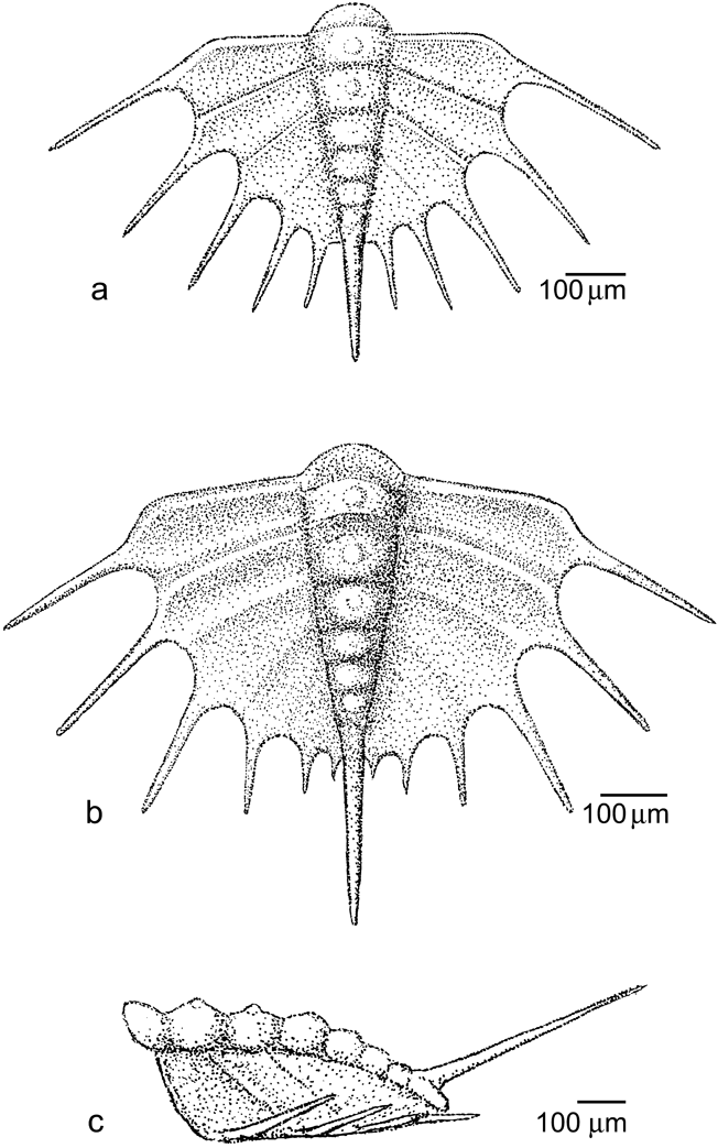
Reconstructions of pygidia of Ctenopyge (Mesoctenopyge) tumida Westergård, Reference Westergård1922, pygidia: (a) pygidium of early meraspid stage; (b–c) pygidium of middle meraspid stage, showing the upward inclination of the spines, dorsal and lateral views.

Early meraspid stage. A few incomplete pygidia, frayed at the edges, but otherwise retaining much structural detail, provide the only available information on the tails of C. (M). tumida. Some other fragments provide additional information. As previous authors have noted, several Ctenopyge species are never found with associated pygidia. Figures 10(a–b, e) and 11 show pygidia retaining long, thin, marginal spines, of which the full length of one of the anterior spines is preserved (Fig. 11g) and others are present, with broken tips. As reconstructed (Fig. 11a), these small pygidia are 320 μm long and 600 μm wide exclusive of spines, and 500 μm long and 1.0 mm wide with spines included. The outline, without the spines, is almost semi-circular, slightly pointed posteriorly. The axis, about 100 µm wide anteriorly and with 5–6 rings, tapers posteriorly, terminating in a strong axial spine. The anterior segments bear an axial node. The anterior pleural field has distinct, rather flattened pleurae, becoming less marked posteriorly and separated by ridges, each with a broad furrow behind, which fade out towards the rear. Each of the pleurae terminates in a long, thin, straight spine, projecting upwards and rearwards at about 20 degrees; each is about as long as the pleura from which it originates. There are five of these marginal spines, decreasing in size posteriorly. Pygidia of this size probably belong to a degree 3 meraspis.
Middle meraspid stage. A few slightly larger pygidia (Figs 10c–d, g, 11b–c) are 400 μm long and 700 μm wide without spines, and 750 μm long and 1.1 mm wide including spines. A transitory pygidium of this size is of similar form to the smaller pygidium and, likewise, has six axial rings, although the marginal spines diminish in size posteriorly. The stout axial spine is seen lying horizontally in Figure 10g, but it has a distinct fracture at the base. It is likely that this spine was originally inclined rearwards and upwards, at about 25 degrees.
3. Concluding remarks
This study has been based on abundant, though generally rather damaged and disarticulated, material from Bornholm, Denmark. It is complementary to previous works on ontogeny of olenid trilobites, and it reveals that the early stages in ontogeny of the Bornholm Sphaerophthalmus alatus are much more spinose than those of S. alatus found in Västergötland, of which the adult forms are very similar. Whether this is taxonomically significant, or is a consequence of local variation, remains an open question; here, we regard the two morphs as variants of the same species. Early ontogeny of C. (M.) tumida, already known in part, is further illuminated by the discovery of very spiny transitory pygidia, the only ones known for this species.
4. Acknowledgements
ENKC would like to thank the SYNTHESYS Foundation for funding a visit to Copenhagen in 2006, and Arne Nielsen for the supply of material, and inviting him to prepare and study it. Assistance with the SEM was given in Copenhagen by Maria Liljeroth, in Lund by Rita Wallén and in Edinburgh by Nicola Cayzer. Brigitte Schoenemann (Cologne) is thanked for helpful discussion; as is Anna Żylińska (Warsaw) for very helpful comments on an earlier draft of this paper. We thank the reviewers Per Ahlberg (Lund) and Alan Owen (Glasgow) for their valuable suggestions.