Introduction
Ecchordosis physaliphora is a congenital, benign lesion originating from notochordal remnants along the craniospinal axis, most frequently located at the level of the clivus and sacrum.Reference Dias, Nakanishi, Mangussi-Gomes, Canuto, Takano and Oliveira1,Reference Lagman, Varshneya, Sarmiento, Turtz and Chitale2 Ecchordosis physaliphora is a rare lesion and can be difficult to recognise and treat.Reference Dias, Nakanishi, Mangussi-Gomes, Canuto, Takano and Oliveira1,Reference Alli, Clark and Mansell3–Reference Galloway and Hayhurst5 We present three cases of previously undiagnosed ecchordosis physaliphora presenting with cerebrospinal fluid (CSF) rhinorrhoea and meningitis. Endoscopic transclival or transsphenoid surgery including three-layer (fat, fascia and nasoseptal flap) reconstruction was used in all cases with complete resolution of the symptoms.
Case reports
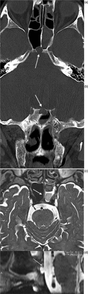
An 81-year-old previously well male presented to the infectious disease department with acute bacterial meningitis. Initial treatment with intravenous antibiotics resulted in improvement, and the patient was discharged a week later, only to be re-admitted 5 days later with recurrent bacterial meningitis. During the second admission, watery rhinorrhoea was noted, and a computed tomography (CT) scan of the sinuses was performed demonstrating opacification of the sphenoid sinus and a well circumscribed bony defect of the midline of the clivus (Figure 1a and b). The patient was subsequently referred to us for otolaryngological evaluation with the diagnosis of sinusitis. Interestingly, the patient had a magnetic resonance imaging (MRI) scan during his first admission, but the typical findings suggesting ecchordosis physaliphora (Figure 1c and d) had been overlooked.
Case 1: showing (a) pre-operative axial and (b) coronal computed tomography scan images showing bone erosion of the posterior wall of the sphenoid sinus and clivus (white arrows); (c) pre-operative axial and (d) sagittal T2-weighted magnetic resonance imaging demonstrating a homogeneous and hyperintense cystic lesion in the posterior wall of the sphenoid sinus that derives from the prepontine cistern.

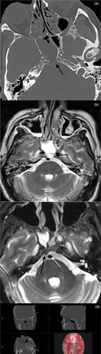
The second patient, a 60-year-old male, was admitted in another hospital with a 15-day history of watery rhinorrhoea, misdiagnosed as a common cold, complicated with meningitis that was treated with intravenous antibiotics. A CT scan of the sinuses was performed and demonstrated a bony defect of the posterior wall of the sphenoid and he was referred to us for further management. The CT and MRI scans demonstrated a well-defined midline cystic mass of approximately 1 cm in diameter, originating in the prepontine cistern, eroding the middle third of the clivus and extending into the posterior sphenoid sinus. The mass had a stalk-like connection to the clivus, was T1-hypointense and T2-hyperintense and did not show enhancement after contrast administration (Figure 2).
Case 2: (a) pre-operative axial computed tomography scan images showing bone erosion (black arrow) of the posterior wall of the sphenoid sinus and clivus between the paraclival carotid arteries (white asterisks); (b and c) pre-operative axial and sagittal T2-weighted magnetic resonance imaging demonstrates a lesion (black arrow) in the posterior wall of the sphenoid sinus which derives from the prepontine cistern, and the lesion appears to be in contact with the basilar artery (white arrow); and (d) intra-operative neuronavigation image demonstrating the navigated suction tip pointing out the lesion (white arrow) between the paraclival carotid arteries and inferior to the sella turcica.

The third patient was a 64-year-old female patient who was referred after an episode of meningitis. The patient had a history of intermittent watery rhinorrhoea since 2013. She was initially diagnosed with allergic rhinitis for which she had been following a local steroid treatment. During hospitalisation, a brain MRI scan and a CT scan were performed showing a T1-hypointense and T2-hyperintense lesion occupying the sphenoid sinus and causing a bone erosion of the clivus and posterior wall of the sphenoid. A nasal fluid sample was also examined and shown to be CSF.
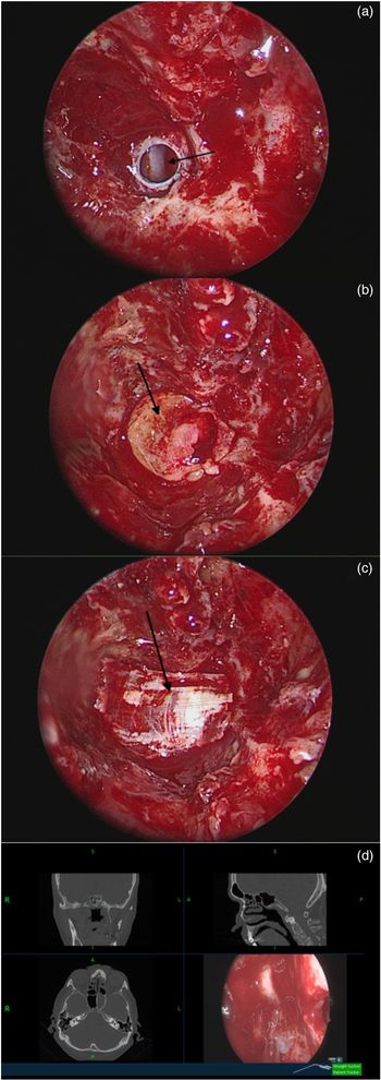
The management and intra-operative findings of all three patients were very similar. All three patients underwent an endoscopic transsphenoid or transclival approach. A nasoseptal (Hadad–Bassagaisteguy) flap was initially harvested and stored in the nasopharynx. Subsequently, the anterior wall, bony septa and the rostrum of the sphenoid were completely drilled out, providing a wide view of the posterior sphenoid wall and clivus. A cystic, whitish, pulsating mass was identified in the upper clivus, protruding into the posterior sphenoid sinus. The mass was well circumscribed, with no adhesions to the bony walls of the clivus and could be dissected from the underlying bony defect and excised, leaving a completely round defect of the dura, just anteriorly to the basilar artery (Figure 3). The surrounding bone of the posterior sphenoid was cleared, and the surrounding mucosa of the posterior sphenoid wall was cauterised and removed. The reconstruction of the defect was performed in a multilayer fashion: initially fat harvested from the thigh was used to fill the bony defect of the clivus, while fascia lata was used as an overlay graft (between the dura and the bone) and finally a nasoseptal (Hadad–Bassagaisteguy) flap was used to cover the defect. Lumbar drainage was not used for any of the three patients.
Case 1: intraoperative images showing (a) endoscopic view by 00 endoscope with round shaped bony and dural defect with exposure of the basilar artery; (b) grafting of fat and (c) fascia lata; and (d) neuronavigation image demonstrating the defect.

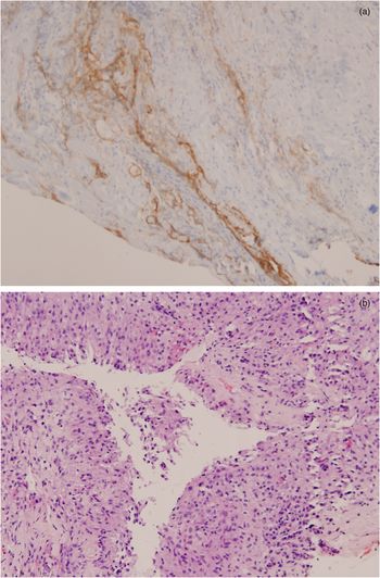
Histological examination of the mass demonstrated its notochordal origin. Immunohistochemistry showed positivity for epithelial membrane antigen and S-100 protein, and focal positivity for cytokeratin AE1 and AE3 and cytokeratin-8. Cluster of differentiation 163 was positive, suggesting multiple histiocytic cells. Vimentin, epithelial membrane antigen and progesterone receptor positivity suggested meningo-epithelial type cells. Cytokeratin AE1 and AE3, cytokeratin-8, cytokeratin 34bE12 and epithelial membrane antigen positivity suggested a covering, respiratory-type epithelium focally (Figure 4). These results, coupled with the radiological findings, confirmed the diagnosis of ecchordosis physaliphora.
Case 1 showing: (a) high power haematoxylin and eosin stain of the excised lesion (note the characteristic, physaliphorous (bubble-like) cells with intracytoplasmic vacuoles); and (b) a high-power image of the excised lesion (note epithelial membrane antigen positivity on immunohistochemistry suggesting meningo-epithelial type cells).

The post-operative course was uneventful, and the patients recovered fully. Mean hospitalisation time was 3 days. Since their surgery, the patients have been free of symptoms for 28, 20 and 7 months, respectively.
Discussion
Herbert Luschka was the first to describe a lesion matching the characteristics of ecchordosis physaliphora at the level of the clivus in 1856. One year later, Rudolf Virchow named a similar lesion ‘ecchordosis physaliphora’, after mistakenly considering that it originates from cartilage tissue. Although Johannes Peter Müller suggested the notochordal origin of the lesion in 1858, it was 40 years until Hugo Ribbert provided experimental evidence to prove it.Reference Choudhri, Feroze, Hwang, Vogel, Ajlan and Harsh6
Previous authors have commented on the differential diagnosis between ecchordosis physaliphora and clival chordoma. Chordomas are malignant, locally invasive tumours, which share the same notochordal origin with ecchordosis physaliphora and represent its main differential diagnosis. Due to the completely different prognosis and management between the two tumours, it is very important to distinguish them. Some imaging features are very helpful in the differential diagnosis between the two lesions. Chordomas are hyperintense in both T1- and T2-weighted MRI scans, whereas ecchordosis physaliphora is hypointense in T1-weighted and hyperintense in T2-weighted MRI, and, most crucially, unlike chordomas, which are highly vascular tumours, ecchordosis physaliphora shows no contrast enhancement after the administration of gadolinium. Furthermore, some previous studies mention the presence of an osseous pedicle connecting the ecchordosis physaliphora retroclival mass to notochordal remnants as a characteristic periodical finding in CT images. In terms of location, ecchordosis physaliphora is predominantly found along the midline craniospinal axis and is mostly found intradurally (Table 1).Reference Park, Lee, Ahn, Suh and Hong7
Comparison of ecchordosis physaliphora and chordoma

MRI = magnetic resonance imaging
Due to limited studies, there are no official diagnostic criteria and classification for ecchordosis physaliphora. In 2016, Lagman et al. conducted a review of the literature and proposed criteria for the diagnosis of ecchordosis physaliphora based on clinical, histopathological and immunohistochemical features.Reference Lagman, Varshneya, Sarmiento, Turtz and Chitale2
In our series however, the main issue was not the differential diagnosis of the lesions but recognition of the defects and the associated lesions in imaging and therefore recognising these as the underlying reason for the patients’ meningitis. This was the case in all three patients because the lesions were rather small (less than 1 cm in maximal diameter) and could be easily missed in a cursory examination of the MRI and CT scans. All patients had watery rhinorrhoea and meningitis; however, the presence of the typical midline defects of the clivus and the associated T2-hyperintense lesions with a stalk on the MRI was missed. This resulted in delayed diagnosis in all cases, with potentially dire consequences.
This is the first series study of patients with ecchordosis physaliphora presenting with CSF rhinorrhoea and associated meningitis. What is interesting is that all three patients had a short history of watery rhinorrhoea concomitant with meningitis, and in two of the patients, the rhinorrhoea preceded the onset of meningitis. In the first case, the rhinorrhoea was evident in between the two episodes of meningitis. Most notably, in all cases, the diagnosis of clival defect and CSF leak was reached during the assessment of meningitis. It seems that unilateral watery rhinorrhoea suggestive of a CSF leak is, unlike meningitis, often underestimated particularly from non-ENT physicians. Radiologically, in all patients, the size and location of the defect was evident in both CT and MRI scans and suggested the diagnosis, which was confirmed following histological examination. As there was no previous imaging of the patients, it was unclear how long the ecchordosis physaliphora had been present. However, the watery rhinorrhoea was only present for a few weeks in the first two patients, raising the possibility that a rupture of the ecchordosis physaliphora wall may have triggered the CSF rhinorrhoea, which rapidly resulted in meningitis in the context of a co-existing upper respiratory tract infection. In the third case, however, the patient described watery rhinorrhoea for a few years, before the episode of meningitis resulted in the recognition of the CSF leak and defect.
Although it appears that ecchordosis physaliphora may be an asymptomatic condition, in all of our cases it presented with a life-threatening complication that was challenging to recognise and treat. It is important to keep this in mind in cases of unexplained CSF leak or meningitis because a small lesion may evade characterisation in CT and MRI scans. Surgically, it is advisable to completely remove the lesion before reconstruction, as we feel that any attempt to ‘drape’ a reconstruction over the ecchordosis physaliphora and the defect may fail. Given the benign behaviour of ecchordosis physaliphora, complete resection should always be balanced with surgical risks and expected outcomes. It is particularly critical in these cases to close the skull base defect.
• Ecchordosis physaliphora is a notochordal lesion that can be associated with clivus defects
• Clivus defects can be associated with cerebrospinal fluid rhinorrhoea and meningitis if they are not recognised and treated
• This study presents three cases where ecchordosis physaliphora was the cause of cerebrospinal fluid rhinorrhoea and meningitis
• All cases were managed successfully with endoscopic transsphenoidal three-layer reconstruction using fat, fascia lata and a nasoseptal flap
In all of our cases, the CSF leak was low flow although leaks from the prepontine cisterns can sometimes be high pressure leaks.Reference Bolzoni-Villaret, Stefini, Fontanella, Bottazzoli, Turri Zanoni and Pistochini8 We performed a three-layer reconstruction with fat and fascia, and we applied a posterior nasoseptal flap as an overlay to further stabilise the reconstruction, reduce the risk of failure, and speed healing and epithelialisation. We generally do not use lumbar drainage as we believe that it does not improve the success rate of reconstruction while being associated with potentially serious complications such as tension pneumocephalus.
Conclusion
This case series study of CSF rhinorrhoea and meningitis associated with ecchordosis physaliphora demonstrates that ecchordosis physaliphora, although histologically benign, is not always a clinically benign condition and should be considered as a possible cause in patients who present with meningitis. These patients should always undergo a thorough endoscopic examination and, most importantly, a thorough neuroradiological evaluation, including an assessment for midline clival defects. The MRI scan features mentioned above (a T2-hyperintense, pedicled lesion originating from the dura with no contrast enhancement) should raise the index of suspicion for a possible ecchordosis physaliphora. In such cases, endoscopic transsphenoidal transclival resection of the lesion with multi-layer reconstruction of the skull base defect should be considered as the treatment of choice.
Competing interests
None declared