I. INTRODUCTION
Large numbers of patients suffer from cardiovascular disease, which remains the leading cause of death in developed countries, despite advances made in balloon angioplasty and conventional surgical interventions.Reference Seifu, Purnama, Mequanint and Mantovani1,Reference Ma, Hu and Ma2 Although large diameter (>6 mm) synthetic grafts such as woven poly(ethylene terephthalate) (Dacron) and extended polytetrafluoroethylene (ePTFE) have been widely used to bypass arteries in high flow regions (e.g., the thoracic and abdominal aorta),Reference Kannan, Salacinski, Butler, Hamilton and Seifalian3 these grafts tend to fail when they are applied to small-diameter (<5 mm) vessels due to thrombosis and intimal hyperplasia.Reference Wang, Lin, Yao and Chen4–Reference Sayers, Raptis, Berce and Miller6 For these reasons, the use of autografts such as the saphenous and umbilical veins and mammary arteries is considered the clinical gold standard for small artery bypass. However, for patients who have had previous bypass surgeries or vascular disease, these veins and arteries are often unsuitable for harvest. In addition, generating a second surgical site can lead to greater risk of infection, swelling, and discomfort. Therefore, there is an urgent need for the development of a functional alternative to autologous grafting.Reference Bujan, Garcia-Honduvilla and Bellon7,Reference Rashid, Fuller, Hamilton and Seifalian8 Tissue engineering technology has emerged as a promising approach to overcoming the shortcomings of current therapies.Reference Nerem and Seliktar9 Vascular tissue engineering attempts to fabricate functional small-diameter vascular grafts by combining cells with natural or synthetic scaffolds under suitable culture conditions to generate a tubular scaffold that can be used in vivo. There are many requirements for a suitable vascular scaffold: biocompatibility, appropriate mechanical properties, and availability in a variety of sizes for grafting applications.Reference Edelman10–Reference Niklason, Gao, Abbott, Hirschi, Houser, Marini and Langer12
Many efforts in vascular tissue engineering have been dedicated toward the optimization of the structural and mechanical properties of scaffolds.Reference Hu, Sun, Ma, Xie, Chen and Ma13–Reference Soletti, Hong, Guan, Stankus, El-Kurdi, Wagner and Vorp17 Several reviews have already been published that discussed different methods for preparing tissue engineering vascular grafts.Reference McClure, Wolfe, Rodriguez and Bowlin18–Reference Naito, Shinoka, Duncan, Hibino, Solomon, Cleary, Rathore, Fein, Church and Breuer20 The main approaches for fabrication of scaffolds include cell self-assembly scaffolds, hydrogels, thermally induced phase separation, and electrospinning. The latter has gained special attention not only because it is an easy processing technique for fabricating vascular scaffolds but also because it provides a fibrous biomimetic environment with fiber diameters in the nanometer to micrometer range.Reference Lee, Liu, Oh, Soker, Atala and Yoo21–Reference Han, Jia, Dai, Yang, Zhao, Zhao, Fan and Yuan25 Numerous review articles are available to illuminate the fundamentals, applications, and historical development of electrospinning.
Poly(ε-caprolactone) (PCL), a biodegradable and biocompatible polyester, has been widely used in various tissue engineering applications. However, it exhibits low hydrophilicity, a slow degradation rate, and few cell recognition sites during cell culture. These attributes greatly limit its application in the biomedical field.Reference Prabhakaran, Venugopal, Chyan, Hai, Chan, Lim and Ramakrishna26 Various kinds of physicochemical and postprocessing surface modification techniques have been used to overcome these shortcomings.Reference Prabhakaran, Venugopal, Chan and Ramakrishna27,Reference Zhu, Gao, Liu and Shen28 Polyethyleneimine (PEI), an organic, cationic polymer with a high density of amino groups, has been widely used as a gene delivery agent due to its high charge density from the protonated amines.Reference Ogris, Brunner, Schuller, Kircheis and Wagner29,Reference Zhou, Laroche, Lamers, Torraca, Voskamp, Lu, Chu, Spaink, Abrahams and Liu30 When the pH value of the medium is lower than 10, the primary amines of the PEI molecular chain can preferentially absorb protons, yielding positive charges on the PEI chains.Reference Andersson and Hatti-Kaul31 PEI is used in the cell culture of weakly anchored cells to increase attachment. The negatively charged outer surfaces of the cells are attracted to dishes coated in PEI, facilitating stronger attachments between the cells and the plate.Reference Vancha, Govindaraju, Parsa, Jasti, Gonzalez-Garcia and Ballestero32 It has been reported that scaffolds with an excessive amount of positive charges are highly toxic in vivo, while lower amounts of cationic charges are favorable. Because PEI was concluded to effectively enhance cell adhesion,Reference Boussif, Lezoualch, Zanta, Mergny, Scherman, Demeneix and Behr33,Reference Forrest, Meister, Koerber and Pack34 one of the goals of this study was to investigate the suitable content of PEI for tissue engineered vascular grafts by electrospinning. Kim et al. reported that PEI as a cationic polymer can enhance the electrospinnability of PCL, and that 3T3 fibroblast cells as cell models were seeded on the fabricated PCL/PEI scaffolds, which were more favorable for 3T3 cell growth than the neat PCL scaffolds.Reference Kim, Choung, Kim, Lim, Son, Choung, Cho and Chung35
In this study, to improve the hydrophilicity and electrospinnability of PCL, the water-soluble cationic polymer PEI was used to blend with PCL at different mass ratios and the resulting blends were used to fabricate tubular scaffolds for vascular tissue engineering applications via electrospinning. The biomechanical properties and biocompatibility of the fabricated scaffolds were evaluated. The results demonstrate the potential application of PEI in vascular tissue engineering applications.
II. MATERIALS AND METHODS
A. Materials
Poly(ε-caprolactone) (PCL, Capa® 6500, M n = 50,000) was purchased from Perstorp (London, UK). Polyethylenimine (PEI, M w = 25,000), chloroform (ACS regent), N,N-dimethyl formamide (DMF) (ACS regent).
B. Scaffold preparation
Vascular scaffolds were fabricated by electrospinning polymer blends of PCL and PEI at different weight ratios (PCL:PEI = 100:0, 99:1, 98:2, and 95:5 by weight ratio) in a mixture of chloroform and DMF (chloroform:DMF = 6:4). The concentration of the prepared solutions was 10% (wt/v). The electrospinning set-up included a syringe pump, a high voltage supply, and a homemade rotating mandrel at spun at 1000 rpm to collect the fibers. The solution was loaded into a 6 mL syringe connected to an 18 gauge blunt needle by Teflon tubing. The desired flow rate was 0.5 mL/h and the applied voltage was kept at 18 kV. The collecting mandrel was an aluminum tube (outer diameter of 3.18 mm). The distance between the syringe tip and the mandrel was 17 cm. In addition, round stainless washers (inner diameter of 8.33 mm × outer diameter of 15.8 mm, McMaster-Carr. Co) were used to collect the fibers for cytocompatibility evaluation.
After electrospinning, the prepared scaffolds were put in a vacuum oven at 45 °C for 3 days to evaporate any residual solvent before characterization.
C. Characterization of the scaffolds
1. Morphological characterization
The morphologies of the scaffolds were observed by scanning electron microscopy (SEM) (LEO GEMINI 1530, Germany) with an accelerating voltage of 3 kV. Prior to SEM imaging, the specimens were sputtered with gold. The average diameter of the fibers was measured from the SEM images using ImageJ software. 100 random filaments of each sample from the images were analyzed. The porosity (ε) was calculated according to the following equation,Reference Vaz, van Tuijl, Bouten and Baaijens36
 $$\varepsilon \left( \% \right) = \left( {1 - {\rho \over {{\rho _0}}}} \right) \times 100\quad ,$$
where ρ is the average density and ρ0 is the standard density.
To investigate the long-term stability of PEI of the scaffolds, the prepared scaffolds were immersed in phosphate buffered saline (PBS, pH = 7.4) for 7 days. Then, the scaffolds were washed with deionized water three times. The same SEM was used to study the morphological changes of the scaffolds after PBS treatment.
2. X-ray photoelectron spectroscopy (XPS)
The surface properties of the prepared scaffolds were investigated by XPS. The XPS N 1s core-level signal was used as a marker for the analysis of PEI on the surface. XPS measurements were performed on an x-ray photoelectron spectrometer with a focused, monochromatic Kα x-ray source and a monoatomic/cluster ion gun (Thermo Scientific). A survey scan spectrum was taken and surface elemental stoichiometries were determined from peak-area ratios.
3. Determination of water contact angle (WCA)
Surface WCAs of various scaffolds were tested using the sessile drop method in air at room temperature by a video contact angle instrument (Data Physics OCA 15, Germany). The droplet size was set at 4 μL. Three samples for each scaffold were tested and the average value was reported with standard deviation (±SD).
D. Biomechanical evaluations
1. Axial and circumferential tensile testing
Mechanical loading tests were performed on the electrospun vascular scaffolds in the axial and circumferential directions using a uniaxial load test machine (Model 5967, Instron Corporation). For the axial direction test, the tubes were 50 mm in length, with a 3.18 mm inner diameter and a 0.5 mm thickness. After the specimens were immersed in PBS for 12 h at room temperature, the tests were performed using the Instron tensile tester equipped with a maximum 250 N load cell at a crosshead speed of 5 mm/min. The ultimate tensile strength and elongation-at-break were obtained from the stress–strain curves. For testing in the circumferential direction, a ring of material was cut from the scaffold with a length (L) of 6 mm. Two custom-made L clamps were used according to the reported method.Reference Ghezzi, Marelli, Muja and Nazhat37 The circumferential test was also performed at 5 mm/min. Due to the difficulty of measuring the real cross-sectional area, the results were reported as load–strain curves and not as the usual stress–strain curves. Five specimens of each group were tested.
2. Suture retention strength
Suture retention tests were conducted in accordance with standard ISO 7198. Samples with 0.6 mm thicknesses were cut into 20 mm long tubes. After being immersed in PBS for 2 h, one end of the tube was fixed to the stage clamp of the Instron tensile tester with a maximum 250 N load cell; the opposite end of the tube was connected to another clamp by a commercial suture (5-0 prolene suture, Ethicon Inc.). The suture was placed 2 mm from the edge of the tube using a tapered noncutting needle. The tube was pulled at a crosshead speed of 5 mm/min until the sample fractured. Suture retention strength, analogous to the fracture strength in this setup, was obtained. Five specimens of each material type were tested.
3. Burst pressure strength
The burst pressure strength for the electrospun scaffolds was measured by increasing the pressure within the tubular vascular scaffold until failure occurred. A syringe pump was connected to one end of the tube to pump water at a speed of 50 mL/h, and a pressure gauge was connected to the tube by a trifurcate tube to record the inflation pressure. The other end of the tube was sealed at the circumference to prevent leaking. The pressure was gradually increased until failure occurred and the pressure change was recorded.Reference Lee, Liu, Oh, Soker, Atala and Yoo21
E. Biocompatibility evaluations
1. Cell seeding
Human umbilical vein endothelial cells (HUVECs) (Lonza) were maintained prior to testing on T75 tissue culture-treated polystyrene (TCPS) flasks (BD Falcon). Cells were fed every other day with an endothelial cell growth medium EGM-2-MV bullet kit (Lonza). Cells were passaged at 1:6 every 4–6 days with TrypLE™ Express (Life Technologies). Maintained cultures were regularly checked for mycoplasma. Before cell seeding, the obtained nanofibers were sterilized using UV light for 30 min on each side of the membrane. The sterilized samples were then positioned in 24-well TCPS plates. The cell suspension was seeded onto the surface of the sterilized scaffolds at a density of 2.5 × 10⁴ cells/well. The medium was replaced every other day.
2. Cell attachment and cell viability
To quantify the degree of cell attachment onto the scaffolds, the cells were stained with green fluorescent calcein-AM, blue fluorescent 4′,6-diamidino-2-phenylindole (DAPI), and red fluorescent ethidium homodimer-1 (EthD-1) 2 h after seeding. A Nikon A1RsiTi-E confocal microscope was used to image the cells and the number of attached cells was counted.
The viability of HUVECs cultured on the scaffolds was evaluated using a Live/Dead Viability/Cytotoxicity Kit (Invitrogen) and MTS assay. The live/dead assay was carried out following the manufacturer's instructions, and the cells were observed using a laser confocal microscope (A1RsiTi-E, Nikon). The MTS assay was performed by following these steps. The Cell Titer 96 Aqueous One Solution Cell Proliferation Assay (Promega) was used to determine the number of cells during culture on the novel materials. Upon testing, cells were treated with 83% media, 17% MTS solution and allowed to incubate for exactly one hour. After incubation, 100 µL of spent media were removed and added to a clear 96-well plate. The absorbance of this plate at 450 nm was then read with a GloMax-Multi+ Multiplate Reader (Promega) and the subsequent number of cells was determined. Cell viability was determined 3 days and 10 days after seeding.
3. Cell–scaffold interactions
The cell morphology and the interaction between cells and scaffolds were studied by SEM at day 3 and 10 time points. The stained samples were tested by SEM to investigate the cell morphology. Samples were rinsed twice with Hanks' balanced salt solution (Thermo Scientific). Paraformaldehyde was diluted with Hy Clone Hy Pure molecular biology grade water (Thermo Scientific) to make a 4% solution. The rinsed samples were then immersed in the solution for 30 min. After that, the samples were dehydrated using a series of ethanol washes (50, 80, 90, and 100 ethanol for 30 min each), and finally the dehydrated samples were dried in a vacuum desiccator for 2 h before gold sputtering for SEM.
4. Cytoskeleton study
HUVEC shape and cytoskeleton organization were determined by phalloidin-tetramethylrhodamine B isothiocyanate (phalloidin–TMRho, Sigma) staining after culturing for 48 h on the scaffolds. For these assays, cells were first fixed in 4% paraformaldehyde (PFA) for 15 min at room temperature. Cells were then washed with PBS and permeabilized with 0.1% Triton-X in PBS for 5 min at room temperature. The cells were washed once again and treated with 0.3 μM phalloidin–TMRho with DAPI for 1 h at room temperature. Samples were then washed with PBS and immediately imaged using a Nikon A1RsiTi-E confocal microscope.
F. Statistical analysis
All of the quantitative results were expressed as mean ± SD. Statistical analyses were carried out by means of a one-way analysis of variance. A p-value less than 0.05 was considered statistically significant.
III. RESULTS AND DISCUSSION
A. Morphological characterization of nanofibrous scaffolds
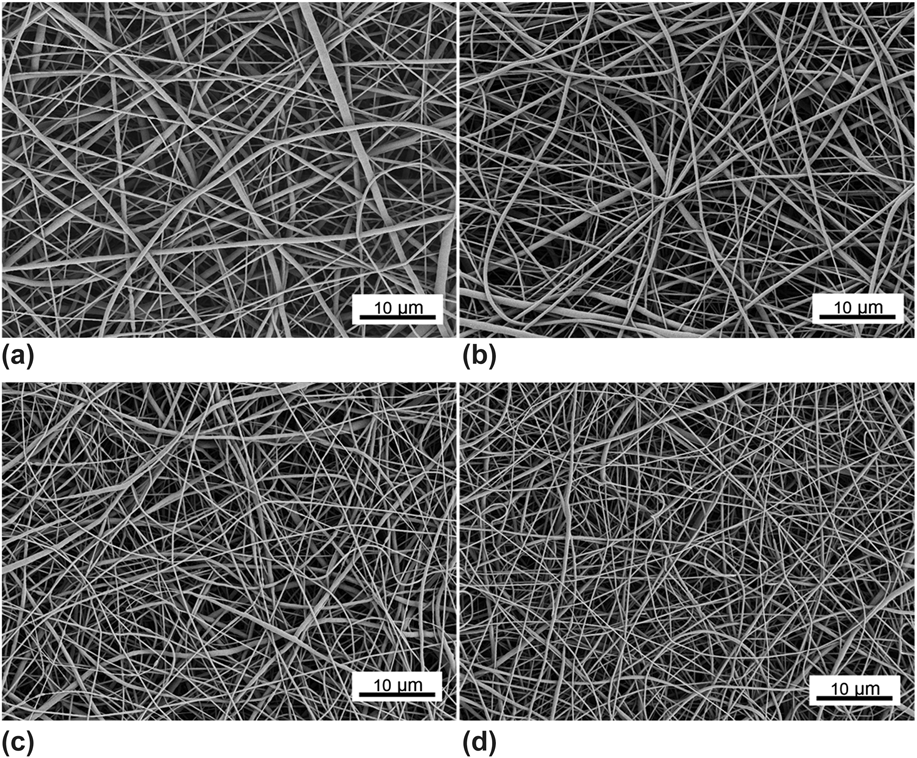
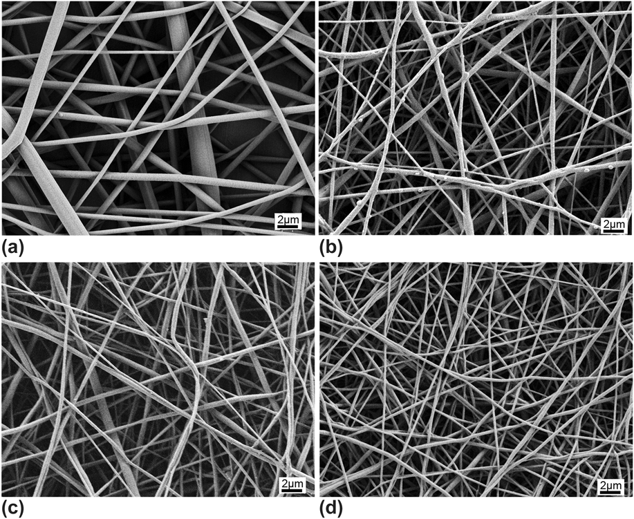
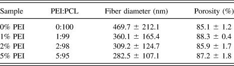
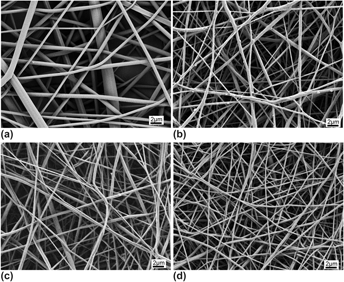
Figure 1 shows the structure of the electrospun PCL/PEI nanofibers at different PEI concentrations. It can be seen that the average diameter of the electrospun nanofibers decreased with increase of PEI in the solution. Accordingly, when the PEI content reached 5%, the fiber diameter decreased from 469.7 ± 212.1 nm for neat PCL fibers (which were named as 0% PEI in the experiments) to 282.5 ± 107.1 nm for 5% PEI. This may be because the cationic characteristic of PEI enhanced the conductivity of the solution. Figure 2 shows the morphological changes of the PCL/PEI scaffolds after immersion in PBS for 7 days. It was found that there was no obvious breakage of fibers in the obtained SEM images. This was because the concentration of PEI was low and the PEI phases were mixed with PCL fibers, which caused slow degradation and morphological changes in the scaffolds.
SEM micrographs of electrospun nanofibers: (a) 0% PEI, (b) 1% PEI, (c) 2% PEI, and (d) 5% PEI (scale bar is 10 μm).

SEM micrographs of electrospun nanofibers: (a) 0% PEI, (b) 1% PEI, (c) 2% PEI, and (d) 5% PEI after immersion in PBS for 7 days (scale bar is 2 μm).

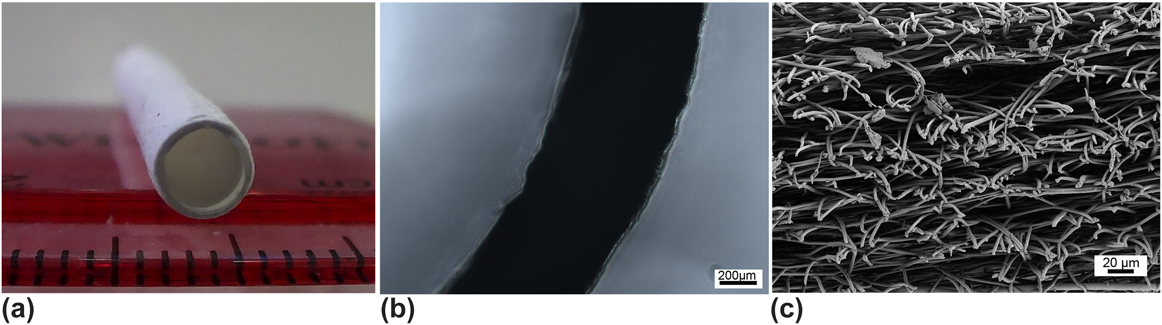
Representative gross and SEM images showing the ultrastructure of the electrospun PCL/PEI scaffold with an inner diameter of 3.18 mm and uniform thickness 0.5 mm as illustrated in Fig. 3. These findings demonstrated the feasibility of using cationic polymers to fabricate vascular scaffolds. In other PCL/PEI scaffolds, the results showed similar trends to that of the 5% PEI scaffold. As shown in Table I, the porosity of the tubular scaffolds was over 80%, which may facilitate the spreading and proliferation of cells, because the preferred porosity of scaffolds used for cellular penetration is generally higher than 60%.Reference Courtney, Sacks, Stankus, Guan and Wagner38
(a) Photograph of 5% PEI tubular scaffold, (b) bright-field microscopic image of circular cross-section, and (c) the morphology of the nanofibers on the circular cross-section.

Morphological characterizations of electrospun nanofibers.

B. Surface properties of PCL/PEI nanofibrous scaffolds
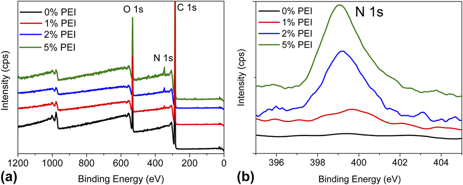
Figure 4(a) shows the whole survey scan of PCL, 1% PEI, 2% PEI, and 5% PEI scaffolds. Compared with neat PCL scaffolds, except C 1s and O 1s components, a third peak that belonged to N 1s was detected on the scaffolds containing PEI, which was located at 399 eV, and the intensity of the nitrogen peak increased with increased PEI loading in the nanofibrous scaffolds [see Fig. 4(b)], suggesting that the PEI was successfully added to the nanofibrous scaffolds.
High-resolution x-ray photoelectron spectra of electrospun PCL nanofibers: (a) survey scan of the PCL/PEI nanofibers and (b) spectra analysis of nitrogen element of PCL/PEI nanofibers.

C. Surface wettability of electrospun nanofibrous scaffolds
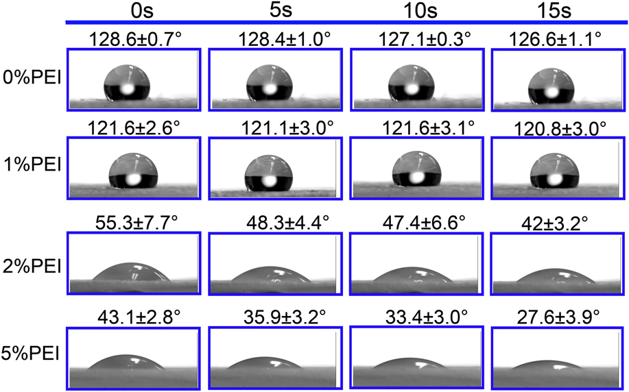
The surface properties of a polymer scaffold play a major role in the interaction between cells and the matrix.Reference Lee, Lim, Lee, Atala and Yoo39 Among many factors influencing the surface chemistry of scaffolds, the balance of hydrophilicity and hydrophobicity of a polymer surface is very important for cell adhesion. WCAs can be used to reflect the hydrophilicity of the scaffolds, since cells may attach and proliferate less well on scaffolds having a wettability that is higher than 90°.Reference Cui, Cheng, Li, Zhou, Zhang and Chang40 As shown in Fig. 5, the incorporation of PEI greatly enhanced the hydrophilicity of the scaffolds; the contact angle of nanofibrous scaffolds changed from 126.6 ± 1.1° for neat PCL to 42 ± 3.2° for 2% PEI and 27.6 ± 3.9° for 5% PEI. These results indicated that the wettability of hybrid scaffolds evolved from hydrophobic to hydrophilic (contact angle <90°). The improved wettability is expected to be beneficial for cell attachment.Reference Mikos, Lyman, Freed and Langer41
WCA of the electrospun nanofibers (n = 3).

D. Biomechanical properties
1. Axial and circumferential tensile testing
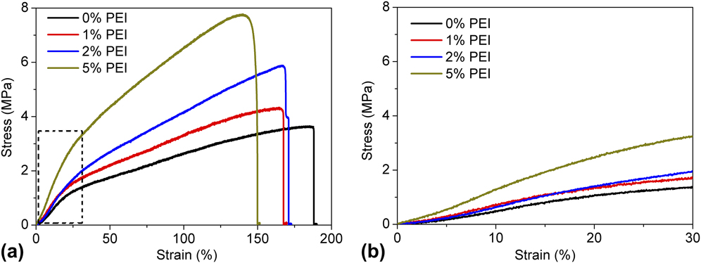
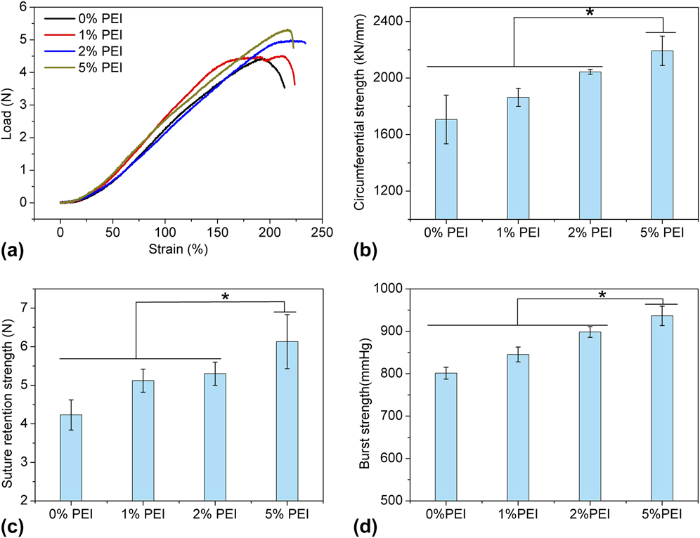
The mechanical properties of the electrospun tubes have been investigated in accordance with the standard ISO 7198 “Cardiovascular Implants–Tubular Vascular Prostheses.” During axial tensile testing, it was shown that the hydrated PCL/PEI scaffolds exhibited initial elastic behavior, followed by stiffening, which is similar to native tissues (as shown in Fig. 6). Based on the stress–strain curves of the electrospun vascular scaffolds, the tensile strength, elongation-at-break, and Young's modulus were calculated (Table II.) The ultimate tensile strength and Young's modulus increased with the amount of PEI added to PCL. The tensile strength of all tubular scaffolds was over 3.5 MPa, which far exceeded that of a porcine artery (1 MPa),Reference Amiel, Komura, Shapira, Yoo, Yazdani, Berry, Kaushal, Bischoff, Atala and Soker42 and was comparable to the left internal mammary artery.Reference Stekelenburg, Rutten, Snoeckx and Baaijens43 However, the elongation-at-break for the tubular scaffolds decreased from 181.1 ± 8.5 for 0% PEI to 148.3 ± 15.5 for 5% PEI due to the brittle properties of PEI. For the circumferential tests, the scaffolds showed similar behavior to those in the axial tests [as shown in Figs. 7(a) and 7(b)), which also demonstrated a “toe” region, followed by a nearly linear region, and a point at which the maximum load is reached. Such trends are typical for connective tissues found in the body.Reference Fung44
Stress–strain curves from tensile testing: (a) representative curves of the tensile tests conducted on the electrospun tubes and (b) the enlarged stress–strain curves (n = 5).

Biomechanical properties of the electrospun scaffolds.

Biomechanical properties of the electrospun PCL/PEI scaffolds: (a) representative curves of the circumferential strength tests conducted on the electrospun tubes, (b) the recorded circumferential strength of the electrospun scaffolds, (c) suture retention strength of the tested scaffolds, and (d) burst strength of the vascular scaffolds (n = 5, *p < 0.05).

2. Suture retention strength
Suture retention is of critical importance in the success of the graft implantation procedure. In this study, the suture retention strength of the fabricated vascular scaffolds remained higher than 4 N [as shown in Fig. 7(c)], which is far beyond the suture strength observed in a human mammary artery (1.4 ± 0.01 N) and a human saphenous vein (1.81 ± 0.02 N).Reference Konig, McAllister, Dusserre, Garrido, Iyican, Marini, Fiorillo, Avila, Wystrychowski, Zagalski, Maruszewski, Jones, Cierpka, de la Fuente and L'Heureux45 After adding PEI to PCL, the suture retention strength was further enhanced. Thus, the suture retention strength of the PCL/PEI vascular scaffolds was considerably higher than the adequate suture retention strength of 2 N.Reference Billiar, Murray, Laude, Abraham and Bachrach46
3. Burst pressure strength
Burst pressure assessment determined that the electrospun vascular scaffolds bore up to 800 mmHg, which is more than six times the upper physiological pressure of 120 mmHg and more than three times the pathological upper pressures of 180–220 mmHg [shown in Fig. 7(d)]. These results and comparisons suggest that the fabricated vascular scaffolds in this study can be developed as a substitute for native blood vessels. However, even though the obtained burst pressure value was higher than the reported value for a female rat femoral artery (503 mmHg),Reference Oskoui, Stadler and Lanzafame47 it was still lower than the typical value of a human carotid artery (5000 mmHg) or a human saphenous vein (2250 mmHg).Reference Sarkar, Salacinski, Hamilton and Seifalian48 This might be because real tissue is composed of highly organized lamellae developed by living cells, which has a tighter and more highly oriented structure. This highly organized structure would have stronger mechanical properties than artificial tubes. Seliktar also reported that tissue engineered blood constructs composed of living cells embedded in collagen-gel scaffolds showed much stronger mechanical behavior with incubation time.Reference Seliktar, Black, Vito and Nerem49
E. Biocompatibility evaluations
1. Cell attachment
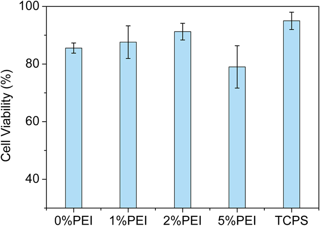
To investigate the effects of added PEI on cell growth, viable cells were monitored after 2 h incubation time. Cell viability was quantified by counting the number of live and dead cells from chosen areas. Figure 8 shows the cell attachment results. It was found that 2% PEI had the highest cell viability, and the number of attached live cells on these scaffolds was the largest among all of the scaffolds, which might be because the hydrophilicity of the scaffold was improved. However, as the PEI content reached 5%, the cell viability and cell numbers decreased. This may be because the high content of PEI was toxic to cells through the introduction of apoptosis/necrosisReference Vancha, Govindaraju, Parsa, Jasti, Gonzalez-Garcia and Ballestero32,Reference Tryoen-Toth, Vautier, Haikel, Voegel, Schaaf, Chluba and Ogier50 ; similar cell behaviors at high PEI contents were also found in bovine knee chondrocytes and osteoblast cells in previous studies.Reference Tryoen-Toth, Vautier, Haikel, Voegel, Schaaf, Chluba and Ogier50,Reference Kuo and Ku51 Therefore, it seems that the appropriate balance between cationic content and hydrophilicity needs to be achieved to attain the highest level of cell attachment and cell viability.
Attachment of HUVECs to the electrospun scaffolds after 2 h incubation (n = 3).

2. Cell viability and proliferation
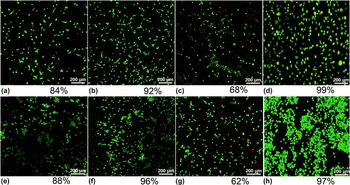
After 3 days and 10 days of in vitro cell culture, fluorescent cell viability assays were performed on the 0% PEI, 2% PEI, and 5% PEI specimens; the results are shown in Fig. 9. The results demonstrated that the HUVECs could survive on the chosen scaffolds after culturing for 3 days. Very few dead cells were observed on the 0% PEI and 2% PEI with cell viabilities of 84% and 92%, respectively, while large numbers of HUVECs on the 5% PEI underwent apoptosis; cell viability on 5% PEI scaffolds was only 68%. On the 10 day cultures, the HUVECs still showed high viability on the 2% PEI, with a cell viability of 96%. However, cells on the 5% PEI showed significantly higher apoptosis compared to other scaffolds. Therefore, these results also confirmed that a high PEI content in the fabricated scaffolds has a negative effect on cell survival.
Day 3 and day 10 live/dead cell assay showing HUVECs cultured on (a, d) 0% PEI, (b, e) 2% PEI, and (c, f) 5% PEI. Live cells showed green fluorescence and dead cells produced red fluorescence (the images on the first and second lines are the results on day 3 and day 10, respectively).

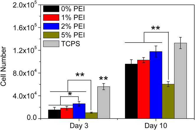
The proliferation of HUVECs on different electrospun nanofibrous scaffolds was evaluated after 3 and 10 days by MTS assay. As shown in Fig. 10, a sustained increase of cell numbers was observed for all scaffolds from day 3 to day 10 of culture, with the cells on the 2% PEI exhibiting the highest amount of cell proliferation after 10 days of culture. These results showed that a higher content of PEI in the fibers influenced the proliferation of HUVECs, with 5% PEI resulting in reduced overall cell numbers. Although in 5% PEI, the scaffold already became hydrophilic, which should be better for cell attachment as known, however, the introduction of apoptosis by PEI sharply counteracted this superiority compared with other scaffolds. Therefore, according to MTS results, it can be concluded that a small addition of PEI to the nanofibers can enhance the hydrophilicity of the PCL vascular scaffolds, which successfully enhanced the adhesion and proliferation of endothelial cells on a PCL nanofiber mesh, however, larger amount of PEI in the scaffolds would introduce obvious cell apoptosis as previous reports.Reference Vancha, Govindaraju, Parsa, Jasti, Gonzalez-Garcia and Ballestero32
Proliferation of HUVECs on 0% PEI, 1% PEI, 2% PEI, and 5% PEI as determined by MTS assay. (n = 3,*p < 0.05, and **p < 0.01).

3. Cell morphology
Figure 11 shows the HUVECs' attachment and growth on the scaffolds at day 3 and day 10. At day 3, only a few cells can be observed on the scaffolds. At day 10, there was an increase in cell density compared to day 3. Additionally, the cells appeared to form continuous monolayers on the scaffolds. However, in the day 3 results, some cells were round-shaped and accumulated with each other to form cell clusters on 0% PEI and 5% PEI, while the cells were largely flat on 2% PEI. It has been reported that cells with a flat shape survive better than cells with a more rounded shape.Reference Chen, Mrksich, Huang, Whitesides and Ingber52 Therefore, the cell morphology results of this study indicated that a small addition of PEI is favorable for cell spreading. The possible reason for that is the cationic PEI could reabsorb some proteins from the cell culture medium, which would be better for cell attachment on the scaffold according to Ref. Reference Zhu, Ji, Tan, Barbosa and Shen53.
Morphology of HUVECs cultured on (a, d) 0% PEI, (b, e) 2% PEI, and (c, f) 5% PEI. (The images on the first and second lines are the results of day 3 and day 10, respectively.)

4. Cytoskeleton organization and phenotype study
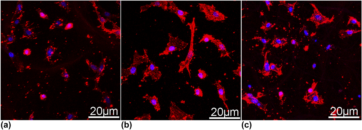
The cytoskeletal organization of HUVECs grown on the nanofibers was assessed by staining the cells with phalloidin. The cell morphology and actin structures of HUVECs on different surfaces were compared. In general, mammalian cells are known to undergo three processes on a substrate including cell adhesion to the matrix, cell spreading, and cytoskeleton development.Reference Hersel, Dahmen and Kessler54 As shown in Fig. 12, cells grown on the 0% PEI and 5% PEI scaffold demonstrated a small spherical or narrow shape, which is a typical nonadherent and nonspreading morphology for HUVECs.Reference Ku and Park55 In contrast, on the 2% PEI scaffold, HUVECs exhibited a fibroblastic morphology with a higher degree of spreading. Furthermore, the cells showed stretched actin bundles when grown on 2% PEI after culturing for 48 h. The increase in cell adhesion on the 2% PEI might be attributed to the adsorption of proteins from the cell culture media by the added PEI and the enhancement in the hydrophilicity of the scaffolds. However, too much PEI might cause instability in the cell culture medium and introduce cell apoptosis, which would be harmful for cell growth and cell proliferation over a longer period of time.
Cytoskeleton study of HUVECs cultured on (a) 0% PEI, (b) 2% PEI, and (c) 5% PEI scaffolds for 48 h.

IV. CONCLUSIONS
A desirable scaffold for vascular tissue engineering should mimic an extracellular matrix exhibiting an architecture that favors cell proliferation as well as the physical properties. In this study, PCL/PEI vascular scaffolds were successfully fabricated by electrospinning PCL/PEI blends at various PEI contents. The biomechanical properties and biocompatibilities of the scaffolds were investigated to explore their potential applications in vascular tissue engineering. It was found that the addition of PEI reduced the fiber diameter and enhanced the tensile strength, modulus, suture retention, and burst pressure of the vascular scaffolds. Results of the in vitro studies demonstrated that the fabricated nanofibers with a relatively low concentration of PEI (e.g., 2%) could promote HUVECs' attachment and proliferation, while scaffolds with a high concentration of PEI (e.g., 5%) showed negative effects on cell viability and attachment, indicating the necessity for an appropriate balance between the cationic content and hydrophilicity of the scaffolds. The scaffolds without PEI were hydrophobic which could not facilitate cell attachment, while the scaffolds with a PEI content of 5% caused cell apoptosis. In summary, the low amount of PEI in the PCL/PEI scaffolds could significantly improve the endothelial cell behavior so that they have potential to be used in vascular tissue engineering applications.
ACKNOWLEDGMENTS
The authors would like to acknowledge the support of the Wisconsin Institute for Discovery (WID), the China Scholarship Council, the financial support of the National Nature Science Foundation of China (No. 51073061, No. 21174044), the Guangdong Nature Science Foundation (No. S2013020013855, No. 9151064101000066), and National Basic Research Development Program 973 (No. 2012CB025902) in China.