Introduction
In order to enhance the health and longevity of the human population, modern nutritionists have focussed on improving food quality and nutritional value. This has brought an increased interest in the reformulation of existing food products and the development of innovative products for better consumer health (Binns and Howlett, Reference BINNS and HOWLETT2009).
In recent years there have been advances in techniques to manipulate fatty acid composition in meat. Since meat is seen as a major source of dietary fat and especially saturated fatty acids, the modification of the lipid content and fatty acid composition may be an effective way to improve consumer's health (Qi et al., Reference QI, CHEN, ZHAO, ZHENG and WEN2010). According to Wood et al. (Reference WOOD, RICHARDSON, NUTE, FISHER, CAMPO and KASAPIDOU2003), the ratio of PUFA to saturated fatty acid (SFA) in some meat is 0.1, yet the recommended PUFA/SFA ratio is >0.4. The consumption of high levels of SFA may lead to cardiovascular disease (Ganji et al., Reference GANJI, KAMANNA and KASHYAP2003). The inclusion of different fat sources in poultry diets affects the meat quality and the proportion of fatty acid composition of abdominal fat and meat (Villaverde et al., Reference VILLAVERDE, BAUCELLS, CORTINAS and BARROETA2006; Taulescu et al., Reference TAULESCU, MIHAIU, BELE, MATEA, DAN, MIHAIU, XANDRA LAPUSAN and CIUPA2010; Royan et al., Reference ROYAN, MENG, OTHMAN, SAZILI and NAVIDSHAD2011; Ramiah et al., Reference RAMIAH, MENG, SHEAU, SWEE and EBRAHIMI2014).
In order to maintain the health and growth of poultry, dietary fats play an important role as a source of energy, essential fatty acids and fat soluble vitamins (Mahesar et al., Reference MAHESAR, SHERAZI, KANDHRO, BHANGER, KHASKHELI and TALPUR2011). Dietary fats can improve the palatability of certain diets, reduced dustiness and act as lubricants in feed preparation (Ziggers, Reference ZIGGERS2005). The composition of dietary fat in poultry feed usually ranges between 2 to 6%. Birds are not able to synthesise all fatty acids and thus, some are essential, such as linoleic (C18:2n-6) and α-linolenic (C18:3n-3) fatty acids (Bionaz et al., Reference BIONAZ, CHEN, KHAN and LOOR2013). Linoleic acid is defined as the only essential fatty acid in poultry feeds (National Research Council, 1994).
Dietary fatty acids in poultry feed
There are many fat sources available for poultry feed requirements; animal and vegetable sources. To date, a number of different fat sources available for poultry feed are from both animal and vegetable sources and from the rendering industry (Sanz et al., Reference SANZ, FLORES, PEREZ, AYALA and LOPEZ-BOTE1999). In monogastric animals such as poultry, the fatty acid profile of meat can easily be modified. The principle site of digestion of dietary fatty acids is the small intestines and fatty acids are incorporated from there and deposited directly into both muscles and fatty tissues (Morel et al., Reference MOREL, MCINTOSH and JANZ2006). This would improve the quality of meat for human consumption. The major sources of n-6 PUFAs are vegetable oils such as corn, safflower, and soybean oil, whereas n-3 PUFA sources include fish such as salmon, trout and tuna, and plant sources such as flaxseed (Betti et al., Reference BETTI, PEREZ, ZUIDHOF and RENEMA2009).
Chickens fed diets rich in PUFAs such as sunflower oil, soybean oil, or linseed oil resulted in a decrease of abdominal fat deposition in broilers with similar metabolisable energy levels but rich in SFA (Villaverde et al., Reference VILLAVERDE, BAUCELLS, CORTINAS and BARROETA2006). Palm oil is a type of saturated fat containing high levels of palmitic (C16:0) acid and is often used to replace animal fat (Smink et al., Reference SMINK, GERRITS, HOVENIER, GEELEN, LOBEE, VERSTEGEN and BEYNEN2008). The higher amount of unsaturated dietary fats decreases the melting point of the fat in the broiler carcass (Bavelaar et al., 2003). This leads to a greater risk of oxidation, causing rancidity and colour deterioration of meat (Eder et al., Reference EDER, GRUNTAL, KLUGELUGE, HIRCHE, SPILKE and BRANDSCH2005).
Fatty acids that act as ligands to PPAR include polyunsaturated fatty acids such as certain omega-3 polyunsaturated fatty acids, e.g. α (alpha)-linolenic acid with C18:3n-3, or docosahexaenoic acid with C22:6n-3), and certain omega-6 polyunsaturated fatty acids such as linoleic acid with C18:2 and arachidonic acid with C20:4 (Varga et al., Reference VARGA, CZIMMERER and NAGY2011). Dietary fatty acids can bind to and activate nuclear receptors and regulate gene expression akin to small molecule endocrine hormones.
Physiological effects of PPAR by fatty acids
PPAR comprise a family of nuclear hormone receptors that act as ligand-activated transcription factors in the regulation of several genes involved in metabolic processes (Evans et al., Reference EVANS, MISYAK, SCHMELZ, GURI, HONTECILLAS and BASSAGANYA-RIERA2010; Monsalve et al., Reference MONSALVE, PYARASANI, DELGADO-LOPEZ and MOORE-CARRASCO2013). The phenomenon of peroxisome proliferation was first reported in the 1960s where increases in hepatic peroxisomes were observed in rats treated with clofibrate (De Duve, Reference DE DUVE1969). In 1990, a group of scientists was working on the mechanism of action of a variety of chemicals such as hypolipidemic drugs on peroxisomal proliferation in the mouse liver and cloned the first PPAR (PPAR α) from the mouse liver (Issemann and Green, Reference ISSEMANN and GREEN1990). Other closely related receptors encoded by different genes were subsequently cloned and named as PPAR β and PPAR γ (Dreyer et al., Reference DREYER, KREY, KELLER, GIVEL, HELFTENBEIN and WAHLI1992; Bionaz et al., Reference BIONAZ, CHEN, KHAN and LOOR2013). However, each PPAR isoform has its own unique physiological functions, probably due to its distinct patterns of tissue distribution, ligands and biochemical properties (Varga et al., Reference VARGA, CZIMMERER and NAGY2011).
The activation of PPAR is by certain natural and synthetic ligands such as PUFA, eicosanoids, and hypolipidemic drugs (fibrates) (Contreras et al., Reference CONTRERAS, TORRES and TOVAR2013). The PPAR ligand binding cavity is three to four times larger than of other receptors and is thus able to accommodate a variety of natural and synthetic lipophilic acids (Grygiel-Górniak, Reference GRYGIEL-GÓRNIAK2014). Upon ligand binding, PPAR regulate transcriptional target genes by binding as heterodimers with the retinoic acid receptor (RXR) to peroxisome proliferator responsive elements (PPRE) through the DNA binding domain (DBD) (Ahmadian et al., Reference AHMADIAN, SUH, HAH, LIDDLE, ATKINS, DOWNES and EVANS2013).
Excessive fat in broiler chickens is a major concern facing the poultry industry (Zhou et al., Reference ZHOU, DEEB, EVOCK-CLOVER, ASHWEL and LAMONT2006) as it reduces meat yield and affects consumer acceptance (Fouad and El-Senousey, Reference FOUAD and EL-SENOUSEY2014). Choct et al. (Reference CHOCT, NAYLOR, HUTTON and NOLAN2000) reported that modern broiler strains contain more than 15 to 20% fat. In the chicken, fatty acids are synthesised in the liver, with most of triglycerides being transported via low density lipoproteins to adipose tissue for storage (Wang et al., Reference WANG, WANG, ZHANG, GU, WANG, WU and LI2010). Therefore, it is important to understand lipid metabolism in chickens at a molecular level in order to further improve its production. Takada et al. (Reference TAKADA, YU, XU, LAMBERT, MONTANA, KLIEWER, EVANS and UMESONO2000) first reported on PPAR subtype genes (α, β, and γ) isolated from 2.5-day-old chicken embryos and adult adipose tissue of chickens. PPAR genes in chickens are different from those present in humans and mice as the latter have two isoforms; PPARγ1 and PPARγ2 (Takada and Kobayashi., 2013). In the chicken, PPAR γ gene is shorter than that in humans and the γ2 isoform is absent (Takada and Kobayashi., 2013) ( Figure 1 ).
Structural organization of chicken (upper panel) and human (lower panel) PPAR γ genes (Takada and Kobayashi, Reference TAKADA and KOBAYASHI2013).

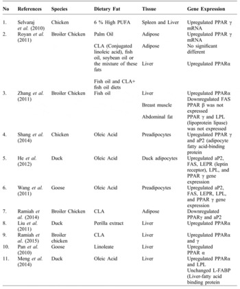
Meng et al. (Reference MENG, LI and WANG2004) reported that PPAR α was mainly expressed higher in the brain, lungs, kidney, heart and intestine, medium in the stomach, liver and adipose tissue, and lower in the spleen of broiler chickens and not detected in the breast muscle. Meanwhile, the expression level of PPAR γ gene was higher in the abdominal adipose tissue, gizzard, spleen, kidney and lower in the heart and was not expressed in the liver, breast muscle, leg muscle, and duodenum in chicken adipocytes (Wang et al., Reference WANG, NA, WANG, WANG, WANG, WANG, LIM and LI2012). Table 1 shows the effect of dietary fat on the expression of PPAR in various poultry species.
The effects of dietary fatty acid on the expression of PPAR in poultry species.

PPAR γ
The 3T3-L1 and 3T3-F442A cells are the most common mammalian preadipocytes cell lines that have been selected to study on adipocyte differentiation (Yagi et al., Reference YAGI, KONDO, OKAZAKI and KANO2004). Matsubara et al. (Reference MATSUBARA, AOKI, ENDO and SATO2013) has indicated that the expression profiles of the adipogenesis-related factors in chickens are different from mammalian adipogenesis. Lipogenesis in chickens and humans are active exclusively in the liver (Ji, Reference JI2013) whereas lipogenesis in dogs, cats and ruminants occurs mainly in the adipose tissue than the liver (Foufelle and Ferre, Reference FOUFELLE and FERRE2004). Several groups of transcriptional factors that promote adipocytes differentiation were identified which include; CCAAT/enhancer-binding protein (C/EBP) gene family and PPAR-γ (Shang et al., Reference SHANG, GUO, WANG, SHI, WANG and LI2014).
Previous studies have indicated that fatty acids have significant effects in reducing abdominal fat deposition in broilers (Wongsuthavas et al., Reference WONGSUTHAVAS, TERAPUNTUWAT, WONGSRIKEAW, KATAWATIN, YUANGKLANG and BEYNEN2008; Ramiah et al., Reference RAMIAH, MENG, SHEAU, SWEE and EBRAHIMI2014). Abdominal fat represents a small part of the total fat deposition in chickens (Smink et al., Reference SMINK, GERRITS, HOVENIER, GEELEN, VERSTEGEN and BEYNEN2010). Several candidate genes that regulate adipogenesis, including PPAR γ, are known to permit, or to be necessary for, the transition of preadipocytes into adipocytes in vitro (MacDougald and Lane, Reference MACDOUGALD and LANE1995). In a study by Matsubara et al. (Reference MATSUBARA, SATO, ISHII and AKIBA2005), PPAR γ mRNA levels in chicken adipocytes markedly increased in the differentiation medium supplemented with fatty acid for up to nine hours, while C/EBP α mRNA levels gradually increased in the first 24 hours of culture. This indicated that the primary step in the induction of chicken adipogenesis was via stimulation of PPAR γ gene expression, rather than C/EBPα expression (Matsubara et al., Reference MATSUBARA, SATO, ISHII and AKIBA2005). Matsubara et al. (Reference MATSUBARA, ENDO and KANO2008) reported that chicken preadipocytes treated with fatty acid mixtures increased PPAR γ expression gradually during days zero to eight after induction of differentiation. Taken together, these data suggest that PPAR is regarded as the ‘master regulator’ of adipocyte differentiation. Furthermore, fatty acids may promote chicken preadipocyte differentiation, at least in part, by activating the molecular program controlled by PPAR γ. Griffin et al. (Reference GRIFFIN, BUTTERWITH and GODDARD1987) noted that the growth of abdominal fat pads in broiler chickens increased rapidly between one and four weeks of age and remains high until 10 weeks of age. In the study by Sato et al. (Reference SATO, FUKAO, SEKI and AKIBA2004), the level of PPAR γ mRNA levels in abdominal adipose tissue in six week old chickens were significantly higher compared with one and two week-old chickens.
LPL enzyme was first discovered by Cooper et al. (Reference COOPER, LU and VISWANATH1992) and it functions in the catabolism of triglyceride from plasma lipoprotein (Liu et al., Reference LIU, WANG, SUN, YU and ZHANG2006). Sato et al. (Reference SATO, AKIBA, CHIDA and TAKAHASHI1999) demonstrated that infusion of anti-lipoprotein lipase antibody into chickens reduced chicken body fat. These results suggested that LPL plays a crucial role in fat deposition in adipose tissue. Griffin et al. (Reference GRIFFIN, BUTTERWITH and GODDARD1987) stated that LPL activity in chicken adipose tissue gradually increases from 4 weeks up to 6 weeks of age. In addition, He et al. (Reference HE, TIAN, LI, SHEN, TAO, FU, NIU and LU2012) and Wang et al. (Reference WANG, LU, YUAN, TIAN, LI, TAO and FU2011) found that oleic acid is able to promote the expression of LEPR, LPL, FAS, and PPAR γ genes in goose and duck adipocytes respectively. Gene expression of LPL in adipose tissue is enhanced by PPAR γ in human preadipocytes (Larsen et al., Reference LARSEN, TOUBRO and ASTRUP2003). Therefore, these results show that the synthesis and translation of chicken PPAR γ mRNA in adipose tissue are accompanied by an increase in LPL mRNA expression.
Adipocyte fatty acid-binding protein (A-FABP) is also referred to as aP2 and adipocyte lipid-binding protein (ALBP) (Shi et al., Reference SHI, ZHANG, WANG, YANG, WANG and LI2011). A study by Wang et al. (Reference WANG, LI, LI, LENG, WANG and TANG2006) stated that A-FABP gene was significantly associated with the fat content in chickens. Furthermore, Shi et al. (Reference SHI, ZHANG, WANG, YANG, WANG and LI2011) stated that A-FABP plays an important role in the lipid metabolism of adipocytes in mammals and poultry. He et al. (Reference HE, TIAN, LI, SHEN, TAO, FU, NIU and LU2012) reported that the A-FABP gene was found to be highly expressed in duck adipose and muscle tissues. The A-FABP gene contains a PPAR-response element (PPRE) (Tontonoz et al., Reference TONTONOZ, HU, GRAVES, BUDAVARI and SPIEGELMAN1994) and CLA is a ligand for PPAR. Ramiah et al. (Reference RAMIAH, MENG, SHEAU, SWEE and EBRAHIMI2014) reported that CLA may have increased A-FABP transcription, which is parallel to PPAR transcription in the adipose tissue of broiler chickens. These observations indicate that CLA could function through PPAR. Wang et al. (Reference WANG, MU and LI2008) showed that A-FABP was altered when PPAR-γ was knocked down in chicken adipocytes. In addition, Shi et al. (Reference SHI, ZHANG, WANG, YANG, WANG and LI2011) presumed that A-FABP in chicken adipocytes was altered by fatty acid and PPAR γ, which neutralised the effect of interference plasmid 12 hour after oleate induction.
In the study by Sato et al. (Reference SATO, FUKAO, SEKI and AKIBA2004), PPAR γ was expressed in chicken ovaries during the onset of egg laying however the mechanism of PPAR γ mRNA in layer chickens is not known. Most studies on PPAR γ in ovarian tissue have been reported in rodents, pigs, and humans (Lambe et al., 1996; Komar et al., Reference KOMAR, BRAISSANT, WAHLI and CURRY2001; Cui et al., Reference CUI, MIYOSHI, CLAUDIO, SIEBENLIST, GONZALEZ, FLAWS, WAGNER and HENNIGHAUSEN2002; Schoppe et al., Reference SCHOPPE, GARMEY and VELDHUIS2002). Long et al. (Reference LONG, SAIRAM and KOMAR2009) explained that PPAR γ expression in rat ovaries is initiated at an early stage during folliculogenesis and is associated with the progressive maturation of granulosa cell (Long et al., Reference LONG, SAIRAM and KOMAR2009). Besides controlling lipid metabolism, PPAR γ are also involved in the regulation of cancer development. Understanding the role of PPAR γ has been investigated in mammals and cancer cell lines (Elstner et al., Reference ELSTNER, MÜLLER and KOSHIZUKA1998; Mueller et al., Reference MUELLER, SARRAF and TONTONOZ1998; Saez et al., Reference SAEZ, TONTONOZ and NELSON1998). Campbell et al. (Reference CAMPBELL, CARLBERG and KOEFFLER2008) concluded that PPAR γ acts as a ligand-activated tumour suppressor that inhibits proliferation and induces differentiation and apoptosis of a wide range of cell types in vitro and in rodent tumour models (Campbell et al., Reference CAMPBELL, CARLBERG and KOEFFLER2008). Jiang et al. (Reference JIANG, REDFERN, BRYCE and MANSEL2000) investigated the role of PPAR γ in human breast cancer cells treated with gamma linolenic acid (GLA). The results revealed that treated tissue cancer cells have lower expression of PPAR γ compared with normal cells (Jiang et al., Reference JIANG, REDFERN, BRYCE and MANSEL2000). This indicates that PPAR γ serves as a receptor for GLA in the regulation of gene expression in breast cancer cells.
PPAR α
PPAR α are highly abundant in tissues (e.g. liver, heart and skeletal muscle) with a high capacity of fatty acid oxidation (Grygiel-Górniak, Reference GRYGIEL-GÓRNIAK2014). PPAR α acts as a master regulator of hepatic lipid catabolism by inducing the expression of numerous genes involved in mitochondrial and peroxisomal fatty acid oxidation and other cellular components (Mandard et al., Reference MANDARD, MÜLLER and KERSTEN2004; Georgiadi and Kersten, Reference GEORGIADI and KERSTEN2012; Honda et al., Reference HONDA, SANEYASU, SUGIMOTO, KURACHI, TAKAGI and KAMISOYAMA2015). Furthermore, the induction of fatty acid oxidation by PPAR α via upregulation of carnitine palmitoyltransferase enzymes and acyl-coenzyme A oxidase has been reported by Royan et al. (Reference ROYAN, MENG, OTHMAN, SAZILI and NAVIDSHAD2011).
In chickens the liver plays a primary role in lipid metabolism (Ramiah et al., Reference RAMIAH, MENG and EBRAHIMI2015) and the adipose tissue serves merely as a storage (O'Hea and Leveille, Reference O'HEA and LEVEILLE1968). Omega-3 fatty acids are the natural ligand of PPAR α. They are highly polyunsaturated and readily undergo oxidation and trigger the activation of PPAR (Grygiel-Górniak, Reference GRYGIEL-GÓRNIAK2014). König et al. (Reference KÖNIG, SPIELMANN, HAASE, BRANDSCH and KLUGE2008) reported that the supplementation of fish oil rich in omega-3 fatty acids increases the expression of PPAR α in the liver of hens, leading to a reduction of plasma triacylglycerol concentration. In the same study, CLA mixtures of cis-9, trans-11 and trans-10, cis-12 CLA did not significantly up-regulate gene expression of PPAR α target genes in hens (König et al., Reference KÖNIG, SPIELMANN, HAASE, BRANDSCH and KLUGE2008). Similarly, Ramiah et al. (Reference RAMIAH, MENG, SHEAU, SWEE and EBRAHIMI2014) observed that PPAR α were triggered by CLA in the liver. Other studies in the rat liver showed that CLA isomers are able to activate PPAR α (Camarena et al., Reference CAMARENA, VAN DEN HEUVEL and BELURY1999).
PPAR β
Wang et al. (Reference WANG, LEE, TIEP, YU, HAM, KANG and EVANS2003) observed that PPAR β deficient mice fed high-fat diets had reduced energy uncoupling and were prone to obesity. This suggests that PPAR β acts as a key regulator of fat burning in peripheral tissues by coordinating fatty acid oxidation and energy uncoupling (Wang et al., Reference WANG, MU and LI2008). Similarly, in an in vitro study, stimulation of PPAR β resulted in the enhancement of fatty acid oxidation, leading to increased energy production in the form of ATP and of energy uncoupling (Sprecher, Reference SPRECHER2007). A study by Sato et al. (Reference SATO, FUKAO, SEKI and AKIBA2004) found that PPAR β in chicken preadipocytes treated with oleate did not play a key role in adipocyte differentiation.
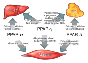
PPAR β plays a critical role in epidermis maturation and skin wound healing, making it a potential target in improving impaired healing in different organs (Tan et al., Reference TAN, MICHALIK, NOY, YASMIN, PACOT, HEIM, FLÜHMANN, DESVERGNE and WAHLI2001; Letavernier et al., Reference LETAVERNIER, PEREZ, JOYE, BELLOCQ, FOUQUERAY, HAYMANN, HEUDES, WAHLI, DESVERGNE and BAUD2005). Tan et al. (Reference TAN, MICHALIK, NOY, YASMIN, PACOT, HEIM, FLÜHMANN, DESVERGNE and WAHLI2001) explained that the stimulation of PPAR β transcriptional activity strongly accelerates the differentiation of keratinocytes and increases their resistance to apoptotic signals. Therefore, the increased proliferation and death of keratinocytes at the edges of epidermal wounds in mice are likely to participate in the healing. The physiological roles of the PPARs have been summarised in Figure 2 .
The physiologic role of the PPARs in the liver, muscle and adipose tissue in humans (Evans et al., Reference EVANS, BARISH and WANG2004).

Conclusions
PPARs belong to the nuclear receptor hormone super family. Various natural fatty acids serve as ligands for the PPAR subtypes. PPARs are a truly fascinating set of molecules as each isoform has its own distinctive systemic and cellular metabolic functions. Taken together, recent studies have clearly demonstrated the essential roles of PPARs; PPAR α act as an activator of mitochondrial and peroxisomal fatty acid β-oxidation in the liver; PPAR β, a regulator of fatty acid oxidation and PPAR γ, an activator of fatty acid synthesis and storage, play divergent roles in metabolism. Studies have proven that fatness in chickens is the same as the predisposition for obesity and diabetes in humans. Furthermore, avian genomes lack of brown adipocytes, which differs from rodents and humans. This trait makes them an ideal animal model to study research on white adipose tissue in the absence of brown adipose tissue. Manipulation of PPAR genes, especially PPAR γ in chickens resulted in leaner meat as PPAR γ is the key regulator of chicken preadipocyte proliferation and differentiation. Advanced research is required to broaden our understanding of the mechanisms underlying the control of PPARs, to define the regulated gene sets and pathways of disease specific contribution in poultry. This will be further fuelled by emerging novel approaches and methodologies in poultry research.
Acknowledgments
The authors are very grateful to the Faculty of Veterinary Medicine, Universiti Putra, Malaysia for their assistance with this paper.