INTRODUCTION
Fascioliasis is caused by trematodes of the genus Fasciola. Within this genus, 2 species, F. hepatica and F. gigantica are the most common species in domestic animals and man (World Health Organization, 1995). F. hepatica has a worldwide distribution but predominates in temperate zones, while F. gigantica is only found in the tropical regions (Andrews, Reference Andrews and Dalton1998). Fascioliasis is recognized as a major public health problem by the World Health Organization (1995) with an estimated 17 million people infected worldwide (Mas-Coma et al. Reference Mas-Coma, Bargues, Esteben and Dalton1999). The routine diagnosis of this disease is done by stool examination. Alternatively, diagnosis can be made by serological analysis.
Serological tests for the diagnosis of fascioliasis have been developed as standard assays using several potential antigens for the detection of total immunoglobulin G (total IgG) antibody responses, such as crude excretory-secretory antigen (Maleewong et al. Reference Maleewong, Intapan, Wongkham, Tomanakan, Daenseekaew and Sukeepaisarnjaroen1996; Intapan et al. Reference Intapan, Maleewong, Wongkham, Tomanakarn, Leamviteevanich, Pipitgool and Sukolapong1998), authentic Fasciola cathepsin L1 (Rokni et al. Reference Rokni, Massoud, O'Neill, Parkinson and Dalton2002) and recombinant cathepsin L (Carnevale et al. Reference Carnevale, Rodriguez, Guarnera, Carmona, Tanos and Angel2001; Cornelissen et al. Reference Cornelissen, Gaasenbeek, Borgsteede, Holland, Harmsen and Boersma2001; Tantrawatpan et al. Reference Tantrawatpan, Maleewong, Wongkham, Wongkham, Intapan and Nakashima2005). Recently, highly sensitive and specific peptide antigens were used for the serodiagnosis of human fascioliasis by detecting total serum IgG antibody (Intapan et al. Reference Intapan, Tantrawatpan, Maleewong, Wongkham, Wongkham and Nakashima2005). Several reports have revealed that IgG4 subclass antibody may play a prominent role in the responses to long-lasting parasitic infections such as schistosomiasis (Iskander et al. Reference Iskander, Das and Aalberse1981; Evengard et al. Reference Evengard, Hammarstrom, Smith, Johansson and Linder1988) and filariasis (Ottesen et al. Reference Ottesen, Skvaril, Tripathy, Poindexter and Hussain1985). Additionally, the diagnostic capability of the serodiagnosis of fascioliasis has been increased by the detection of IgG4 subclass antibody using F. hepatica or F. gigantica antigen (O'Neill et al. Reference O'Neill, Parkinson, Strauss, Angels and Dalton1998, Reference O'Neill, Parkinson, Dowd, Strauss, Angels and Dalton1999; Strauss et al. Reference Strauss, O'Neill, Parkinson, Angels and Dalton1999; Hassan et al. Reference Hassan, Mostafa, Ramadan, Nassar, Hassounah and Omar2000, Reference Hassan, Matar, Mowafy, Saleh, el-Badrawy el, Awad and Ramadan2002; Wongkham et al. Reference Wongkham, Tantrawatpan, Intapan, Maleewong, Wongkham and Nakashima2005). The present study was designed to develop a peptide-based ELISA for the detection of serum specific IgG4 subclass antibody and to compare the effectiveness of 2 peptides for the serodiagnosis of human fascioliasis.
MATERIALS AND METHODS
Peptides derived from Fasciola cathepsin L1
Two peptides corresponding to the antigenic regions of Fasciola cathepsin L, based on the length of peptide 7 with 20 amino acids, were synthesized as described by Cornelissen et al. (Reference Cornelissen, Gaasenbeek, Boersma, Borgsteede and van Milligen1999) with some modifications. The first peptide, derived from amino acids 110–129 of the enzymatic part of F. hepatica cathepsin L1 (Wijffels et al. Reference Wijffels, Panaccio, Salvatore, Wilson, Walker and Spithill1994) (GenBank Accession number L33771) as well as of F. gigantica cathepsin L1-B (Grams et al. Reference Grams, Vichasri-Grams, Sobhon, Upatham and Viyanant2001) (GenBank Accession number AF239264) or from amino acids 3–22 of F. gigantica cathepsin L1-E (Grams et al. Reference Grams, Vichasri-Grams, Sobhon, Upatham and Viyanant2001) (GenBank Accession number AF239267), was synthesized as a carboxamide at the C-terminus and acetyl at the N-terminus: acetyl-DKIDWRESGYVTEVKDQGNC-carboxamide (namely, peptide V). A second peptide, consisting of amino acids 110–129 from the enzymatic part of F. gigantica cathepsin L1 (Grams et al. Reference Grams, Vichasri-Grams, Sobhon, Upatham and Viyanant2001) (GenBank Accession number AF112566), was synthesized as a carboxamide at the C-terminus and acetyl at the N-terminus: acetyl-DKIDWRESGYVTELKDQGNC-carboxamide (namely, peptide L). The valine at position 14 of the peptide V was substituted with leucine in the peptide L sequence. The peptides were synthesized by Bio-Synthesis, Lewisville, TX.
Human sera
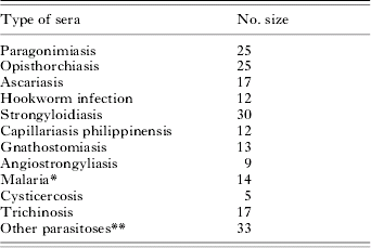
Sera were obtained from serum banks of different departments of the Faculty of Medicine, Khon Kaen University (1992–2003). Each serum was aliquoted and stored at −70°C until used. The study protocol was approved by the Human Research Ethics Committee of Khon Kaen University. Informed consent was obtained from all adult participants or the parents or legal guardians of minors. Twenty-five sera were obtained from parasitologically confirmed cases of F. gigantica infection. To evaluate potential cross-reactivity, 212 serum samples from individuals with parasitic diseases other than fascioliasis were obtained from parasitologically confirmed cases (Table 1). Thirty-two sera were obtained from patients who were residents of areas endemic for Opisthorchis viverrini, whose pathological features were compatible with a diagnosis of bile duct cancer or cholangiocarcinoma (9 of them were proven opisthorchiasis-associated cholangiocarcinoma). Negative control sera were obtained from 57 healthy adults whose stool examinations were performed at the same time of the blood collections using the formalin-ether concentration method (Erdman, Reference Erdman1981). No evidence for intestinal parasitic infections was found. The human sera were pooled as positive and negative reference sera by combining equal volumes of proven fascioliasis and healthy control sera, respectively.
The type of sera of parasitic diseases other than fascioliasis

* 8 with Plasmodium falciparum; 5 with Plasmodium vivax and 1 mixed infection with P. falciparum and P. vivax.
** 5 patients infected with Echinostoma spp.; 4 patients infected with Taenia spp.; 4 patients infected with Trichuris trichiura; 3 patients infected with Entamoeba histolytica; 2 patients infected with Giardia lamblia; 4 patients infected with T. trichiura and Ascaris lumbricoides; 3 patients infected with T. trichiura and hookworm; 4 patients infected with T. trichiura, A. lumbricoides and hookworm; 1 patient infected with T. trichiura, Strongyloides stercoralis and hookworm; 3 patients infected with T. trichiura, A. lumbricoides, S. stercoralis and hookworm.
Peptide-based enzyme-linked immunosorbent assay (peptide-based ELISA)
The method was performed as previously described (Intapan et al. Reference Intapan, Tantrawatpan, Maleewong, Wongkham, Wongkham and Nakashima2005) with some modifications. Each well of the ELISA plate was sensitized with 0·5 μg of each peptide in 0·1 ml of 0·01 m phosphate buffer (pH 7·5) at 4°C overnight. The wells were washed 5 times with 10 mm PBS (pH 7·4) containing 0·05% Tween 20 (PBS/T) and blocked with 2% BSA in PBS/T for 1 h at room temperature. After washing with PBS/T, the wells were incubated at 37°C for 1 h with 0·1 ml of human sera diluted 1:100 with 1% BSA in PBS/T. After another washing step with PBS/T, a peroxidase-conjugated anti-human IgG4 subclass (Zymed, South San Francisco, CA) diluted 1:10 000 with 1% BSA in PBS/T was used as secondary antibody. The wells were then washed with PBS/T and incubated with 0·1 ml orthophenylene diamine (OPD) as substrate for 30 min. The reaction was stopped with 0·05 ml of 8 m H2SO4 and the optical density (OD) was measured at 492 nm using a microplate ELISA reader (Tecan, Salzburg, Austria).
Data analysis
The statistical analysis was performed by Student's t-test using the statistical software of Sigma Stat (San Rafael, CA). The mean OD value+7 s.d. of the healthy group sera was used as the lower limit of positivity. The diagnostic sensitivity, specificity, accuracy and predictive values were calculated and expressed using the method of Galen (Reference Galen1980).
RESULTS
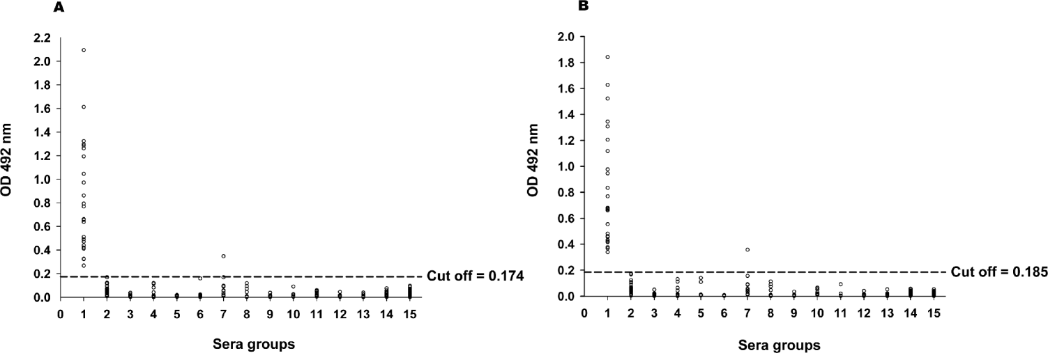
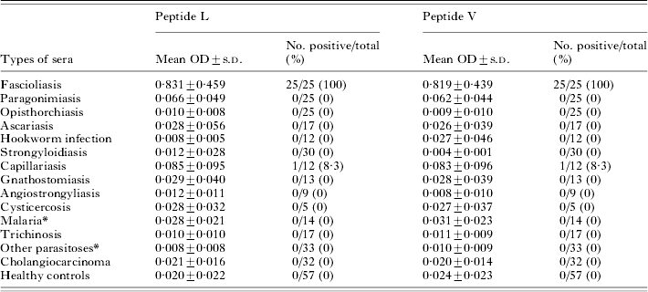
The mean OD values and the number of positive sera for the specific IgG4 subclass antibody against the two synthetic peptides by ELISA for all serum groups are shown in Fig. 1 and Table 2. The mean OD values against both peptides were significantly higher in the proven fascioliasis group than in the group of parasitic diseases other than fascioliasis, the cholangiocarcinoma patients and the healthy controls (P<0·001). When the mean OD value plus 7 s.d. of the negative control sera from the healthy control group was used as the cut-off limit between positivity and negativity for fascioliasis, values of 0·174 and 0·185 absorbance units were obtained for peptide L and peptide V, respectively. The sensitivity, specificity, accuracy and positive and negative predictive values of both peptide antigens were similar and calculated as 100%, 99·7%, 99·7%, 96·2% and 100%, respectively. Only 1 serum sample from a heterologous infection cross-reacted with both peptide antigens. It originated from a patient infected with Capillaria philippinensis. We assessed the reproducibility of the ELISA by using pooled positive and negative conference sera. Coefficients of between-day variations showed that both peptide L- and peptide V-based ELISAs have no day-to-day variations (data not shown).
Absorbance of the ELISA (at 492 nm) using peptide L (A) and peptide V (B) antigens for the detection of specific IgG4 subclass antibody in sera from cases with proven fascioliasis, parasitic diseases other than fascioliasis, cholangiocarcinoma and healthy controls. Sera groups: 1=fascioliasis; 2=paragonimiasis; 3=opisthorchiasis; 4=ascariasis; 5=hookwork infection; 6=strongyloidiasis; 7=capillariasis; 8=gnathostomiasis; 9=angiostrongyliasis; 10=cysticercosis; 11=malaria; 12=trichinosis; 13=other parasitoses; 14=cholangiocarcinoma; 15=healthy controls.

Absorbance values (at 492 nm) and number of positive sera for IgG4 subclass antibody against peptide L and peptide V antigens by indirect ELISA

* See Table 1.
DISCUSSION
Many investigators reported that the detection of specific IgG isotype antibody could improve the sensitivity and specificity of diagnostic tests for various parasitic infections such as schistosomiasis (Iskander et al. Reference Iskander, Das and Aalberse1981; Jassim et al. Reference Jassim, Hassan and Catty1987), filariasis (Ottesen et al. Reference Ottesen, Skvaril, Tripathy, Poindexter and Hussain1985; Chanteau et al. Reference Chanteau, Plichart, Spiegel, Martin and Cartel1991; Egwang et al. Reference Egwang, Nguiri, Kombila, Duong and Richard-Lenoble1993), strongyloidiasis (Genta and Lillibridge, Reference Genta and Lillibridge1989), trichuriasis (Lillywhite et al. Reference Lillywhite, Bundy, Didier, Cooper and Bianco1991), gnathostomiasis (Anantaphruti et al. Reference Anantaphruti, Nuamtanong and Dekumyoy2005), angiostrongyliasis (Intapan et al. Reference Intapan, Maleewong, Sawanyawisuth and Chotmongkol2003) and hookworm infection (Loukas et al. Reference Loukas, Opdebeeck, Croese and Prociv1996). Interestingly, the detection of specific IgG4 antibody could increase the diagnostic values for the serodiagnosis of fascioliasis using both native and recombinant Fasciola cathepsin L (O'Neill et al. Reference O'Neill, Parkinson, Strauss, Angels and Dalton1998, Reference O'Neill, Parkinson, Dowd, Strauss, Angels and Dalton1999; Strauss et al. Reference Strauss, O'Neill, Parkinson, Angels and Dalton1999; Wongkham et al. Reference Wongkham, Tantrawatpan, Intapan, Maleewong, Wongkham and Nakashima2005). In the present study we report the development of a highly specific and sensitive peptide-based immunoassay by the detection of specific IgG4 antibody and its use for the serodiagnosis of human fascioliasis.
Recently, we also revealed that the sensitivity, specificity, accuracy, positive predictive value and negative predictive value of both peptide L and peptide V antigens for the detection of specific total IgG antibody were 100%, 97·3%, 97·5%, 61·9% and 100%, respectively (Intapan et al. Reference Intapan, Tantrawatpan, Maleewong, Wongkham, Wongkham and Nakashima2005). The cut off values for peptide L and peptide V by the detection of IgG4 antibody were lower than those calculated for the detection by total IgG ELISA and also showed a better discrimination between positivity and negativity. The sensitivity and negative predictive value of this study were similar to our previous report, whereas the specificity, accuracy and positive predictive value were increased with both peptide antigens. These results agree with those of previous reports on specific IgG4 antibody detection for the serodiagnosis of fascioliasis (O'Neill et al. Reference O'Neill, Parkinson, Strauss, Angels and Dalton1998, Reference O'Neill, Parkinson, Dowd, Strauss, Angels and Dalton1999; Strauss et al. Reference Strauss, O'Neill, Parkinson, Angels and Dalton1999; Hassan et al. Reference Hassan, Mostafa, Ramadan, Nassar, Hassounah and Omar2000, Reference Hassan, Matar, Mowafy, Saleh, el-Badrawy el, Awad and Ramadan2002; Wongkham et al. Reference Wongkham, Tantrawatpan, Intapan, Maleewong, Wongkham and Nakashima2005). The increased specificity of the assay may also result from the assumptions that clinical manifestations of human fascioliasis are only present in chronic cases and that specific IgG4 antibody against peptide L and peptide V antigens are especially prominent in the total IgG responses when antigenic exposure is chronic or parasitic infections are long-lasting (Ottensen et al. Reference Ottesen, Skvaril, Tripathy, Poindexter and Hussain1985). Hence, IgG4 was the predominant reactive antibody in human fascioliasis sera when selected B-cell epitopes of F. gigantica cathepsin L1 were used to capture specific IgG subclasses.
However, one serum from a C. philippinensis infected individual gave cross-reactions with the peptide L and peptide V antigens, which could have either resulted from an antibody response to a subclinical infection of Fasciola spp. or from the relative similarity of the two antigenic repertoires of the Fasciola spp. and Capillaria spp. It could be argued, however, that cross-reaction was rather unlikely to be the cause of the positive result, since only 1 out of 12 capillariasis samples gave a positive result. In any case, these IgG4-peptide ELISAs present a great advantage as they can differentiate human fascioliasis from paragonimiasis, gnathostomiasis, and trichinosis, all diseases with a high prevalence in Asia (Blair et al. Reference Blair, Xu and Agatsuma1999; McCarthy and Moore, Reference McCarthy and Moore2000; Takahashi et al. Reference Takahashi, Mingyuan and Waikagul2000; Pozio, Reference Pozio2001; Moore et al. Reference Moore, McCroddan, Dekumyoy and Chiodini2003).
The peptide L and peptide V antigens differed by only 1 amino acid, their cut off values were slightly apart, but their diagnostic values were equal. This equal outcome may be explained by the fact that the antigenic sites of peptide L and peptide V which reacted with the specific IgG4 antibody are made of amino acid residues at position 14 other than leucine or valine residues. These results suggest that both peptides can be used as a potential antigen in IgG4-ELISAs for the serodiagnosis of human fascioliasis. In addition, the use of peptide antigens in serological tests is of advantage because synthetic peptide is relatively cheap and identical batches of synthetic peptide can be generated indefinitely resulting in easy reproducibility, in contrast to biological products which tend to vary from batch to batch.
Considering both peptide L and V antigen IgG4-ELISAs as presented in this study, we conclude that these new tests could enhance the specificity, accuracy and positive predictive value without effect on sensitivity and negative predictive value. Furthermore, these IgG4-peptide ELISAs are reliable and could be developed into a serodiagnostic test for human fascioliasis and be used in future large-scale epidemiological studies of human fascioliasis throughout Southeast Asia where this disease is prevalent.
We are indebted to the Graduate Study Office, Faculty of Medicine, Thammasat University, Rangsit Campus, Pathumthani, Thailand for the use of all facilities for the preparation of this manuscript. This research was supported by a grant from Khon Kaen University, Ministry of University Affairs, Thailand. We also thank Dr Mark Roselieb for improving the English-language presentation of the manuscript.