Takayasu arteritis is a granulomatous chronic panarteritis of unknown aetiology, affecting the aorta along its length and its main branches and can also involve the pulmonary and coronary arteries. Rare in children, the incidence in all ages is estimated at 2.6/1,000,000.Reference Kerr, Hallahan and Giordano 1
Vessel inflammation shows adventitial thickening, leucocytic infiltration of the tunica media, and intimal hyperplasia leading to fibrosis, stenosis, and thrombus formation.
The diagnosis is difficult to make during the acute early phase, in part because patients show non-specific symptoms such as fever, headache, hypertension, muscle pain, arthralgia, and weight loss, and because of the lack of specific biomarkers. Therefore, the disease often goes unrecognised in this phase.Reference Brunner, Feldman and Tyrrell 2
Without proper treatment, the vessel involvement generally progresses to stenosis causing ischaemia in the irrigated territory with significant morbidity and mortality in young patients.Reference Morales, Pineda and Martinez-Lavin 3 , Reference Watson, Brogan and Peart 4
The aim of this article was to review the current management of Takayasu arteritis in children.
Epidemiology and etiopathogenesis
Initially described in the Asian population, Takayasu arteritis is a worldwide disease with an incidence of 1.2–2.6/million per year, depending on the different geographical areas.Reference Fries, Hunder and Bloch 5 , Reference Jain, Sharma and Singh 6
The pattern of involvement of the aorta also shows ethnic differences. Japanese have predominant aortic arch involvement, whereas Asian Indian and American patients show preponderance of thoracic and abdominal aorta involvement. Although rare in children, Kerr et al reported a paediatric population of 30% in their study.Reference Kerr, Hallahan and Giordano 1
The aetiology of Takayasu arteritis is unknown; however, it has been accepted that the inflammatory process begins at the vaso vasorum in the adventitia.
T-cells and macrophages invade the outer layer of the media, progressing from the adventitial side to the intimal side of the vessel. The result is a thickened intima secondary to the depositions of mucopolysaccharides, smooth muscle cells, and fibroblasts. The elastic fibres are replaced by fibrosis, resulting in thickening of all three layers. The chronic process causes vessel stenosis, occlusion, and aneurysmal formation. Autoimmune mechanisms controlled by the HLA and MIC genes appear to accelerate the chronic inflammation, which promotes atherosclerotic changes in the intima.Reference Tann, Tulloh and Hamilton 7 , Reference Numano 8
Genetic aspects
There are different patterns of arterial involvement, expression, and prognosis. Several reports have pointed out that genetic factors might play an important role in the severity of the disease. In 2007, Jeeva et al reported a multiplex family with Takayasu arteritis supporting the role of as-yet-undefined genetic factors.Reference Jeeva, Sajid and Ali 9
HLA-B*52 is the most significant HLA region associated with the disease in Japanese patients and has also been confirmed in cases from Korea, India, Thailand, and Turkey. Interestingly, this allele particularly is associated with a higher incidence of left ventricular wall abnormalities, aortic regurgitation, and an earlier disease onset, whereas the HLA-B*39 allele is associated with the development of renal artery stenosis and HLA B52:01 with Takayasu arteritis and ulcerative colitis.Reference Saruhan-Direskeneli, Hughes and Aksu 10
Diagnosis and assessment of disease activity
The diagnosis of Takayasu arteritis is based on clinical criteria. Several criteria of differing sensitivity and specificity have been proposed.Reference Ishikawa 11 – Reference Ozen, Ruperto and Dillon 13
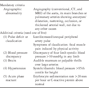
In 2010, the European League Against Rheumatism (EULAR)/Pediatric Rheumatology International Trials Organization (PRINTO)/Pediatric Rheumatology European Society (PRES) validated the Takayasu arteritis criteria for childhoodReference Ozen, Pistorio and Iusan 14 (Table 1).
European League Against Rheumatism (EULAR)/Pediatric Rheumatology International Trials Organization (PRINTO)/Pediatric Rheumatology European Society (PRES) classification criteria of Takayasu arteritis and additional criteria.

Diagnosis requires the presence of vascular imaging abnormalities, such as aneurysms and narrowing, occlusion, or thickening of the arterial wall by angiography, CT scan, MRI, or positron emission tomography in the aorta or in its main branches, plus at least one of the following criteria: decreased peripheral artery pulse(s) or claudication of extremities; discrepancy of four limb blood pressure >10 mmHg; bruits or thrills over the aorta and/or its major branches; hypertension; and abnormal acute phase reactants, erythrocyte sedimentation rate, or C-reactive protein.
In 1996, Numano developed a new classification for a more precise angiographic diagnosis (Table 2).
Angiographic classification of Takayasu arteritis.

Source: Hata et al.Reference Hata, Noda, Moriwaki and Numano 15
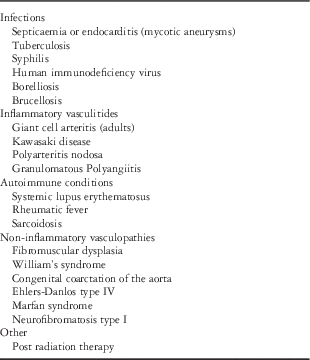
Nevertheless, the diagnosis of Takayasu arteritis in children continues to be challenging due to several factors and a vast spectrum of differential diagnoses (Table 3). In the acute phase of the disease the clinical picture is not specific and sensitive biomarkers for Takayasu arteritis are scarce. Symptoms such as fever, night sweats, weight loss, arthralgia, myalgia, abdominal pain, headache, anaemia, and increased erythrocyte sedimentation rate are common, but without clinical awareness and suspicion this phase usually remains undiagnosed.
Differential diagnosis for Takayasu arteritis.

This delay in the diagnosis results in an insidious progression to a chronic stenotic phase with progressive arterial narrowing causing tissue ischaemia.Reference Kerr, Hallahan and Giordano 1 – Reference Morales, Pineda and Martinez-Lavin 3
Mathew et al have reported that one-third of children present with significant sequelae with apparently inactive disease.Reference Mathew, Goel and Kumar 16 According to a different series, the time from symptom onset to diagnosis ranges between 2 and 11 years and is four-fold longer than in adult Takayasu arteritis patients.
Clinical features at presentation differ between adults and children, although in both arterial hypertension is the most frequent sign, accounting for 82% in the paediatric population.Reference Morales, Pineda and Martinez-Lavin 3 , Reference Eleftheriou, Batu and Ozen 17 In children, other symptoms are headaches (31%), fever (29%), dyspnoea (23%), weight loss (22%), vomiting (20%), and arthralgia-myalgia (14–65%).Reference Morales, Pineda and Martinez-Lavin 3 , Reference Watson, Brogan and Peart 4 , Reference Eleftheriou, Batu and Ozen 17 According to the majority of the series, musculoskeletal symptoms account for only 14% in children; however, in Morales et al in a study in Mexico described an incidence of up to 65%, describing arthritis and heart failure provoking confusion with rheumatic fever.Reference Morales, Pineda and Martinez-Lavin 3
Other uncommon manifestations are retinopathy and skin lesions such as nodules, rashes, and lymphadenopathy. Skin ulcers have not been reported in children.
Adult series report bruits (48%) and claudication (27%), but rarely musculoskeletal symptoms. Decreased pulses and murmurs are common in advanced Takayasu arteritis.
The chronic or late occlusive phase poses the challenge of defining whether acute vasculitis activity is absent or minimal and deciding treatment options for each patient.
In the laboratory tests, acute phase reactants may be elevated in the active phases of the disease. It has been demonstrated that erythrocyte sedimentation rate is more sensitive than C-reactive protein; however, both still have poor sensitivity and specificity.Reference Moriwaki and Numano 18 , Reference Dagna, Salvo and Tiraboschi 19 The novel biomarker pentraxin-3 is a protein rapidly produced in response to an inflammatory reaction, especially by endothelial cells.Reference Ciéslik and Hrycek 20 Plasma biomarker pentraxin-3 levels higher than 1 ng/ml are more accurate than normal thresholds of C-reactive protein or erythrocyte sedimentation rate to distinguish active from inactive disease, as demonstrated by Dagna et al, although this marker should not be used in the clinics until these findings are confirmed in larger series of patients.Reference Dagna, Salvo and Tiraboschi 19
Currently, an increase in the number of CD8 cells with reversal of T cell CD4:CD8 ratio is considered an important marker of disease activity.Reference Uppal and Verma 21
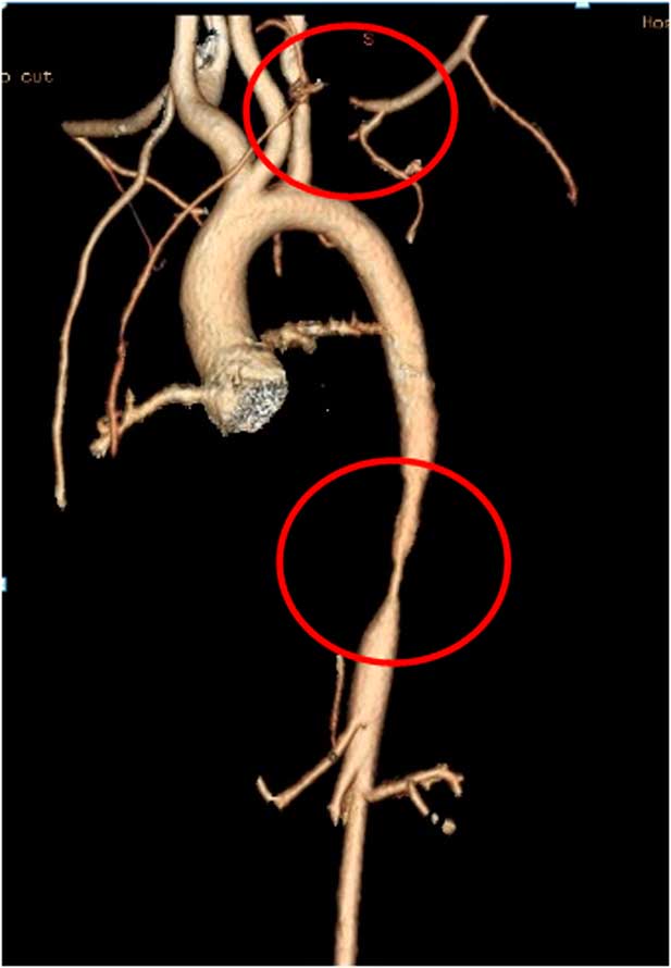
For the diagnosis as well as follow-up of Takayasu arteritis in children a combination of angiography, CT, MRI, or positron emission tomography is used to detect lumen and vascular wall abnormalitiesReference Brunner, Feldman and Tyrrell 2 , Reference Perera, Mason and Wolfe 22 (Fig 1).
3 D reconstruction multislice computed tomography angiography. Aortogram demonstrating severe involvement of left subclavian artery and abdominal aorta.

Cardiac imaging assessment begins with echocardiography evaluating ventricular function, hypertrophy, aortic valve function, and aortic and coronary arteries, followed by a cardiac MRI.Reference Eleftheriou, Varnier and Dolezalova 23
Although digital subtraction arteriography is considered the gold standard for “lumenography”, MRI and computed angiotomography are useful to assess lumen vessel characteristics.Reference Eleftheriou, Batu and Ozen 17 , Reference Mavrogeni, Dimitroulas, Chatziioannou and Kitas 24
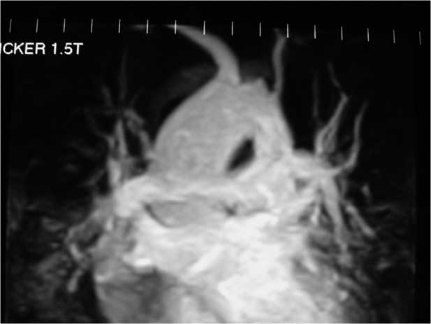
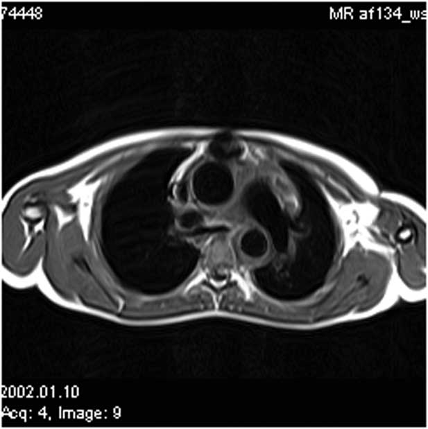
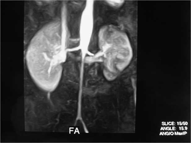
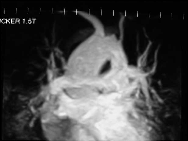
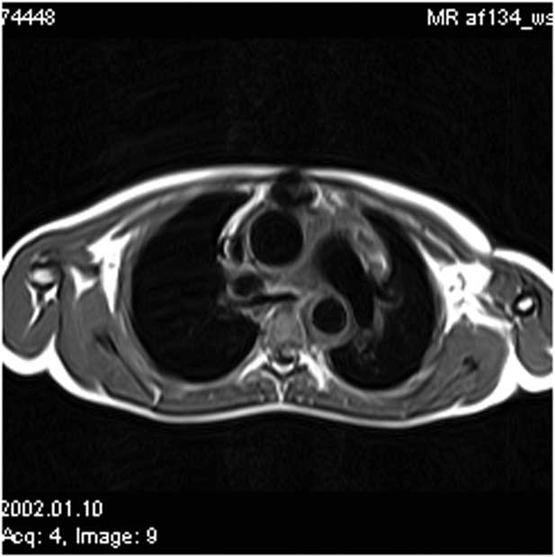
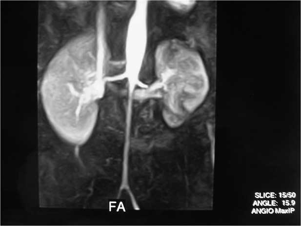
MRI is a sensitive and accurate method that allows imaging of the aorta and its major branches without radiation. Contrast-enhanced MRI can detect structural abnormalities of the vessel walls, such as thickening, oedema, and increased vascularity, and is an important tool not only for the diagnosis of Takayasu arteritis but for disease follow-up as wellReference Mavrogeni, Dimitroulas, Chatziioannou and Kitas 24 – Reference Mavrogeni, Servos and Smerla 26 (Figs 2–4). In the recent years recommendations on the role of MRI in connective tissue diseases have been published, pointing out the need for tools that enable early diagnosis of cardiovascular involvement and the potential utilisation of cardiac MRI to clarify aspects of cardiac pathophysiology in connective tissue diseases.Reference Mavrogeni, Markousis-Mavrogenis and Koutsogeorgopoulou 27
Contrast-enhanced MRA showing severe left carotid artery stenosis involving its origin.
Axial T1 weighted magnetic resonance image. The descending aorta shows a thickened wall.
Contrast-enhanced MRA showing significantly decreased caliber and irregularity of the descending abdominal aorta from infra-renal abdominal aorta involving the iliac arteries
Some studies have suggested a close association between wall thickness, oedema, and enhancement of the vessel wall by MRI and disease activity, but no consistent correlation has been confirmed.Reference Choe, Han, Koh, Do and Lee 28 , Reference Andrews, Al-Nahhas and Pennell 29 Tso et al highlighted that sustained vessel wall oedema reflects both activity and damage. MRI revealed vessel wall oedema during active disease in 94% of patients and in 56% of those considered in clinical remission.Reference Tso, Flamm, White, Schvartzman, Mascha and Hoffman 30
MRI is a time-consuming study, needs specific expertise, and is expensive. Its main disadvantages include overestimation of stenotic arteries as occluded (2%) and poor visualisation of small vessels and vascular calcification.Reference Mavrogeni, Dimitroulas, Chatziioannou and Kitas 24
18F-fluorodeoxyglucose positron emission tomography provides information on large-vessel vasculitis with a reported sensitivity and specificity of 73–100% and 83–92%, respectively, in adults.Reference Karapolat, Kalfa and Keser 31 , Reference Fuchs, Briel and Daikeler 32 Nevertheless, the radiation dose is high, the modality is available at few medical centres, and a negative result does not rule out inflammatory activity.Reference Eleftheriou, Varnier and Dolezalova 23
A newer modality, the positron emission tomography/MRI is expected to improve the diagnosis and monitoring of Takayasu arteritis, but meanwhile digital subtraction arteriography remains the gold standard for diagnosis.
Currently, however, there is no gold standard for monitoring disease activity. In an autopsy study of 10 patients with Takayasu arteritis in India, Sharma et al identified persistent disease activity with no detectable clinical signs.Reference Sharma, Jain and Radotra 33 Kerr et al and Cong et al also reported active vasculitis in aortic biopsies of 44 and 58.3% of patients, respectively, with clinically inactive Takayasu arteritis.Reference Kerr, Hallahan and Giordano 1 , Reference Cong, Dai and Feng 34 Clearly, acute phase reactants are unable to detect low levels of inflammatory activity.
Unlike in children, in adults the Indian Takayasu Arteritis Activity Score (ITAS2010) and the Disease Extent Index-Takayasu (DEI Tak) have been validated to assess Takayasu arteritis disease activity and extent, although their usefulness has been reported.Reference Aydin, Yilmaz and Akar 35 , Reference Misra, Danda and Rajappa 36 For children the Pediatric Vasculitis Activity Score (PVAS) is the only validated score.Reference Dolezalova, Price-Kuehne and Özen 37
Treatment
In Takayasu arteritis early treatment is crucial to resolve or alleviate the inflammation and prevent complications and disease progression. Corticosteroids are the first-line treatment. A large section of patients requires the use of combined therapies. Eleftheriou et al described that more than one-half (54%) of the patients required treatment with biological agents. The reasons were either steroid-related complications and inadequate disease control or to prevent relapses when steroid dose is lowered or discontinued.Reference Eleftheriou, Varnier and Dolezalova 23
Options for add-on medications are immunosuppressant drugs and biological agents.
The use of immunosuppressant drugs, such as methotrexate, cyclophosphamide, azathioprine, and mycophenolate mofetil, has been shown to be safe and effective as a second agent to achieve sustained remission, improve vascular lesions, and decrease steroid doseReference Misra, Sharma, Kadhiravan and Negi 38 ; however, side effects are not minor and should be taken into account when indicating these drugs.
Recently, Misra et al, reviewing the use of non-biologic disease modifying anti-rheumatic drugs in the management of large-vessel vasculitis, emphasised the paucity of high-quality evidence to guide the use of these drugs in Takayasu arteritis, as most studies have small sample sizes and heterogeneous outcome measures. Overall, methotrexate, leflunomide, azathioprine, mycophenolate mofetil, and cyclophosphamide were effective in Takayasu arteritis with low-quality evidence.Reference Misra, Sharma, Kadhiravan and Negi 38 , Reference Clemente, Hilário and Lederman 39
Biological agents, such as anti-tumour necrosis factor agents (infliximab, etanercept, adalimumab) and anti-IL6 therapy (tocilizumab) have been used with variable effectiveness. Osman et al published a review and meta-analysis pointing out that all the evidence supporting the use of biological agents for large-vessel vasculitis comes from case series (in contrast to randomised-controlled trials) suggesting that tocilizumab and infliximab may be effective in the management of large-vessel vasculitis and refractory Takayasu arteritis, respectively.Reference Stern, Silva, Reiff, Ramos, Marzan and Terreri 40 , Reference Osman, Pagnoux, Dryden, Storie and Yacyshyn 41
There have been important therapeutic advances in Takayasu arteritis, but neither meta-analyses nor randomised-controlled trials are available to guide treatment in children. The primary objective of treatment is inducing and maintaining remission of the disease. Medical treatment with glucocorticoids combined with antihypertensive and antiplatelet drugs have shown good efficacy in most cases. Children with corticosteroid resistance should receive high-dose corticosteroids combined with another immunosuppressant agent, usually methotrexate, in order to avoid irreversible vessel damage. In summary, methotrexate, azathioprine, mycophenolate mofetil, and cyclophosphamide can be used in children as first- or second-line agents. Biological agents such as tocilizumab and tumour necrosis factor antagonists appear to be effective, particularly in adult patients with a severe disease course or those who are refractory to corticosteroids and/or conventional immunosuppressive drugs. In children the results are promising but long-term multicenter studies are lacking.
Surgical and endovascular repair
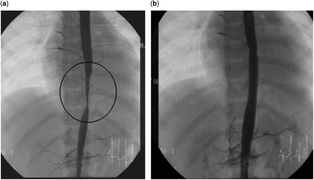
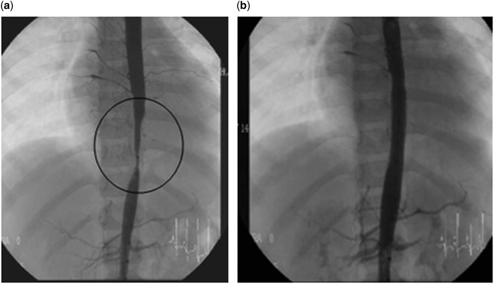
Many patients show symptoms related to stenotic or occlusive lesions in different organs causing uncontrolled hypertension secondary to renal artery stenosis, symptomatic coronary ischaemia, cerebrovascular disease, severe aortic regurgitation, limb ischaemia, and aneurysms at risk for rupture. Reported incidences of aneurysms in children, especially in the abdominal aorta, ranged from 41 to 65%. Performing surgical or endovascular procedures for revascularisation is essential (Fig 5).
(A) Aortic angiogram : Long, diffuse stenosis of abdominal aorta before stent angioplasty. (B) Aortic angiogram: note marked improvement in the diameter of the aorta immediately after stent angioplasty
Although both surgical and endovascular procedures can be performed, surgical intervention has shown better long-term outcomes.Reference Lee, Jeon and Do 42 Nevertheless, the goal is to control the inflammatory activity previous to either procedure.
In a multicenter study of 79 patients, of whom 62.7% underwent surgery and 37.3% endovascular repair, Saadoun et al observed the following complications: restenosis in 67%, thrombosis in 8.8%, bleeding in 7.6%, and stroke in 5% after a follow-up of 6.5 years. Complication rate in the surgical procedure group was 37.5% compared with 62% in the endovascular repair group. The authors pointed out that biological inflammation increased the likelihood of complications after revascularisation.Reference Saadoun, Lambert, Mirault and Resche-Rigon 43
Furthermore, Miyata et al demonstrated that in patients with major complications and progressive disease surgery increased long-term survival.Reference Miyata, Sato, Koyama, Shigematsu and Tada 44
The technique of percutaneous transluminal angioplasty is evolving. More effective and safe materials adapted to small patients make it a valid option to treat these lesions. Nevertheless, in Takayasu arteritis the vessels are more firm, scarred, and fibrotic and carry the risk for severe complications especially when interventions are performed on the cerebral arch vessels.Reference Singh, Hughes and Sebire 45 Comparing the results after endovascular treatment and restenosis rate in atherosclerotic disease and Takayasu arteritis showed more variability and restenosis in the latter.
Tiagy et al described percutaneous transluminal balloon angioplasty for stenosis of the aorta due to aortic arteritis in 45 lesions in 41 children. The procedure was successful in 91.1% with a restenosis rate of 19%. The authors concluded that restenosis was more common in patients with long-segment (>3 cm) than in those with short-segment (<3 cm) stenosis (30 versus 9.1%). Whereas surgical experience of complex vascular lesions is poor in paediatrics, and while the bypass graft rapidly becomes inadequate because of growth of the child, the authors state that balloon angioplasty appears to be the treatment of choice, especially for discrete stenosis of the aorta.Reference Tyagi, Verma and Gambhiret 46
Follow-up and prognosis
Takayasu arteritis is associated with significant morbidity and mortality in young patients. Mortality rates vary according to several factors: geographical location, severity and extension of the lesions, treatment strategies, time of follow-up, and whether early or late series are described.
While most publications show mortality rates ranging from 16 to 40%, Perera et al described mortality rates ranging from 3% in the United States of America to 11% in India, and Eleftheriou et al analysed a case series of 11children with a mortality rate of 27% related to younger age at presentation.Reference Morales, Pineda and Martinez-Lavin 3 , Reference Jain, Sharma and Singh 6 , Reference Eleftheriou, Batu and Ozen 17 , Reference Tyagi, Verma and Gambhiret 46
Using a combination of prognostic factors – major complications, progressive course, and low erythrocyte sedimentation rate (<20 mm/h) – Ishikawa and Maetani published a classification establishing three stages, where patients with both major complications and a progressive course (stage 3) had the worst prognosis (43% survival at 15 years), whereas in those without a progressive course but with an elevated erythrocyte sedimentation rate (stage 1), the survival rate was 100% at 15 years.Reference Jeeva, Sajid and Ali 9
In summary, over the past decade the prognosis of Takayasu arteritis has improved. Probably, greater awareness among clinicians and the development of non-invasive diagnostic imaging tools have led to earlier diagnosis. The development of a new generation of immunosuppressive drugs and biological agents combined with classical steroids have been an important contribution to the control of disease activity. Advances in surgical technology and perioperative care in addition to the role of endovascular treatment, which has increasingly become a less invasive option, have improved the overall results. However, in spite of these developments, the management of childhood Takayasu arteritis remains a challenge as diagnosis is still often late and currently there is no sensitive and specific gold standard for the monitoring of inflammatory disease.
Acknowledgements
None.
Financial Support
This research received no specific grant from any funding agency, commercial or not-for-profit sectors.
Conflicts of Interest
None.