Introduction
Wegener's granulomatosis is a potentially life-threatening systemic autoimmune condition that characteristically involves the lungs, kidneys and upper respiratory tract; however, any organ system may be involved.
Wegener's granulomatosis, together with microscopic polyangiitis and Churg–Strauss syndrome, are considered anti-neutrophil cytoplasmic antibody (ANCA) associated vasculitides that specifically involve small to medium-sized vessels.Reference Oristrell, Bejarano, Jordana, Monteagudo, Marí and Casanovas1 Histologically, a necrotising vasculitis with granulomata occurs without immune deposits.Reference Del Pero, Chaudhry, Jones, Sivasothy, Jani and Jayne2 The exact cause of Wegener's granulomatosis is unknown, but it seems to have characteristics of an autoimmune disease.
Cytoplasmic ANCAs are considered the serological hallmark of Wegener's granulomatosis. According to Popa et al., cytoplasmic ANCAs may be formed against complementary peptide sequences to proteinase-3.Reference Popa and Tervaert3 It is thought these ‘anti-sense’ antibody sequences may arise endogenously or be introduced exogenously via an infectious source.Reference Popa and Tervaert3Staphylococcus aureus has been implicated as the most likely infectious source as it exhibits a protein with a similar peptide sequence to that of proteinase-3.
The clinical presentation of Wegener's granulomatosis can vary from a slow insidious onset occurring over a few months to a less common fulminant and possibly fatal onset over a few days. Seventy-three per cent of patients will initially present with head and neck manifestations.Reference Erickson and Hwang4 In 25 per cent of cases, Wegener's granulomatosis will present in a more limited form affecting one system only, usually the upper respiratory tract or lungs. Patients with this limited form of the disease are generally younger, have a greater likelihood of recurrence and are less likely to be ANCA-positive.Reference Khan, Lawson, Quinn, Isdale and Green5
Current first-line induction therapy for Wegener's granulomatosis consists of cyclophosphamide with glucocorticoids, usually prednisolone. For most patients (70–90 per cent), this regimen will be successful.Reference Sanchez-Cano, Callejas-Rubio and Ortego-Centeno6 However, cyclophosphamide is associated with multiple side effects, and long-term glucocorticoid therapy is associated with increased morbidity.Reference Stone, Merkel, Spiera, Seo, Langford and Hoffman7
Once the disease is in remission, methotrexate or azathioprine, and low dose prednisone are used to prevent relapses.Reference de Groot, Rasmussen, Bacon, Tervaert, Feighery and Gregorini8, Reference Jayne, Rasmussen, Andrassy, Bacon, Tervaert and Dadoniené9 Most patients respond to this regimen, but resistant cases are problematic.
Recent literature has reported cases of Wegener's granulomatosis treated with rituximab. Rituximab is a biological agent currently approved for the treatment of lymphoma and rheumatoid arthritis.Reference Oristrell, Bejarano, Jordana, Monteagudo, Marí and Casanovas1 It has been successfully used in the treatment of various autoimmune disorders that are refractory to conventional therapy and has recently been introduced as a form of treatment for Wegener's granulomatosis.
We present a patient with the acute, resistant fulminant form of Wegener's granulomatosis who displayed a spectacular response to the use of rituximab.
Case report
A 28-year-old previously healthy man was referred to our unit with apparent complicated sinusitis. He presented with a three-week history of nasal obstruction, purulent rhinorrhoea and frontal headaches that did not respond to oral antibiotics. Examination revealed mild right proptosis, purulent nasal discharge and inflamed nasal mucosa. Bilateral antral washouts and frontal trephines were performed. The patient's post-operative recovery was unremarkable.
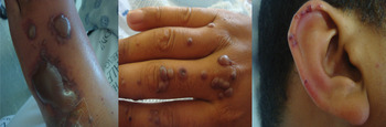
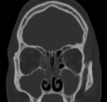
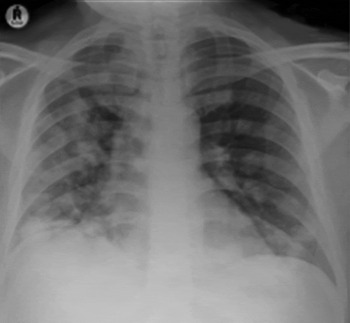
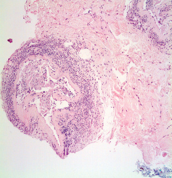
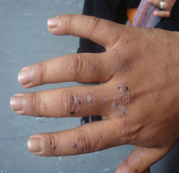
At the follow up one week later, the patient was severely ill; he was wheelchair-bound, with rigors and spiking fever. Examination revealed a generalised skin rash and polyarthritis. Photographs of the patient, which show suspected necrotising vasculitis involving multiple skin sites, are presented in Figure 1. His nose was completely obstructed with inflamed polypoid mucosa. Computed tomography of the sinuses confirmed bilateral sinus opacification (Figure 2). Blood tests revealed an elevated inflammatory response (C-reactive protein = 219 mg/l, erythrocyte sedimentation rate = 98 mm/h) and positive cytoplasmic anti-neutrophil cytoplasmic antibody levels (87 IU/ml). A chest X-ray showed diffuse bilateral, multinodular infiltrates (Figure 3). Skin biopsy confirmed necrotising vasculitis (Figure 4).
Photographs of the patient showing necrotising vasculitis involving multiple skin sites.

Coronal computed tomography image of the sinuses showing bilateral sinus opacification.

Chest X-ray showing diffuse bilateral multinodular infiltrates.

Photomicrograph of the skin biopsy confirming necrotising vasculitis (H&E; ×40).

Treatment with high dose prednisone and cyclophosphamide was commenced immediately. Despite the treatment, the patient had multiple relapses and developed renal failure and subglottic stenosis. His condition deteriorated over the next four weeks. Funding for a single course of rituximab (375 mg/m2) was eventually approved, and within two weeks of treatment his symptoms and signs improved remarkably (Figure 5). The disease was still in remission at 24 months' follow up.
Photograph of the patient following two weeks' treatment with rituximab; his symptoms and signs showed remarkable improvement.

Discussion
In 1936, Friederich Wegener first described a disease characterised by necrotising granulomata of the upper and lower respiratory tract, focal glomerulonephritis, and necrotising systemic vasculitis. The annual incidence of Wegener's granulomatosis has been estimated to be 8–10 cases per million people.Reference Ntatsaki, Watts and Scott10 Wegener's granulomatosis is a multi-systemic disease and can therefore present with various clinical manifestations aside from the classic triad (lungs, kidneys and upper respiratory tract) mentioned earlier.
Wegener's granulomatosis generally has a gradual onset, and symptoms have often been present for a few months prior to presentation. The acute fulminant form of the disease is rare, and its incidence, unsurprisingly, has not been reported in the literature. All the cases mentioned in the literature were potentially life-threatening and the diagnosis was often delayed. The clinical presentations of the acute form were often atypical and varied, ranging from epistaxis, myocardial infarction and diffuse alveolar haemorrhage, to acute respiratory and renal failure.Reference Thapar, George and Pfleiderer11–Reference Miszalski-Jamka, Szczeklik, Sokołowska, Miszalski-Jamka, Karwat and Grzadziel13 Our case is unique in that it is the first published case to present in the fulminant form and masquerade as an acute complicated sinusitis. We believe it is important that most clinicians are made aware of this possibility.
The aims of treatment for Wegener's granulomatosis are firstly to induce remission (induction therapy) and secondly to keep the patient in remission (maintenance therapy). Prednisone and cyclophosphamide are used as first-line agents for induction therapy. Cyclophosphamide impairs DNA replication and transcription, and has numerous side effects including cystitis, opportunistic infection, malignancy, infertility and cytopenia.Reference Erickson and Hwang4 To decrease the side effect profile of daily cyclophosphamide treatment, some authors have attempted an intermittent pulse-dosing regimen. This has resulted in similar success to that of remission induction therapy, but using only half the total dose of the daily regimen.Reference Haubitz, Schellong, Gobel, Schurek, Schaumann and Koch14, Reference Guillevin, Cordier, Lhote, Cohen, Jarrousse and Royer15 Randomised controlled trials by De Groot et al. and Jayne et al. suggest that exposure to cyclophosphamide could be further decreased by substituting it with methotrexate or with azathioprine.Reference de Groot, Rasmussen, Bacon, Tervaert, Feighery and Gregorini8, Reference Jayne, Rasmussen, Andrassy, Bacon, Tervaert and Dadoniené9 In their studies, methotrexate was used as an induction agent, but only in patients with limited, mild forms of Wegener's granulomatosis. Azathioprine was used for maintenance therapy after just three months of induction therapy with cyclophosphamide. Maintenance therapy utilises either azathioprine or methotrexate, with tapered doses of prednisone to prevent relapses. Although most patients respond to the above regimen, 75 per cent will have a flare-up of the disease. This has motivated the study of alternative forms of treatment, such as leflunomide, intravenous immunoglobulin and rituximab.Reference Fortin, Tejani, Bassett and Musini16, Reference Henes, Fritz, Koch, Klein, Horger and Risler17
The presence of B lymphocytes is essential for the regulation of the immune response and the production of antibodies. They have multiple functions, which include expressing co-stimulatory molecules, producing cytokines, functioning as antigen-presenting cells, and regulating the activation and differentiation of T lymphocytes and dendritic cells.Reference Keogh, Ytterberg, Fervenza, Carlson, Schroeder and Specks18 They are also responsible for the production of anti-neutrophil cytoplasmic antibodies (ANCAs), which have multiple pro-inflammatory effects that cause tissue injury and vasculitis.Reference Stone, Merkel, Spiera, Seo, Langford and Hoffman7, Reference de Groot, Rasmussen, Bacon, Tervaert, Feighery and Gregorini8, Reference Keogh, Ytterberg, Fervenza, Carlson, Schroeder and Specks18 Rituximab is a chimeric monoclonal antibody directed against the CD20 antigen, which is a cell surface antigen expressed on B lymphocytes. The binding of the antibody to CD20 results in the selective depletion of B lymphocytes by a variety of different mechanisms. Rituximab has become an important component of standard treatment regimens for non-Hodgkin's B-cell lymphoma.Reference Oristrell, Bejarano, Jordana, Monteagudo, Marí and Casanovas1, Reference Del Pero, Chaudhry, Jones, Sivasothy, Jani and Jayne2, Reference Khan, Lawson, Quinn, Isdale and Green5, Reference Sanchez-Cano, Callejas-Rubio and Ortego-Centeno6, Reference Jayne, Rasmussen, Andrassy, Bacon, Tervaert and Dadoniené9 The antibody showed early success as a treatment for autoantibody-mediated diseases. This was later followed by promising results achieved through B lymphocyte depletion in autoimmune conditions such as rheumatoid arthritis, which until recently was thought to be predominantly T lymphocyte mediated.Reference Edwards, Szczepanski, Szechinski, Filipowicz-Sosnowska, Emery and Close19 Treatment with rituximab is administered as a weekly infusion for four weeks, with a dose of 375 mg per square metre of body surface area.
• Wegener's granulomatosis is generally considered to be a chronic indolent disease
• The acute fulminant form is rare and may be fatal if diagnosis is delayed
• Rituximab is a relatively new treatment for resistant Wegener's granulomatosis
• It has fewer side effects compared with conventional treatment and seems more effective in inducing remission
• This paper reports a rare case of fulminant Wegener's granulomatosis, and illustrates the dramatic effects of rituximab
Rituximab is a relatively new treatment for resistant Wegener's granulomatosis. Approximately 80 cases of Wegener's granulomatosis treated with rituximab have been reported in the literature, most of which are case reports and small case series. In 2010, two randomised controlled trials reported similar remission rates for cases treated with cyclophosphamide and rituximab, and rituximab was shown to be as effective as cyclophosphamide. However, rituximab has been suggested to be superior in resistant cases and in those patients in whom the side effects of cyclophosphamide were to be avoided.Reference Stone, Merkel, Spiera, Seo, Langford and Hoffman7, Reference Jones, Tervaert, Hauser, Luqmani, Morgan and Peh20 Most published reports have revealed that rituximab has an overall positive response, with minimal side effects and infrequent relapses. Despite the decrease in B lymphocytes associated with rituximab use, there is no convincing evidence that rituximab increases the frequency of severe infections when used in the treatment of ANCA-associated vasculitis.Reference Guerry, Brogan, Bruce, D'Cruz, Harper and Luqmani21
Conclusion
The case presented here provides a number of key learning points. Firstly, Wegener's granulomatosis should be included as a differential diagnosis in patients presenting with atypical upper airway symptoms (even acute ones) who do not respond to conventional therapy. Secondly, this paper emphasises the fact that Wegener's granulomatosis may present in an acute life-threatening form; early diagnosis and treatment is essential. Thirdly, the paper highlights the successful experience with rituximab and adds to the evidence in support of this agent.