

Introduction
In recent years, the administration of alkalinizing agents to horses arose with the intent to buffer the decrease in plasma pH that occurs as a consequence of high-intensity exercise. Commonly known as the practice of ‘milkshaking’, this illegal practice has become more sophisticated and is a threat to the health of the horse as well as the integrity of racingReference Rose and Lloyd1–Reference Lloyd and Rose5. Research has addressed this concern by developing methods to detect the administration of alkalinizing agentsReference Irvine2, Reference Lloyd, Reilly and Rose3, Reference Schott and Hinchcliff6–Reference Slocumbe, Huntington, Lind and Vine8. Extensive data have been published to establish plasma total carbon dioxide concentration ([tCO2]) as a valid determinate for the detection of an alkalinizing agentReference Lloyd and Rose5, Reference Lorimer9–Reference Kauffman, Kline, Foreman and Lyman13. According to Lloyd et al. Reference Lloyd, Reilly and Rose3, plasma [tCO2] represents the combined measure of plasma bicarbonate and dissolved carbon dioxide. The actual bicarbonate ion concentration is approximately 95% of the tCO2 valueReference Lloyd, Reilly and Rose3.
As a result of this research and subsequent studies, New Jersey established the practice of testing [tCO2] a minimum of 1 h after the completion of a raceReference Lorimer9. Threshold limits in New Jersey were determined after a study involving 269 horses demonstrated that the mean plasma [tCO2] measured in fit Standardbred horses, tested on a non-race day (where it was highly unlikely that they were given an alkalinizing agent), was 30 mmol l− 1 with the highest [tCO2] reading of 34 mmol l− 1 Reference Lorimer9. This observed mean was used to set the threshold for a non-lasix horse at 37 mmol l− 1, and a value of four standard deviations from the mean was set as the threshold to insure against false positives. Even though plasma [tCO2] is a tightly regulated physiological variable, it can be inadvertently influenced by management factors like feeds that contain small amounts of bicarbonate and other alkalinizing ingredients. The possible effects that external factors may have on plasma [tCO2] have raised concerns from regulators, veterinarians and owners/trainers who wish to avoid anything that may inadvertently cause a horse to exceed the established threshold for a ‘milkshake’ positive. To that end, many studies have been conducted to confirm that external factors do not have a substantial effect in increasing [tCO2] closer to threshold. For example, Kauffman et al. Reference Kauffman, Kline, Foreman and Lyman13 demonstrated that while horses kept on pasture had higher [tCO2] compared with those housed in stalls, the difference was minor. They also demonstrated that three diets, differing in forage/concentrate ratios and forage composition, did not alter plasma [tCO2]Reference Kauffman, Kline, Foreman and Lyman13. Slocumbe et al. Reference Slocumbe, Huntington, Lind and Vine8 demonstrated that transportation had no effect on [tCO2], while fluctuations in breathing had only a minor effect. Excitement was demonstrated to have no effect on plasma [tCO2]Reference Irvine2, Reference Slocumbe, Huntington, Lind and Vine8. IrvineReference Irvine2 also demonstrated that sex or age had no effect on [tCO2]. As regards management factors, Szucsik et al. Reference Szucsik, Baliskonis and McKeever14 demonstrated that plasma [tCO2] is not affected by electrolyte supplements that do not contain alkalinizing materials. Another set of studies demonstrated that pelleted feed supplements, not containing materials like bicarbonate, did not affect plasma [tCO2]Reference McKeever, Szucsik, Balaskonis, Betros, Kearns and Malinowski15. Interestingly, while trained horses appear to have lower plasma tCO2 concentrations compared with untrained horses, there appears to be no effect of short-term detraining analogous to the 3-day quarantine offered by some states as a way to screen whether a horse has a ‘naturally high’ plasma [tCO2]Reference McKeever, Szucsik, Balaskonis, Betros, Kearns and Malinowski15.
Unfortunately, anything that affects whole-body acid–base status, including acid-blocking therapies commonly given to racehorses to treat or prevent gastric ulcers, falls under the category of external factors that could potentially alter [tCO2]. Equine gastric ulcer syndrome (EGUS) affects up to 93% of racehorses, 60% of performance horses and 57% of foals as either a primary illness or secondary to another disorderReference Murray, Grodinskey, Anderson, Radue and Schmidt16–Reference Andrews, Sifferman, Bernard, Hughes, Holste and Daurio18. Omeprazole and ranitidine are two drugs that have been found to be efficacious and practical for maintaining gastric pH when given to horses with EGUS. Ranitidine, commonly known as Zantac, falls under the category of histamine-2 receptor antagonistsReference Murray, Grodinskey, Anderson, Radue and Schmidt16–Reference Andrews, Sifferman, Bernard, Hughes, Holste and Daurio18. Omeprazole, distributed by Merial (Iselin, NJ, USA) under the name Gastrogard, is an effective inhibitor of basal gastric acid secretionReference Murray, Grodinskey, Anderson, Radue and Schmidt16–Reference Andrews, Sifferman, Bernard, Hughes, Holste and Daurio18. Unlike ranitidine, omeprazole is classified as a proton pump blocker. Specifically, it blocks the K+/H+-ATPase pump that catalyses the last step of hydrochloric acid production by parietal cells regardless of a stimulusReference Murray, Grodinskey, Anderson, Radue and Schmidt16–Reference Andrews, Sifferman, Bernard, Hughes, Holste and Daurio18. Both omeprazole and ranitidine are used often in the racing industry to treat EGUS; however, it is unknown whether their effects on gastric acid secretion affect whole-body acid–base as reflected in plasma [tCO2]. This study investigated the effects of these two acid-blocking therapies in potentially increasing plasma [tCO2] using the standards and procedures established by the New Jersey Racing Commission. Specifically, we tested the hypothesis that the administration of omeprazole and ranitidine affects plasma [tCO2] in Standardbred mares.
Materials and methods
Animals
Twelve untrained, healthy Standardbred mares (~520 kg, 9–18 years) accustomed to laboratory procedures and surroundings were used to test the above hypothesis. The Rutgers University Institutional Review Board for the Care and Use of Animals reviewed and approved all the methods and procedures used in this study. During the course of the study, all horses were housed, fed and administered drugs while on pasture. Each mare was fed 3 kg alfalfa and grass hay in addition to 1.5 kg commercial grain twice daily. Water and trace mineral blocks were available ad libitum. Routine maintenance such as hoof trimming occurred prior to beginning the study. One month prior to drug administration, each mare underwent an endoscopic examination that confirmed that none of the mares used in the study had gastric ulcers. Two weeks prior to administering the ulcer-treating drugs, all mares performed a graded incremental exercise test (GXT) to determine the treadmill speed that would elicit maximal oxygen consumption, VO2maxReference McKeever, McKeever, Albeirci, Gordon and Manso Filho19. Oxygen uptake was measured using an open-flow calorimeterReference McKeever, McKeever, Albeirci, Gordon and Manso Filho19.
Experimental design
Each mare received one of the three treatments three times daily (TID) at 06.30, 12.30 and 18.30 hours. The three treatments administered were omeprazole (Merial), ranitidine (Geneva Pharmaceuticals, Inc., Broomfield, CO, USA) and apple sauce as the control. Omeprazole paste was administered orally at a 4 mg kg− 1 daily dose daily [strong ion difference (SID)] in the morning. Horses on omeprazole received control at the later dosing times to encourage good behaviour. Ranitidine was administered TID at 6 mg kg− 1 crushed and mixed in 20 ml control. Horses on control were administered 20 ml sweetened apple sauce TID. Each mare received one of the three treatments for 1 week prior to treadmill exercise. After exercise, they received an equal volume of the control for 48 h prior to starting the next treatment in an effort to prevent any interference between treatments. Drugs, doses and the 48 h withdrawal times were based on recommendations by the respective manufacturers and published literature. Treatments were administered in a random crossover fashion over the course of 5 weeks, with the investigators blind to the treatment given. The 12 mares were divided into three groups for logistic purposes so as to conduct the GXTs between 09.00 and 11.00 hours. Sequence of the testing was maintained through the rotation of treatments.
After being on a drug for 1 week, each horse ran a simulated race test (SRT) on a treadmill (Sato I, Lexington, KY, USA). During each SRT, the horses ran on the treadmill up to a fixed 6% incline for 2 min at a warm-up speed of 4 m s− 1, 2 min at the velocity required to reach VO2max and 2 min at a cool-down speed of 4 m s− 1. The SRT was repeated until each of the horses had received each of the three treatments. On the morning of an SRT, the horses were brought into their stalls 30 min after the drugs had been administered. Each horse received half a bucket of water and one quarter of an alfalfa hay flake. Catheters were inserted percutaneously into the jugular veins using sterile techniques and local lidocaine anaesthesia. The horses then stood quietly for a minimum of 45 min before running the SRT.
Blood sampling
Plasma samples (14 ml) were collected at the following times: pre-exercise; immediately post-exercise; at 60 and 90 min post-exercise. Pre-exercise samples were obtained 15 min prior to exercise. Samples were collected in tubes containing lithium heparin (Vacutainer; Becton Dickinson, Parsippany, NJ, USA) and were stored on ice for not more than 4 h until the time of analysis.
Assay procedures
Plasma was obtained from one 7 ml tube per sampling interval via centrifugation (Model TJA-6; Beckman-Coulter, Brea, CA, USA) at 1500 × g at 4°C for 10 min. An ion-selective analyser (Synchron ELISE, Beckman) was used to determine the plasma concentrations of tCO2, sodium, potassium and chloride ions. Plasma lactate (LA) concentration was measured using an LA analyser (1500 Sport; Yellow Springs Instrument Company, Yellow Springs, OH, USA). The second 7 ml tube of blood collected during each sampling interval was used to measure packed cell volume (PCV) using the microhaematocrit technique and total protein (TP) concentration (mg dl− 1) via refractometry. All samples were analysed in duplicate.
Statistical analysis
Two-way analysis of variance for repeated measures was used to analyse the data for main effects and interactions (SigmaStat, version 2.0; SPSS, Chicago, IL, USA). When significant, the post hoc separation of means utilized the Student–Newman–Keuls and Dunnett's post hoc tests. The null hypothesis was rejected when P < 0.05. Power of the analysis was greater than 70% for all measures.
Results
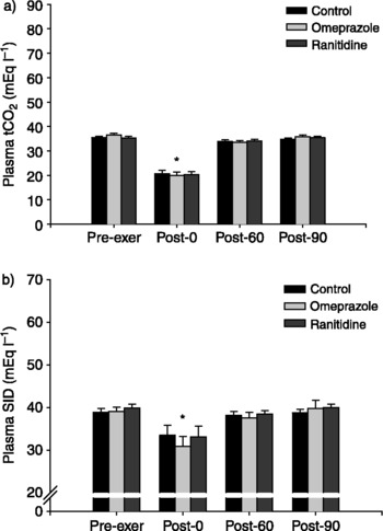
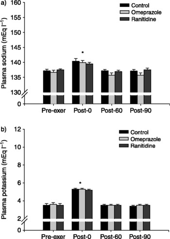
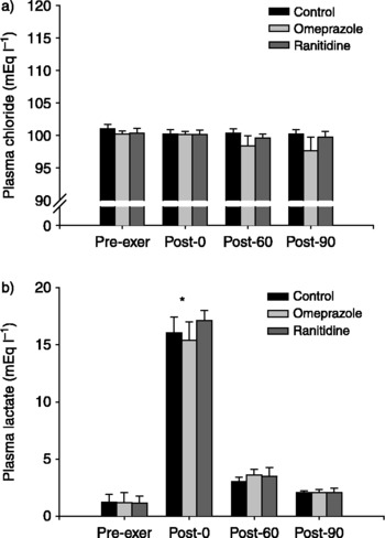
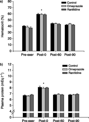
Data are presented in a series of bar graphs representing mean ± SE. There were no significant differences between treatment groups (P>0.05) across sampling intervals for plasma tCO2 (Fig. 1a) and SID (Fig. 1b). Similarly, there were no differences (P>0.05) between treatments across any of the five sampling intervals for [Na+], [K+], [Cl− ], [LA], [TP] and PCV (Figs 2a, 4b). However, differences (P < 0.05) were evident in post-exercise samples immediately following exercise for [tCO2], SID, [Na+], [K+], [Cl− ], [LA], [TP] and PCV compared with pre- and post-exercise samples. All measurements returned to normal resting levels (P>0.05) by the 60 and 90 min samples.
Data present the effect of control, omeprazole or ranitidine and exercise on (a) plasma tCO2 concentration and (b) plasma SID (mean ± SE). No difference (P>0.05) was detected between treatments across each of the four sampling intervals. An asterisk (*) indicates that plasma tCO2 concentration and SID decreased (P < 0.05) for all treatment groups from the pre-exercise (pre-exer) to post-0 intervals and returned to pre-exer levels by 60 min post-exercise

Data present the effect of control, omeprazole or ranitidine and exercise on (a) plasma [Na+] and (b) plasma [K+] (mean ± SE). No significant difference (P>0.05) was detected between treatments across each of the four sampling intervals. An asterisk (*) indicates that plasma [Na+] and [K+] increased (P < 0.05) for all treatment groups from the pre-exercise (pre-exer) to post-0 intervals and returned to pre-exer levels by 60 min post-exercise

Data present the effect of control, omeprazole or ranitidine and exercise on (a) plasma [Cl− ] and (b) plasma LA [LA] concentrations. No significant difference (P>0.05) was detected between treatments across each of the four sampling intervals. An asterisk (*) indicates that plasma [Cl− ] and plasma [LA] increased (P < 0.05) for all treatment groups from the pre-exercise (pre-exer) to post-0 intervals and returned to pre-exer levels by 60 min post-exercise

Data present the effect of control, omeprazole or ranitidine and exercise on (a) plasma [TP− ] and (b) PCV (mean ± SE). An asterisk (*) indicates that plasma [TP] and PCV increased (P < 0.05) for all treatment groups from the pre-exercise (pre-exer) to post-exercise interval and returned to pre-exercise levels by 60 min post-exercise. Treatment did not alter plasma [Cl− ] (P>0.05) at any of the four sampling intervals examined in this investigation. Exercise did not alter plasma [Cl− ]

Discussion
The major findings in this study were that neither omeprazole nor ranitidine altered plasma [tCO2] or SID in horses that did not have gastric ulcers. We are unaware of any data to suggest that horses with gastric ulcers would respond differently; however, that is a question for future studies. Nevertheless, many horses are treated for gastric ulcers and then placed on a maintenance dose of the drug, and thus the data from the present study have direct application to a large part of the racing population of horses. The significant decrease in tCO2 following SRT is a normal response to high-intensity exercise, resulting from anaerobic metabolism. The prominent upward spike in plasma LA in the post-0 sampling interval coincides with the observed decrease in tCO2. LA and H+ ion production are optimal during anaerobic exercise as by-products of lactic acid production. These findings provide practical data in response to speculation that pre-race administration of the medication would evoke responses similar to alkalinizing agents. In the state of New Jersey, post-race drug testing at harness tracks includes a plasma tCO2 test for alkalinizing agents with a threshold allowance of 37.0 mmol l− 1 for horses excluded from the ‘furosemide list’. Horses on this list are cleared to receive furosemide 4 h prior to racing to treat exercise-induced pulmonary haemorrhage (EIPH). The medication has been scientifically documented as an alkalinizing agentReference Freestone, Carlson, Harrold and Church20, and so horses on this list are allowed 39.0 mmol l− 1 tCO2. Many controlled and field studies with large sample populations have endorsed this limit as being more than adequate. LorimerReference Lorimer9 reported average plasma tCO2 for 269 Standardbreds in training to be 30.0 ± 1.8 mmol l− 1. Measurements were made on an out-of-competition day, when it was highly unlikely for a horse to be given a milkshake. However, some feed ingredientsReference Freestone, Carlson, Harrold and Church20, electrolyte supplementsReference Freestone, Carlson, Harrold and Church14, Reference McKeever and Pagan21 and other substances may contain alkalinizing ingredients that have the potential to increase circulating [HCO3− ] and consequently increase tCO2. Thus, these findings are important to the horse racing industry, particularly trainers and owners who seek methods of circumventing gastrointestinal ulcers, while competing within the rules and regulations of the sport.
The major electrolytes (strong ions) were analysed to quantitatively measure physiochemical factors affecting acid–base status in the horse via Stewart's approachReference Stewart22, Reference Constable23. This method involves measuring the interactions between relative concentrations of strong ions (Na+, Cl− , K+ and LA− ), weak acids (protein) and the PCO2 of a solution, in this case venous blood plasmaReference Stewart22, Reference Constable23. The equation states that the summation of cationic strong ions (Na+ and K+) minus the difference between Cl− and LA− concentrations will give SID (SID = [Na+]+ [K+] − [Cl− ] − [LA− ]). Increases in pH, such as the case for horses administered NaHCO3, are associated with a higher SID than normal horses. In this investigation, there were no significant differences between SID of the treatment versus the experimental group. With changes in SID across time consistent with the loss of electrolytes in sweat and compartmental electrolyte shifts, and the prominent spike in circulating LA during exercise, responses to NaHCO3 have shown alkalosis by a major increase in [Na+], while [K+], [Cl− ] and [LA] all remained constant versus a control. The response to treatments in this study did not show similar interactions to NaHCO3 in response to exercise based on SID.
Since plasma [tCO2] has been shown to be representative of plasma bicarbonate concentrations, the extent to which the horse is able to buffer the decrease in plasma pH that occurs with high-intensity exercise can also be shownReference Lloyd, Reilly and Rose3–Reference Slocumbe, Huntington, Lind and Vine8, Reference Frey, Kline, Foreman, Brady and Cooper10, Reference Frey, Kline, Foreman, Lyman and Butadom11. During high-intensity exercise, the metabolic pathways used in muscle cells shift toward anaerobic glycolysis. This shift results in the production and accumulation of hydrogen ions and LA ions. These ions diffuse into the blood causing increased plasma LA concentrations, as the animal approaches the point of fatigue. However, hydrogen ions and LA ions accumulate within the muscle, decreasing intracellular pH and decreasing the activity of glycolytic enzymes. This ultimately causes the shift in the metabolic pathways used to fuel exerciseReference Miller and Lawrence24, Reference Hodgson and Rose25. The bicarbonate system is one aspect of the buffering capacity of the muscle cell. This system functions to neutralize the metabolic acidosis that is produced with exercise. This metabolic acidosis is marked by increased LA concentrations, a decrease in intracellular and extracellular pH and a decrease in bicarbonate concentrations, which is reflected by decreases in plasma [tCO2]Reference Hodgson and Rose25. The changes described in the present study, where exercise was the only factor contributing to a decrease in plasma [tCO2], were consistent with the results obtained in other studiesReference Szucsik, Baliskonis and McKeever14, Reference McKeever, Szucsik, Balaskonis, Betros, Kearns and Malinowski15 (Fig. 1). Since plasma [tCO2] was consistent between each therapy, we can conclude that neither omeprazole nor ranitidine alters plasma [tCO2] in horses.
The pre-ex [tCO2] reported for this study is slightly greater than the 30 mmol l− 1 average plasma [tCO2] for fit racehorses, which was used to establish the threshold concentration for post-race testingReference Slocumbe, Huntington, Lind and Vine8 in New Jersey. This observation, however, is consistent with a recent studyReference McKeever, Szucsik, Balaskonis, Betros, Kearns and Malinowski15 that showed that unfit horses had significantly higher plasma [tCO2] (34.4 mmol l− 1) than a subject group that underwent 12 weeks of moderate training (31.4 mmol l− 1). These results suggest that the subject group in this study had higher plasma [tCO2] due to their level of unfitness and not due to the administration of the acid-blocking therapies.
Plasma [Na+], plasma [Cl− ] and plasma [K+] were not altered due to the administration of omeprazole or ranitidine. Plasma [Na+] is maintained within very narrow limits, roughly within the range of 134–144 mmol l− 1. With short-term, high-intensity exercise, a slight increase in plasma sodium concentration can be seen, though not normally exceeding the aforementioned rangeReference Hyyppa and Poso26. The findings of this study were consistent with this range and trend, though not highly significant (Fig. 2). Since plasma [Na+] was consistent between each therapy, we can conclude that neither omeprazole nor ranitidine altered plasma [Na+] in horses. Plasma [Cl− ] is normally maintained within a range of 94–104 mmol l− 1. After short-term, high-intensity exercise, no significant change in plasma [Cl− ] has been notedReference Hyyppa and Poso26. Plasma [Cl− ] obtained in this study was within this range and remained relatively constant with respect to each sampling interval and therapy administered (Fig. 3). Thus, we can conclude that neither omeprazole nor ranitidine altered plasma [Cl− ] in horses. Plasma [K+] is held within a normal resting range of 3.2–4.2 mmol l− 1. With short-term, high-intensity exercise, a significant hyperkalaemia is evident; in Thoroughbreds, concentrations have been shown to increase to approximately 10 mmol l− 1. This increase has been found to increase proportionally with increasing exercise intensity. Upon exercise cessation, plasma [K+] reduces to, at times, levels below pre-exercise concentrations, as the ion is pumped back into the muscle cell mainly due to the action of Na+/K+-ATPaseReference Hyyppa and Poso26. Plasma [K+] obtained in this study fell within these ranges and, thus, we can conclude that neither omeprazole nor ranitidine altered plasma [K+] in horses.
According to Hodgson and RoseReference Hodgson and Rose25, resting haematocrits fall in a range of 32–46%. Haematocrits have been found to increase proportionally to exercise intensity primarily due to the initial effects of splenic contraction and red cell reserve mobilization, followed by a progressive decrease in plasma volume associated with increases in hydrostatic forces proportional to exercise intensity. This can cause haematocrit to increase to a range of 60–65% with high-intensity exerciseReference McKeever, Hinchcliff, Reed and Robertson27, Reference McKeever, Hinchcliff, Kaneps and Geor28. Administration of either omeprazole or ranitidine appeared not to alter haematocrit percentages in horses. The increase in total plasma protein seen in the subject group is consistent with changes brought about by the effects of exercise and not due to the administration of either omeprazole or ranitidine.
For horse owners, trainers, clinicians and racing officials, the important conclusion of the present study is that the administration of omeprazole and ranitidine does not alter plasma [tCO2] in Standardbred mares, despite their action in buffering gastric contents. The second major observation in the present study that there were no changes in any parameter due to treatment, and only changes associated with exercise that subsequently return to baseline by 60 min post-SRT adds to a growing list of studies that have demonstrated the same response to other treatments such as electrolyte supplementation and treatment with drugs such as sucralphateReference Szucsik, Baliskonis and McKeever14, Reference McKeever, Hinchcliff, Kaneps and Geor28, Reference Caltibilota, Milizio, Malone, Kenney and McKeever29. These observations are important because some have tried to confound efforts to detect the administration of alkalinizing agents by suggesting that one must measure the concentrations of the major electrolytes as well as pH and other factors along with plasma [tCO2]. Data from this study and others demonstrate that there is no need to focus on a long list of extraneous factors, especially when those treatments do not affect the major electrolytes independently of the physiological pressure of exertion.
Acknowledgements
The authors thank the undergraduate animal science research students, as well as Anthony Sacchetti, William Kovach and Thomas Lehman of Cook College Animal Care, for their hard work in collecting and recording data. Research support was provided by the New Jersey State Initiative on Equine, the New Jersey Agricultural Experiment Station, Rutgers University, the George H. Cook Scholars Program and the Aresty Research Council. Conflict of interest. None of the authors of this paper has a financial or personal relationship with other people or organizations that could inappropriately influence or bias the content of the paper.