1. Introduction
Silurian deposits of the Central Precordillera Basin, in San Juan Province, Argentina, comprise the La Chilca and the Los Espejos formations, which combined constitute the Tucunuco Group. Beginning in the Hirnantian (uppermost Ordovician), the Tucunuco Group encompasses the entire Silurian, culminating at its top in the Ludow, Pridoli and even lowest Devonian (Lochkovian) at the northern outcrops.
These deposits have been intensely studied with various geologic disciplines, such us geodynamics, tectonics, sequence stratigraphy, facies analysis and palaeontology, in order to understand the evolution of the basin (i.e. Cuerda, Reference Cuerda1969; Hünicken & Sarmiento, Reference Hünicken and Sarmiento1988; Sánchez, Waisfeld & Benedetto, Reference Sanchez, Waisfeld and Benedetto1991; Benedetto et al. Reference Benedetto, Racheboeuf, Herrera, Brussa and Toro1992; Astini, Benedetto & Vaccari, Reference Astini, Benedetto and Vaccari1995; Astini & Mareto, Reference Astini and Maretto1996; Rickards et al. Reference Rickards, Brussa, Toro and Ortega1996; Albanesi, Ortega & Hünicken, Reference Albanesi, Ortega and Hünicken2006).
Even though the Silurian of the Precordillera has yielded a very rich and diverse invertebrate fauna, including brachiopods, trilobites, bivalves and gastropods among others, fossils of recognized stratigraphic value, i.e. graptolites and conodonts, are sparse, particularly in the Upper Silurian strata.
Traditionally, the La Chilca Formation has been interpreted as Hirnantian–Llandovery in age and the overlying Los Espejos Formation probably as old as Wenlock to locally as young as Lochkovian. The age of the Los Espejos Formation is based mainly on correlation with known brachiopod faunas. However, the scarcity of biostratigraphic index fossils in the upper stratigraphic unit hinders accurate age determination, particularly for its contacts with the underlying La Chilca Formation and the overlying Talacasto Formation.
Palynological studies, mainly those concerning marine organic-walled phytoplankton, shed light on the age of this unit, thus allowing the interpretation of an age not older than Ludlow for the base of the Los Espejos Formation in different sections (Rubinstein & García Muro, Reference Rubinstein and García Muro2011). These palynomorphs, however, did not confirm the Pridoli and Lochkovian ages for the uppermost part of the Los Espejos Formation (Rubinstein, Reference Rubinstein, Fombella Blanco, Fernández González and Valencia Barrera2001; Rubinstein & García Muro, Reference Rubinstein, García Muro and Steemans2011, Reference Rubinstein and García Muro2013). Therefore, palynological data indicates a Late Silurian age for the formation.
The organic-walled phytoplankton assemblages from the Los Espejos Formation are abundant and diverse; terrestrial palynomorphs are, in comparison, subordinate (Rubinstein & García Muro, Reference Rubinstein and García Muro2011) through all the Los Espejos Formation, in the studied sections. Local exceptions occur in levels close to the upper boundary of the unit.
The occurrence of trilete spores in the Los Espejos Formation was first mentioned by Pöthe de Baldis (Reference Pöthe de Baldis1981). She documented four species from the basal portion of the Los Azulejitos Section. Later, Rubinstein Reference Rubinstein(1992) recorded trilete spores from the middle and upper parts of the Los Espejos Formation in the Las Aguaditas and Loma de Los Piojos sections (Rubinstein, Reference Rubinstein1995), with fewer than ten identified species.
The incorporation of detailed palynological studies from more sections of the Los Espejos Formation allows us to recognize the presence of miospore assemblages (trilete spores and cryptospores) in almost all of them, from the base to the top, with varying abundances, diversity and quality of preservation (Rubinstein, Le Hérissé & Steemans, 1996; Rubinstein & Brussa, Reference Rubinstein, Brussa, Tongiorgi and Playford1999; Rubinstein, García Muro & Steemans, Reference Rubinstein, García Muro and Steemans2010, Reference Rubinstein, García Muro and Steemans2011; Rubinstein & García Muro, Reference Rubinstein and García Muro2011, Reference Rubinstein and García Muro2013).
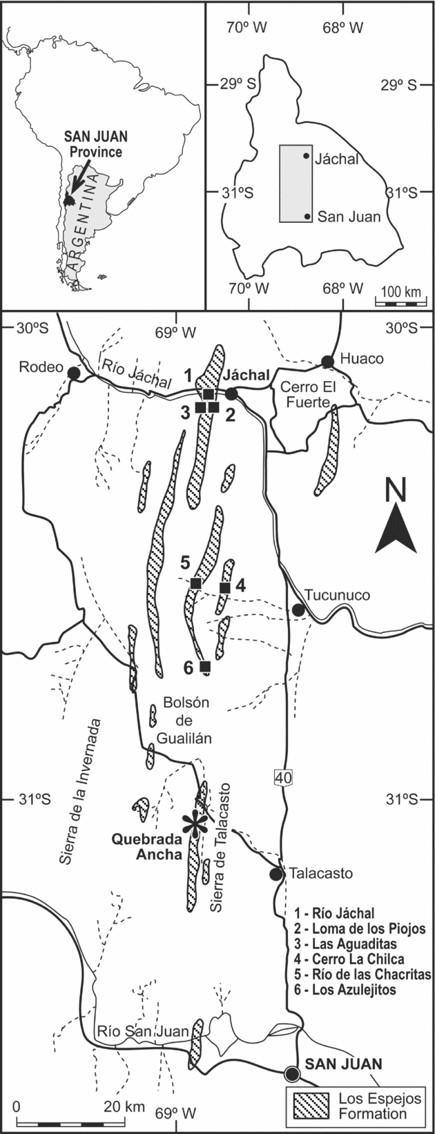
Recent studies expanded our knowledge of the Los Espejos miospore assemblages by recording many taxa from levels corresponding to the lower, middle and upper parts of the unit at the Quebrada Ancha, Cerro La Chilca, Río Jáchal and Río de Las Chacritas sections. To date, only the palynomorphs of the Río de Las Chacritas section have been illustrated and analysed in detail (Rubinstein & García Muro, Reference Rubinstein and García Muro2011). The most significant miospore assemblage owing to its diversity, abundance and preservation comes from the Quebrada Ancha area, near the Talacasto locality (Fig. 1). Therefore, the goal of this contribution is to evaluate the cryptospores and trilete spores coming from 14 productive levels from the Quebrada Ancha area, including a taxonomic reappraisal, evolution of the diversity and correlation with coeval microfloras worldwide as well as their biostratigraphic, palaeoenvironmental and palaeogeographic significance.
Location map of the Silurian Precordillera Basin showing the miospore assemblage localities sampled by the authors. The black asterisk indicates the section reported on here.

2. Geology and palaeontology
The evolution of the Siluro-Devonian Argentinean Precordillera Basin differs from other South American basins because it was affected by accretion of the Cuyania terrane with the Gondwana margin during the Mid–Late Ordovician (Benedetto, Reference Benedetto2010). One of the main consequences of the collision was development of a flexural bending forming the Talacasto–Tambolar arch in the Central Precordillera (Astini, Benedetto & Vaccari, Reference Astini, Benedetto and Vaccari1995), which underwent recurring relaxation and contraction events. Towards this arch, stratigraphic units become wedged and important interruptions of sedimentation occur. Therefore, the Siluro-Devonian succession of the Precordillera constitutes a typical foreland deposit, with a sequence stratigraphic arrangement interpreted as a consequence of lithosphere rheology and eustasy (Astini, Benedetto & Vaccari, Reference Astini, Benedetto and Vaccari1995).
In the Precordillera Basin, the Silurian siliciclastic shelf consists of storm-dominated shallow-marine deposits, composed mainly of intercalated pelites and sandstones. The La Chilca and the Los Espejos formations both show evidence of a transgressive to sea-level highstand history. This interpretation is based on the presence of a thin ferruginous oolite layer and phosphate-rich chert conglomerates at the two units’ bases, succeeded by shaly intervals with a gradual upward thickening and coarsening. Shaly-predominant sediments of the lower part of the Los Espejos Formation represent a low-energy open shelf environment, followed towards the top by an increasing amount of sandstone beds, representing storm-dominated shelf and shoreface environments. Shell-bearing storm beds tend to dominate from the middle to the upper part of the unit. The upper third of the Los Espejos Formation shows evidence of subaerial exposures at the top of the regression cycles. The internal cyclic stacking patterns are considered to have been generated by forced regressions (Sanchez, Waisfeld & Benedetto, Reference Sanchez, Waisfeld and Benedetto1991; Astini & Mareto, Reference Astini and Maretto1996).
The age of the Los Espejos Formation is mainly based on brachiopod faunas (Benedetto et al. Reference Benedetto, Racheboeuf, Herrera, Brussa and Toro1992). Four faunas were assigned to the Wenlock?, Ludlow and Pridoli, and the younger one, at the top of the unit next to Jáchal (Fig. 1), to the early Lochkovian. Graptolite and conodont records are limited and they do not further constrain the overall age of the formation. Graptolites such as Monograptus uncinatus notouncinatus Cuerda, Monograptus leintwardensis var. incipiens Elles & Wood and Monograptus argentinus Cuerda have been identified near the middle part of the unit, allowing the Los Espejos Formation to be assigned to the late Llandovery – early Wenlock to the early–mid Ludlow (Cuerda, Reference Cuerda1969). Rickards et al. Reference Rickards, Brussa, Toro and Ortega(1996) recorded graptolites from Cerro del Fuerte, near Jáchal (Fig. 1), corresponding to the Neodiversograptus nilssoni – Lobograptus scanicus zones in the upper third of the unit, which indicate an early Ludlow age. Hünicken & Sarmiento (Reference Hünicken and Sarmiento1988) recorded conodonts of the upper part of the Polygnathoides siluricus Zone and the lower part of the Pedavis latialata Zone, in the middle to upper part of the Los Espejos Formation in the Quebrada Ancha area, suggesting an age not older than Ludfordian for these levels. On the other hand, Albanesi, Ortega & Hünicken (Reference Albanesi, Ortega and Hünicken2006) documented the Kockelella variabilis variabilis Zone of early Ludlow (Gorstian) age in shell beds of the middle–upper part of the Los Espejos Formation in the same area. The latter authors considered that the species recorded by Hünicken & Sarmiento (Reference Hünicken and Sarmiento1988) do not confirm the proposed age. According to Benedetto et al. Reference Benedetto, Racheboeuf, Herrera, Brussa and Toro(1992), the lower to middle part of the Los Espejos Formation, in the Quebrada Ancha area, cannot be dated by means of brachiopods, whereas the uppermost part of the unit contains Coelospira extensa Benedetto & Toro that indicates a Pridoli age (Fig. 2). Summarizing, the previously studied fauna does not provide a precise age for the lower part of the formation while the top could probably be as young as Pridoli. Meanwhile, palynological studies carried out from the Los Espejos Formation allow its age to be constrained to the Ludlow (Rubinstein, Reference Rubinstein, Fombella Blanco, Fernández González and Valencia Barrera2001; Rubinstein & García Muro, Reference Rubinstein and García Muro2011, Reference Rubinstein and García Muro2013).
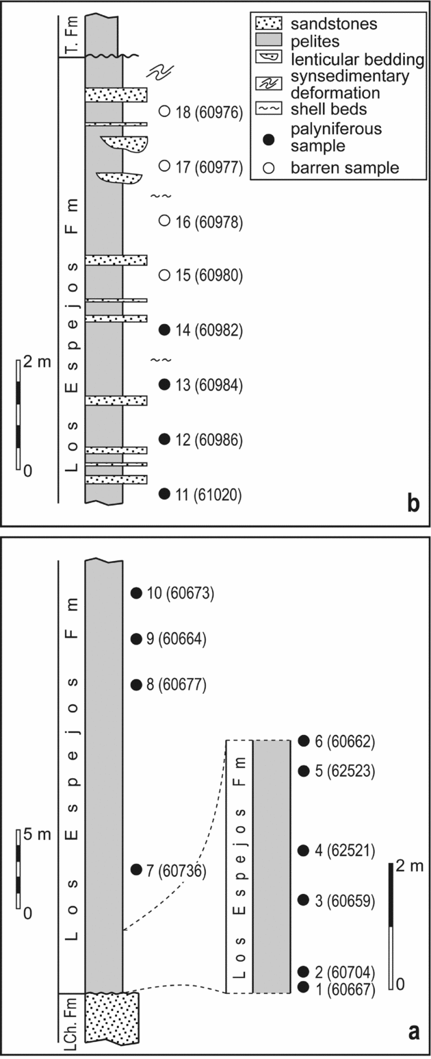
Stratigraphic sections of the studied of Los Espejos Formation in the Quebrada Ancha area. (a) Lower section. (b) Uppermost section. Location and identification numbers of palynological samples in both sections, and the position of the third faunal assemblage of Benedetto et al. Reference Benedetto, Racheboeuf, Herrera, Brussa and Toro(1992) – mainly represented by Coelospira extensa – are indicated.

In the Quebrada Ancha area, the Los Espejos Formation is partially exposed in different sections and reaches a total thickness of 235 m. In this contribution, two sections representing the lower and the uppermost parts of the stratigraphic unit (Fig. 2) have been sampled and are considered the most significant for constraining the age and recognizing the differences with the underlying Lower Silurian La Chilca Formation and the overlying Lower Devonian Talacasto Formation.
The marine phytoplankton, including acritarchs and chlorophytes, is currently under investigation. Nevertheless, owing to the presence of taxa unknown below the Ludlow, from the lower levels of the Los Espejos Formation (i.e. Fimbriaglomerella divisa Loeblich & Drugg, Reference Loeblich and Drugg1968, Ozotobrachion palidodigitatus (Cramer) Playford, Reference Playford1977 and Stellinium rabians (Cramer) Eisenack, Cramer & Díez, Reference Eisenack, Cramer and Díez1976) an age not older than Ludlow is interpreted for the unit. In the upper section, the diversity of the marine phytoplankton decreases significantly, and taxa considered indices of the Pridoli have not been recorded. Consequently, marine palynomorphs restrict the age to the Ludlow.
3. The miospore biostratigraphy of the Upper Silurian
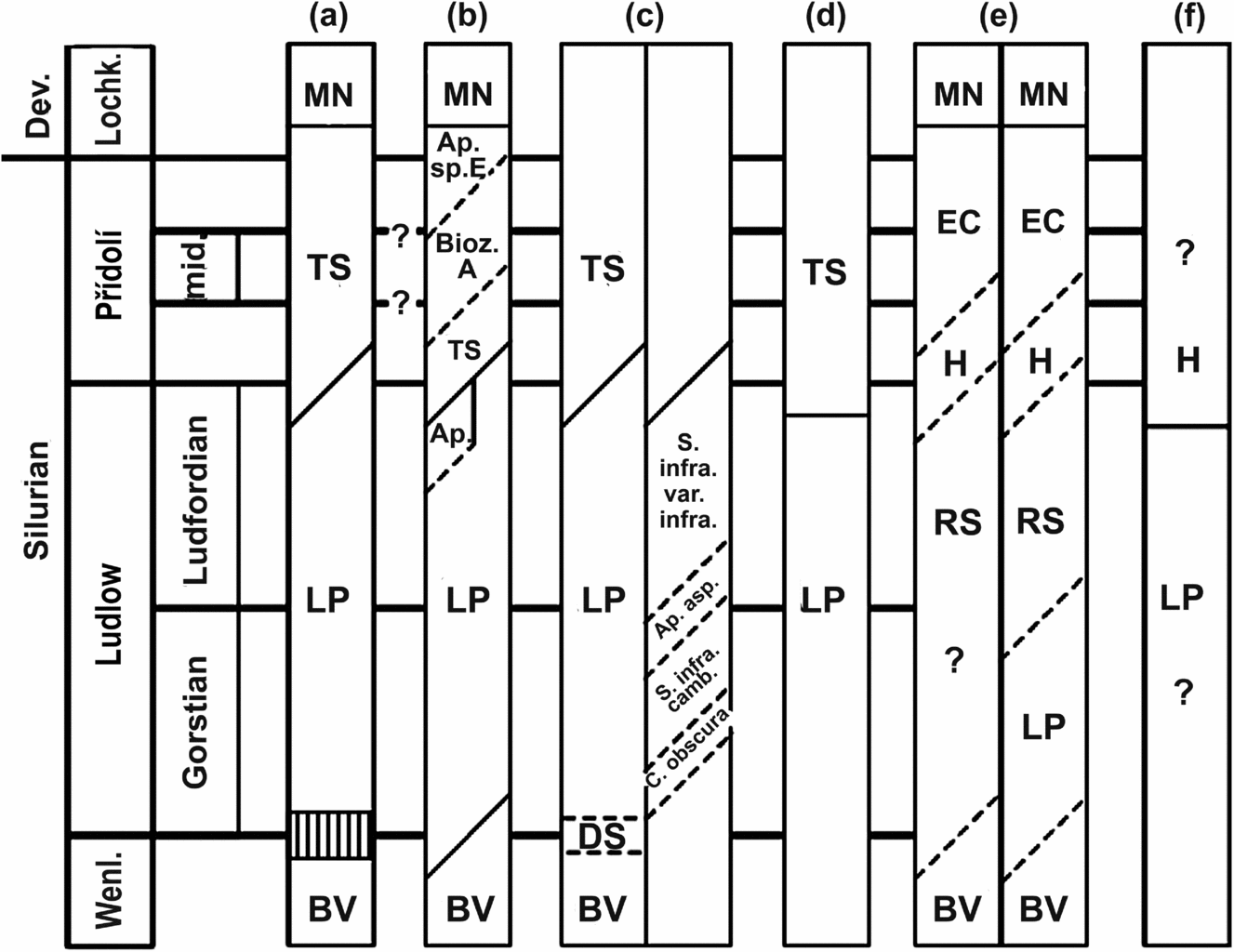
Numerous studies on the stratigraphic distribution of miospores from the Upper Silurian and Siluro-Devonian boundary are available (Hassan Kermandji, Reference Hassan Kermandji2007; Steemans, Rubinstein & Melo, Reference Steemans, Rubinstein and Melo2008 and references therein; Spina & Vecoli, Reference Spina and Vecoli2009 and references therein; Mehlqvist, Vajda & Steemans, Reference Mehlqvist, Vajda and Steemans2012). Most of these are geographically concentrated on northwestern Gondwanan (North Africa) sections, with the remaining few from Euramerica. Among them, there are few sections complete enough to allow the creation of biozonations. Attempts have been made in combining data from Euramerica and Gondwana (Richardson & McGregor, Reference Richardson and McGregor1986; Richardson & Edwards, Reference Richardson, Edwards, Holland and Bassett1989), or on Euramerican samples from the UK (Burgess & Richardson, Reference Burgess and Richardson1995), on material from the Peri-Gondwanan terrane of Spain (Richardson, Rodríguez & Sutherland, Reference Rubinstein, Fombella Blanco, Fernández González and Valencia Barrera2001), or on Libyan (Rubinstein & Steemans, Reference Rubinstein, Steemans, Steemans, Servais and Streel2002) and Algerian (Hassan Kermandji, Reference Hassan Kermandji2007) miospore assemblages from northwestern Gondwana (Fig. 3). The application of these biostratigraphic zonations to new studies remains somewhat problematic because of the lack of independent biostratigraphic control and differences in the composition of the assemblages depending on palaeogeography.
Miospore biostratigraphic zonal correlations in the Upper Silurian and the Lochkovian. (a) Richardson & McGregor (Reference Richardson and McGregor1986); (b) Richardson & Edwards (Reference Richardson, Edwards, Holland and Bassett1989); (c) Burgess & Richardson (Reference Burgess and Richardson1995); (d) Rubinstein & Steemans (Reference Rubinstein, Steemans, Steemans, Servais and Streel2002); (e) Richardson, Rodríguez & Sutherland (Reference Richardson, Rodríguez and Sutherland2001); (f) Hassan Kermandji (Reference Hassan Kermandji2007). Diagonal lines indicate boundaries revised herein. The exact position of biozones and sub-biozones and their correlation with series and stage boundaries remain doubtful because of the lack of reliable independent biostratigraphic control. BV – brevicosta–verrucatus; DS – downiei–sagittarius; LP – libycus–poecilomorphus; RS – reticulata–sanpetrensis; H – hemiesferica; TS – tripapillatus–spicula; EC – elegans–cantabrica; MN – micrornatus–newportensis.

The boundary between the cf. protophanus–verrucatus Zone (brevicosta–verrucatus in Burgess & Richardson, Reference Burgess and Richardson1995) and the libycus–poecilomorphus (LP) Zone (Richardson & McGregor, Reference Richardson and McGregor1986) was interpreted to correspond, more or less, with the base of the Ludlow in Libya (Richardson & Ioannides, Reference Richardson and Ioannides1973). However, unequivocal evidence to confirm this correlation is unavailable. Burgess & Richardson (Reference Burgess and Richardson1995) clearly put the base of the LP Biozone in the lower part of the Gorstian in Wales, somewhere between the uppermost nilssoni and the lowermost incipiens graptolite zones, i.e. somewhere in the lower part of the Gorstian. Contrary to the biostratigraphic zonations established in Libya, the brevicosta–verrucatus (BV) Zone is not directly overlain by the LP Zone in Wales but by a new biozone defined by Burgess & Richardson (Reference Burgess and Richardson1995), the downiei–sagittarius (DS) Zone, and above the LP Zone. The base of this zone is correlated with the latest ludensis or the early nilssoni zones (i.e. latest Homerian or earliest Gorstian). Burgess & Richardson (Reference Burgess and Richardson1995) noted that the DS Zone is only found in approximately 20 m of strata from the Rumney Borehole, and in North Africa there is a sampling gap of over 100 m at the critical part of the sequence.
The upper part of the LP Zone has been subdivided into an Apiculiretusispora spp. Subzone by Richardson & Edwards (Reference Richardson, Edwards, Holland and Bassett1989). The LP Zone has also been subdivided into four Subzones by Burgess & Richardson (Reference Burgess and Richardson1995): C. obscura, S. inframurinata var. cambrensis, A. asperata and S. inframurinata var. inframurinata subzones. Independent biostratigraphic control is too imprecise to clearly correlate the Gostian–Ludfordian boundary with this miospore stratigraphic zonation, which probably lies in between the base of the S. inframurinata var. cambrensis and S. inframurinata var. inframurinata subzones.
In Libya, the LP Zone is overlain by the tripapillatus–spicula (TS) Zone (Richardson & McGregor, Reference Richardson and McGregor1986). In the UK, its base is immediately above the Ludlow Bone Bed (Richardson & Lister, Reference Richardson and Lister1969). The age of its base is thought to correspond to the Ludlow–Pridoli boundary and its top spans the Pridoli–Lochkovian boundary. However, the exact position of the base of the Pridoli in Britain cannot be confidently established. Conodont evidence from the Whitcliffe Formation in the UK suggests that the base of the Pridoli may be slightly higher than the base of the Downton Castle Sandstone Formation represented at its base by the Ludlow Bone Bed (Aldridge & Schönlaub, Reference Aldridge, Schönlaub, Holland and Bassett1989; Miller, Reference Miller1995). This is confirmed by independent dating based on chitinozoans (Jaglin & Paris, Reference Jaglin and Paris2002) from Libya where a miospore assemblage correlated with the TS Zone is attributed to the latest Ludlow (Rubinstein & Steemans, Reference Rubinstein, Steemans, Steemans, Servais and Streel2002).
In Spain, Richardson, Rodríguez & Sutherland (Reference Richardson, Rodríguez and Sutherland2001) correlated the top of the LP Zone with the base of the overlying reticulata–sanpetrensis (RS) Zone in the Geras and the Argovejo outcrops. But they also correlated the top of the BV Zone with the base of the RS Zone in the La Peral outcrop creating a confusing stratigraphic correlation since the BV Zone had been defined as underlying the LP Zone in Richardson & McGregor (Reference Richardson and McGregor1986) and in Burgess & Richardson (Reference Burgess and Richardson1995). Richardson, Rodríguez & Sutherland (Reference Richardson, Rodríguez and Sutherland2001) considered that at least part of the RS Zone is of late Ludfordian age. The base of the overlying H Zone is observed above the first incoming of Ramochitina villosa Laufeld, Reference Laufeld1974 in the Geras outcrop, a chitinozoan appearing close to the Gorstian–Ludfordian boundary (Nestor, Reference Nestor2009), and in the four outcrops studied in Spain below the first incoming of typical Pridoli chitinozoans such as Urnochitina urna Eisenack, Reference Eisenack1934 (figs 10–13 in Richardson, Rodríguez & Sutherland, Reference Richardson, Rodríguez and Sutherland2001). Thus, the latter authors believe that the base of the H Zone is Pridoli in age because the chitinozoans observed below the first incoming of U. urna may span the Pridoli. However, contrary to Richardson, Rodríguez & Sutherland (Reference Richardson, Rodríguez and Sutherland2001), we consider that a Ludfordian age cannot be excluded. The boundary between the H and the elegans–cantabrica (EC) zones is present in the four outcrops above the appearance of U. urna and in the La Vid section below the first incoming of Margachitina elegans Taugourdeau & de Jekhowski, Reference Taugourdeau and de Jekhowsky1960, which is characteristic of the mid Pridoli. The base of the Aneurospora Subzone in the upper part of the EC Zone is observed in layers containing M. elegans. Therefore, a mid Pridoli age cannot be excluded for its lower boundary. The micrornatus–newportensis (MN) Zone is observed in the lowermost Lochkovian characterized by Eisenackitina bohemica Eisenack, Reference Eisenack1934. The base of the Devonian is around 30 m below the contact of the San Pedro and La Vid formations in the Argovejo outcrop. Priewalder Reference Priewalder(1997) considered that the presence of M. elegans, 10 m below the top of the San Pedro Formation in the La Vid outcrop (data from Cramer, Reference Cramer1967), constrained the age of the chitinozoan-bearing levels to be mid Pridoli, thus concluding that the San Pedro Formation does not extend into the Lochkovian. This result seems to be in contradiction with the one from Richardson, Rodríguez & Sutherland (Reference Richardson, Rodríguez and Sutherland2001), but perhaps there is a hiatus in the La Vid outcrop or the formation is diachronous. More analyses are required to solve this apparent contradiction. In their conclusions, Richardson, Rodríguez & Sutherland (Reference Richardson, Rodríguez and Sutherland2001) pointed out that there are some discrepancies between the age provided by the chitinozoans and those based on miospores. It must be remembered that miospores are absent, or of poor utility, in the Silurian stratotypes, which are marine. Therefore, miospore biozones should be calibrated with marine fossils present in the stratotypes, like chitinozoans. Hence, we cannot exclude an older age for the boundaries of the different biozones than those provided by Richardson, Rodríguez & Sutherland (Reference Richardson, Rodríguez and Sutherland2001).
The sanpetrensis–triangulatus is a spore zone established for the Ludlow by Hassan Kermandji (Reference Hassan Kermandji2007) in Algeria. This biozone is overlain by the H Zone of Richardson, Rodríguez & Sutherland (Reference Richardson, Rodríguez and Sutherland2001) and by a newly created radiate–synorea Zone. No independent age control is available to date these Algerian miospore biozones.
In the UK and other areas of the Euramerican continent, the MN Zone overlays the TS Zone in the lowermost Lochkovian (Richardson & McGregor, Reference Richardson and McGregor1986; Steemans, Reference Steemans1989; Streel et al. Reference Streel, Higgs, Loboziak, Riegel and Steemans1987). Richardson & Edwards (Reference Richardson, Edwards, Holland and Bassett1989) have created, intermediate between the TS and the MN zones, a Zone A and an Apiculiretusispora sp. E Zone separated by a ‘no records’ interval (in Wellman & Richardson, Reference Wellman and Richardson1996, text-fig. 7). Richardson, Rodríguez & Sutherland (Reference Richardson, Rodríguez and Sutherland2001) have defined in Spain a newportensis–argovejae (NA) Subzone in the lower part of the MN Zone characterized by the incoming of Streelispora newportensis and Leonispora argovejae, and the absence of Emphanisporites micrornatus. The MN Zone is well known on the Euramerican plate. It has also been observed on Peri-Gondwanan terranes, in Spain (Richardson, Rodríguez & Sutherland, Reference Richardson, Rodríguez and Sutherland2001) and in Brittany (Steemans, Reference Steemans1989). Only one specimen of Streelispora newportensis has been identified in Gondwana, in Libya (Loboziak et al. Reference Loboziak, Steemans, Streel and Vachard1992). The definition of the Siluro-Devonian boundary based upon miospore zones is therefore problematic on this palaeocontinent.
4. Preparation and techniques
Eighteen samples were collected from two sections in the Quebrada Ancha area (Fig. 2a, b), representing the lower part (ten samples) and the uppermost part (eight samples) of the Los Espejos Formation. Fourteen samples were productive while the four samples closest to the upper boundary were barren.
The samples were processed in the University of Liège palynology laboratory (Belgium), using standard HCl–HF–HCl acid maceration techniques. The residues were oxidized with a Schulze solution (HNO3+KClO3) and then screened on a 12 μm sieve.
Miospore assemblages from different palaeogeographies were compared to the Quebrada Ancha assemblage through a cluster analysis, using Infostat 2009®.
The palynological slides are housed in the palynological collection of the IANIGLA, CCT CONICET Mendoza, Argentina. Specimen locations in the slides are located by England Finder coordinates.
5. Palynology
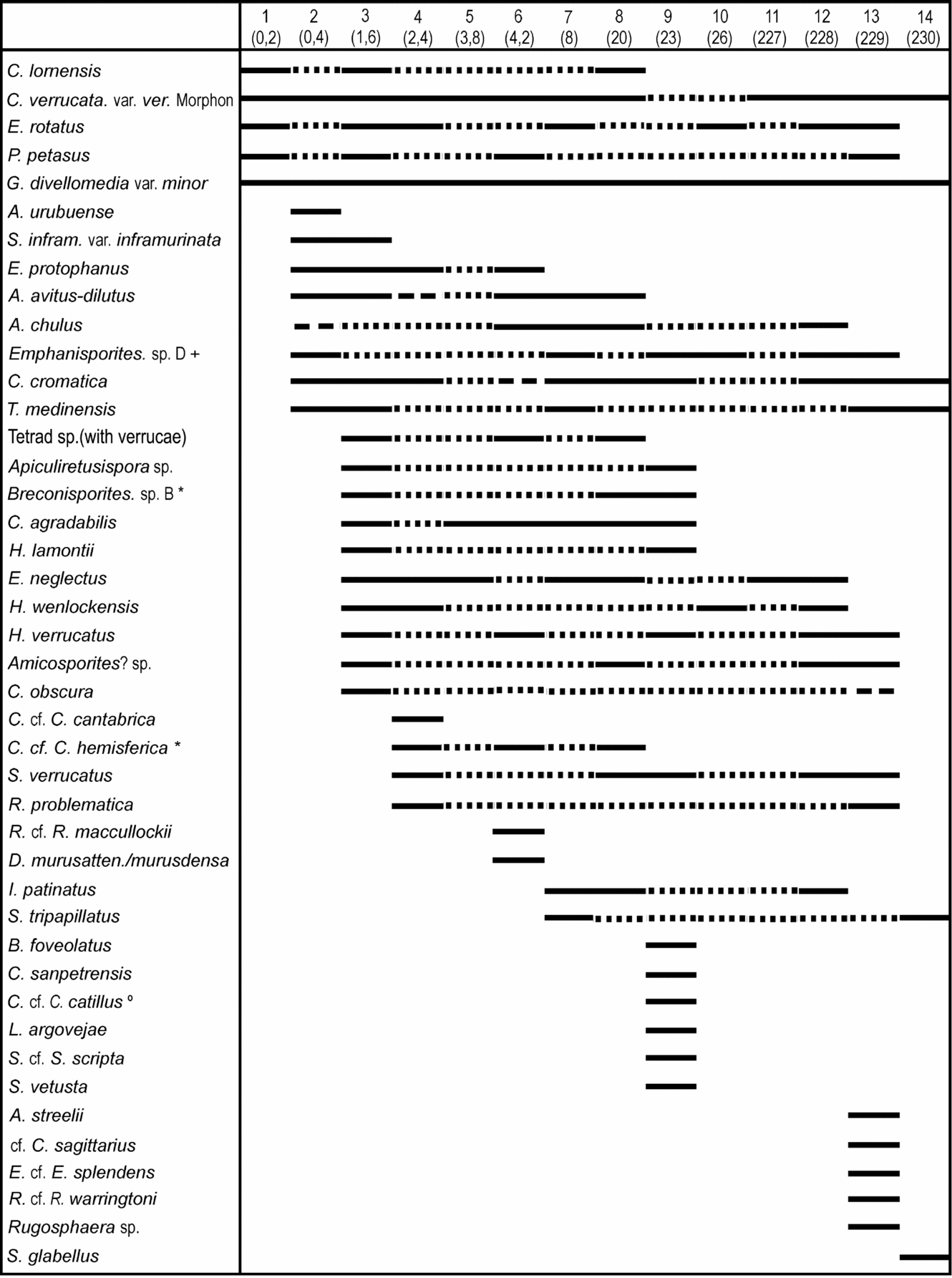
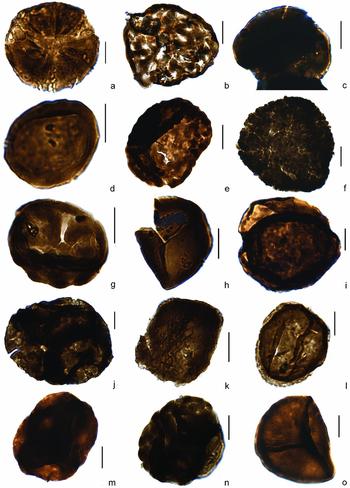
The miospore assemblage consists of 43 miospore species (29 trilete spores and 14 cryptospores). The lower part of the Los Espejos Formation is richer in trilete spore species (25 species) compared with the upper part (14 species), while cryptospore diversity undergoes a smaller decrease from 12 to 9 species (Fig. 4).
Stratigraphic distribution of trilete spore and cryptospore species in the studied levels. The numbers between brackets represent the metres from the base of the formation. Identified species are indicated in continuous lines; cf. species in dashed lines; supposed stratigraphic range in dotted lines. + in Richardson, Rasul & Al-Ameri (Reference Richardson, Rasul and Al-Ameri1981); * in Richardson, Rodríguez & Sutherland (Reference Richardson, Rodríguez and Sutherland2001); º in Richardson & Lister (Reference Richardson and Lister1969).

5.a. Cryptospores
Artemopyra urubuense Steemans, Rubinstein & Melo, Reference Steemans, Rubinstein and Melo2008 Figure 5a
Remarks. This spore is known only from Gondwana. The other two localities where this species has been identified are from the Ludlow of Libya (Richardson & Ioannides, Reference Richardson and Ioannides1973) and from the lower Pridoli of Brazil (Steemans, Rubinstein & Melo, Reference Steemans, Rubinstein and Melo2008).
(a) Artemopyra urubuense Steemans, Rubinstein & Melo, Reference Steemans, Rubinstein and Melo2008, diameter 43 μm, slide 60704, K35/3–4. (b) Chelinohilates lornensis Wellman & Richardson, Reference Wellman and Richardson1996, slide 60677, Q36/1-2. (c) Dyadaspora murusattenuata/murusdensa morphon Steemans, Le Hérissé & Bozdogan, Reference Steemans, Le Hérissé and Bozdogan1996, slide 60662, P39/1. (d) Gneudnaspora divellomedia (Chibrikova) Balme, 1988 var. minorBreuer et al. Reference Breuer, Al-Ghazi, Al-Ruwaili, Higgs, Steemans and Wellman2007, slide 60984, J32/1-3. (e) Hispanaediscus lamontii Wellman, Reference Wellman1993, slide 60664, S46/1. (f) Hispanaediscus wenlockensis Burgess & Richardson, Reference Burgess and Richardson1991, slide 60659, K47/1. (g) Hispanaediscus verrucatus (Cramer) Burgess & Richardson, Reference Burgess and Richardson1991, slide 60984, N31/1. (h) Imperfectotriletes patinatus Steemans, Higgs & Wellman, 2000, slide 60986, S39/1. (i) Pseudodyadospora petasus Wellman & Richardson, Reference Wellman and Richardson1993, slide 60667, O42/0. (j) Rimosotetras problematica Burgess, Reference Burgess1991, slide 62521, P43/0. (k) Rugosphaera sp., 60984, Q47/4. (l) Sphaerasaccus glabellus Steemans, Higgs & Wellman, Reference Steemans, Higgs, Wellman, Al-Hajri and Owens2000, slide 60982, N36/1. (m) Tetrahedraletes medinensis (Strother & Traverse) Wellman & Richardson, Reference Wellman and Richardson1993, slide 60704, G43/4. (n) Tetrad with coarse verrucae, slide 60659, S29/1. (o) Stellatispora inframurinata var. inframurinata (Richardson & Lister) Burgess & Richardson, Reference Burgess and Richardson1995, slide 60659, S28/0. The scale bar represents 20 μm.

Chelinohilates lornensis Wellman & Richardson, Reference Wellman and Richardson1996
Figure 5b
Dyadospora murusattenuata/murusdensa morphon Steemans, Le Hérissé & Bozdogan, Reference Steemans, Le Hérissé and Bozdogan1996
Figure 5c
Gneudnaspora divellomedia var. minor (Chibrikova) Breuer et al. Reference Breuer, Al-Ghazi, Al-Ruwaili, Higgs, Steemans and Wellman2007
Figure 5d
Hispanaediscus lamontii Wellman, Reference Wellman1993
Figure 5e
Hispanaediscus verrucatus (Cramer) Burgess & Richardson, Reference Burgess and Richardson1991
Figure 5g
Remarks. This is one of the eponymous species of the BV Zone, Wenlockian in age (Burgess & Richardson, Reference Burgess and Richardson1995; Richardson & McGregor, Reference Richardson and McGregor1986).
Hispanaediscus wenlockensis Burgess & Richardson, Reference Burgess and Richardson1991
Figure 5f
Remarks. This species has been identified in the type Wenlock area, England (Burgess & Richardson, Reference Burgess and Richardson1991) and could be reworked here.
Imperfectotriletes patinatus Steemans, Higgs & Wellman, Reference Steemans, Higgs, Wellman, Al-Hajri and Owens2000
Figure 5h
Pseudodyadospora petasus Wellman & Richardson, Reference Wellman and Richardson1993
Figure 5i
Rimosotetras problematica Burgess, Reference Burgess1991
Figure 5j
Rugosphaera sp.
Figure 5k
Sphaerasaccus glabellus Steemans, Higgs & Wellman, Reference Steemans, Higgs, Wellman, Al-Hajri and Owens2000
Figure 5l
Remarks. This species has been identified from the Ordovician of the UK (Wellman, Reference Wellman1996), the NW of Argentina (Rubinstein et al. Reference Rubinstein, García Muro and Steemans2010) and the Llandovery of Saudi Arabia (Steemans, Higgs & Wellman, Reference Steemans, Higgs, Wellman, Al-Hajri and Owens2000).
Tetrahedraletes medinensis (Strother & Traverse) Wellman & Richardson, Reference Wellman and Richardson1993
Figure 5m
Tetrad sp. (ornamented with verrucae)
Figure 5n
5.b. Trilete spores
Ambitisporites avitus/dilutus sensu Steemans,
Le Hérissé & Bozdogan, Reference Steemans, Le Hérissé and Bozdogan1996
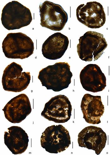
Figure 6a
Amicosporites streelii Steemans, Reference Steemans1989 partim
Figure 6c
Remarks. The specimens of A. streelii figured in Steemans Reference Steemans(1989) include several with distal verrucae and specimens with only one large verrucae centred at the distal pole. It would seem that these represent two new species. In the Lower Devonian of Saudi Arabia (Breuer & Steemans, Reference Breuer and Steemans2013), only specimens with one distal verrucae are known. Similar specimens are also known from the Saudi Arabian Upper Silurian (P. Breuer, pers. com. 2012).
(a) Ambitisporites avitus/dilutus sensu Steemans, Le Hérissé & Bozdogan, Reference Steemans, Le Hérissé and Bozdogan1996, slide 60659, K45/3. (b) Amicosporites? sp., slide 60986, M45/0. (c) Amicosporites streelii Steemans, Reference Steemans1989, slide 60984, S29/1. (d) Apiculiretusispora sp., slide 60664, F41/2. (e) Archaeozonotriletes chulus (Cramer) Richardson & Lister, Reference Richardson and Lister1969, slide 60986, L29/4. (f, g) Breconisporites sp. B in Richardson, Rodríguez & Sutherland, Reference Richardson, Rodríguez and Sutherland2001; (f) slide 60659, T295/1-2; (g) slide 60664, O40/1. (h) Brochotriletes foveolatus Naumova, Reference Naumova1953, slide 60664, H27/1. (i, j) Concentricosisporites agradabilis (Rodriguez) Rodriguez, Reference Rodriguez1983, (i) slide 60664, T39/0; (j) slide 60664, V30/4. (k) cf. Concentricosisporites agradabilis (Rodriguez) Rodriguez, Reference Rodriguez1983, slide 60677, H38/0. (l) cf. Concentricosisporites sagittarius (Rodriguez) Rodriguez, Reference Rodriguez1983, slide 600984, M37/2. (m–o) Coronaspora cromatica (Rodríguez) Richardson, Rodríguez & Sutherland, Reference Richardson, Rodríguez and Sutherland2001; (m) slide 62521, L39/4 (distal face); (n) slide 60664, G37 (proximal face); (o) slide 60984, J28/4 (proximal face). The scale bar represents 20 μm.

Amicosporites? sp.
Figure 6b
Remarks. Several poorly preserved specimens are close to Concentricosisporites agradabilis. However, it has not been possible to see if there are radial muri on the proximal face. For this reason they have been tentatively placed in the genus Amicosporites.
Apiculiretusispora sp.
Figure 6d
Archaeozonotriletes chulus (Cramer) Richardson & Lister, Reference Richardson and Lister1969
Figure 6e
Breconisporites sp. B in Richardson, Rodríguez & Sutherland, Reference Richardson, Rodríguez and Sutherland2001
Figure 6f, g
Remarks. Richardson, Rodríguez & Sutherland (Reference Richardson, Rodríguez and Sutherland2001) included in Breconisporites? spp. specimens with curvaturae extending equatorially onto a possible zona. These authors also observed rare laevigate or proximally rugulate zonate spores, more clearly bizonate, that they positively assigned to the genus Breconisporites (as Breconisporites sp. B, pl. 4, fig. 4), from the NA Subzone of the MN Zone in Spain of early Lochkovian age. They did not describe Breconisporites sp. B but from its illustration it appears very similar to our species.
Brochotriletes foveolatus Naumova, Reference Naumova1953
Figure 6h
Remarks. This species first appears in the Lochkovian and it is widely distributed over portions of Euramerica and Gondwana. The only previous older record is from the lower Pridoli of Brazil (Steemans, Rubinstein & Melo, Reference Steemans, Rubinstein and Melo2008).
Chelinospora cf. C. cantabrica Richardson, Rodríguez & Sutherland, Reference Richardson, Rodríguez and Sutherland2001
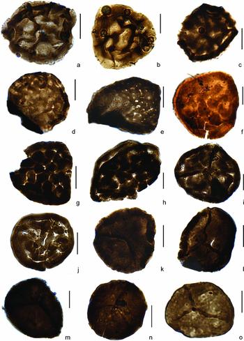
Figure 7c
Chelinospora cf. C. hemiesferica (Cramer & Diez) in Richardson, Rodríguez & Sutherland, Reference Richardson, Rodríguez and Sutherland2001
Figure 7a, b
Remarks. C. hemiesferica is the eponymous species for the H Zone (Richardson, Rodríguez & Sutherland, Reference Richardson, Rodríguez and Sutherland2001) of Ludfordian to Pridoli age, and C. cf. C. hemieferica first appears within the H Zone.
(a, b) Chelinospora cf. C. hemisferica in Richardson, Rodríguez & Sutherland, Reference Richardson, Rodríguez and Sutherland2001; (a) slide 60677, K33/3; (b) 62521, S31/2. (c) Chelinospora cf. C. cantabrica Richardson, Rodríguez & Sutherland, Reference Richardson, Rodríguez and Sutherland2001, slide 62521, O30/3. (d) Chelinospora obscura Burgess & Richardson, Reference Burgess and Richardson1995, slide 60659, J25/2. (e) Chelinospora sanpetrensis (Rodríguez) Richardson, Rodríguez & Sutherland, 2001, slide 60664, W45/2. (f–j) Chelinospora verrucata var. verrucata morphon; (f) slide 60984, H39/2; (g) slide 60704, V37/2; (h) slide 60659, U43; (i) slide 60677, V34/0–3; (j) slide 60984, H26/1. (k, l) Cymbosporites cf. C. catillus in Richardson & Lister, Reference Richardson and Lister1969; (k) slide 60664, X27/1; (l) slide 60664, P27/2. (m) Retusotriletes cf. R. maccullockii Wellman & Richardson, Reference Wellman and Richardson1996, slide 60662, F25/1. (n) Retusotriletes cf. R. warringtoni Richardson & Lister, 1989, slide 60984, K28/1. (o) Retusotriletes sp., slide 60984, R29/1. The scale bar represents 20 μm.

Chelinospora obscura Burgess & Richardson, Reference Burgess and Richardson1995
Figure 7d
Chelinospora (Clivosispora) verrucata (McGregor) comb. nov.
Figure 7f–j
Basionym. Clivosispora verrucata McGregor, Reference McGregor1973, pp. 54–5, pl. 7, figs 4, 5, 10. The following synonymy list includes only those references with descriptions and/or illustrations of Silurian specimens. Several other specimens have also been recorded from the Lower Devonian and are also listed here.
Synonymy and references
-
1973 Clivosispora verrucata McGregor, pp. 54–5, pl. 7, figs 4, 5, 10.
-
1973 Lophozonotriletes? poecilomorphus Richardson & Ioannides, pp. 279–80, pl. 7, figs 9–15; pl. 8, figs 1, 4–6.
-
1975 Lophozonotriletes? poecilomorphus Richardson & Ioannides; Cramer & Diez, p. 342, pl. 1, fig. 1; pl. 2, fig. 33.
-
1976 Clivosispora verrucata var. verrucata McGregor & Camfield, p. 15, pls 3, 11–14.
-
1976 Clivosispora verrucata var. convoluta McGregor & Camfield, p. 15, pls 2, 13–21.
-
1995 Lophozonotriletes? poecilomorphus Richardson & Ioannides; Burgess & Richardson, pp. 22–3, pl. 9, figs 10–13.
-
1995 Lophozonotriletes? poecilomorphus Richardson & Ioannides; Steemans, pl. 3, fig. 9.
-
2001 Lophozonotriletes? poecilomorphus Richardson & Ioannides; Beck & Strother, p. 151, pl. 6, figs 8(?), 9,10, 12, 13.
-
2001 Chelinospora (Lophozonotriletes?) poecilomorpha Richardson, Rodríguez & Sutherland, p. 159, pl. 10, figs 6, 7.
-
2002 Lophozonotriletes? poecilomorphus Richardson & Ioannides; Rubinstein & Steemans, pl. III, fig. 14.
-
2002 Clivosispora verrucata McGregor var. convoluta McGregor & Camfield; Rubinstein & Steemans, pl. II, fig. 6.
-
(?)2007 Chelinospora poecilomorpha Richardson, Rodríguez & Sutherland; Hassan Kermandji, pl. 1, figs 1, 4.
-
2007 Clivosispora verrucata McGregor; Breuer et al. figs 5, 13–14.
-
2008 Chelinospora poecilomorpha Richardson, Rodríguez & Sutherland; Steemans, Rubinstein & Melo, fig. 7(3–4).
-
2008 Clivosispora verrucata McGregor var. convoluta McGregor & Camfield; Steemans, Rubinstein & Melo, fig. 7(7).
-
2008 Clivosispora verrucata McGregor var. verrucata McGregor & Camfield; Steemans, Rubinstein & Melo, fig 7(8).
-
2009 Chelinospora poecilomorpha Richardson, Rodríguez & Sutherland; Spina & Vecoli, pl. III, fig. 5.
Remarks. We consider Chelinospora (Lophozonotriletes?) poecilomorpha (Richardson & Ioannides, Reference Richardson and Ioannides1973) Richardson, Rodríguez & Sutherland (Reference Richardson, Rodríguez and Sutherland2001) as a junior synonym of Chelinospora (Clivosispora) verrucata (McGregor, Reference McGregor1973) comb. nov.; the latter was published in July 1973 and the former in April of the same year. Both show exactly the same type of ornamentation and patina. The diameter ranges in the original descriptions show differences between both taxa (23–45 μm for C. poecilomorpha and 39–80 μm for C. verrucata). Even though C. verrucata can be larger, it could represent phenotypic (particular environmental) variation. Subsequent literature mentions smaller specimens of C. verrucata (e.g. Gao, Reference Gao1981; Wellman et al. Reference Wellman, Thomas, Edwards and Kenrick1998; Rubinstein & Steemans, Reference Rubinstein, Steemans, Steemans, Servais and Streel2002; Wellman, Reference Wellman2006; Steemans, Rubinstein & Melo, Reference Steemans, Rubinstein and Melo2008; Spina & Vecoli, Reference Spina and Vecoli2009). Rare specimens of C. verrucata show a cingulum with radial depressions resembling coalescent verrucae, which is why McGregor Reference McGregor(1973) assigned this species to Clivosispora. Personal observations (P. Steemans) of this species from various Silurian and Devonian samples suggest this character is most probably an artefact of the thick distal verrucae observed through the patina at the equator. A similar artefact may also be due to specimen compression during fossilization (see pl. 10, fig. 7 in Richardson, Rodríguez & Sutherland, Reference Richardson, Rodríguez and Sutherland2001).
Chelinospora verrucata var. verrucata morphon
Remarks. A morphon, Chelinospora verrucata var. verrucata morphon, is created on the basis of the distal ornamentation for these miospores. It includes the varieties verrucata var. verrucata and verrucata var. convoluta of McGregor & Camfield (Reference McGregor and Camfield1976). These taxa are very similar having a subtriangular to subcircular amb. Equatorial thickenings are well developed, ranging from 2–15 μm in width (most not exceeding 4 μm), laevigate or with rounded humps and intervening radial depressions that may resemble coalescent verrucae. The trilete mark, when present, extends three-quarters to the total radius of the spore. All spores included in this morphon have a proximal face that is thin, laevigate and folded along the trilete mark. The distal face has coarse verrucae 1–15 μm wide, separated by depressions with a maximum width of 3 μm (Fig. 6f), but commonly they are more closely packed, sometimes even fused, giving the appearance of coarse convoluted muri (see Fig. 6i, j). In some specimens the verrucae overlap the cingulum.
The specimens found in Quebrada Ancha have a triangular to subtriangular amb. Equatorial crassitudes range from 2–4 μm in width. In most specimens the trilete mark is not preserved; when it is preserved it extends over the equatorial crassitude. The distal face has coarse verrucae of 2–6 μm wide, frequently collapsed, and often overlapping the equatorial crassitude.
Dimensions. 28–52 μm (36 μm average; 17 Quebrada Ancha specimens measured).
Occurrence. Ludlow to Emsian from localities on the Gondwana, Avalonia, Armorica and Baltica palaeoplates.
Chelinospora sanpetrensis (Rodriguez) Richardson, Rodríguez & Sutherland, Reference Richardson, Rodríguez and Sutherland2001 Figure 7e
Concentricosisporites agradabilis (Rodriguez) Rodriguez, Reference Rodriguez1983
Figure 6i–k
Remarks. Poorly preserved specimens may be confused with Amicosporites sp.
cf. Concentricosisporites sagittarius (Rodriguez) Rodríguez, 1983
Figure 6l
Coronaspora cromatica (Rodríguez) Jansonius & Hills, Reference Jansonius and Hills1979
Figure 6m–o
Cymbosporites cf. C. catillus in Richardson & Lister, Reference Richardson and Lister1969
Figure 7k–l
Emphanisporites neglectus Vigran, Reference Vigran1964
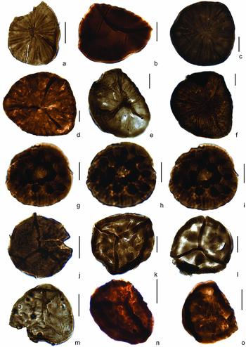
Figure 8a
Emphanisporites protophanus Richardson & Ioannides, Reference Richardson and Ioannides1973
Figure 8b
Emphanisporites rotatus (McGregor) McGregor, Reference McGregor1973
Figure 8c–e
Emphanisporites cf. E. splendens Richardson & Ioannides, Reference Richardson and Ioannides1979
Figure 8g–i
Description. Amb subtriangular to subcircular; proximal face shows an annular thickening formed by joined circular to elongated verrucae, 5 μm wide. Separated verrucae, up to 5 μm wide, are distributed on the proximal face forming a ring between the annular thickening of joined verrucae and the equatorial outline. Radial muri extend from almost the proximal pole to the equatorial margin.
(a) Emphanisporites neglectus Vigran, Reference Vigran1964, slides 60986, W26/1. (b) Emphanisporites protophanus Richardson & Ioannides, Reference Richardson and Ioannides1973, slide 60984, V36/4. (c) Emphanisporites rotatus (McGregor) McGregor, Reference McGregor1973, slide 60659, Q41. (d, e) Emphanisporites cf. E. rotatus (McGregor) McGregor, Reference McGregor1973; (d) slide 60673, P26/3; (e) slide 60736, B32/4. (f) Emphanisporites sp. D in Richardson, Rasul & Al-Ameri, Reference Richardson, Rasul and Al-Ameri1981, slide 60736, M31/1. (g–i) Emphanisporites cf. E. splendens Richardson & Ioannides, Reference Richardson and Ioannides1979, slide 60984, J47/1; (g) proximal focus; (h) distal focus; (i) intermediate focus. (j) Leonispora argovejae Cramer & Diez, Reference Cramer and Diez1975, slide 60664, E26/4. (k) Synorisporites verrucatus Richardson & Lister, Reference Richardson and Lister1969, slide 60986, V30/3. (l) Synorisporites tripapillatus Richardson & Lister, Reference Richardson and Lister1969, slide 60736, O32/0–2. (m) Scylaspora cf. S. scripta in Richardson, Rodríguez & Sutherland, Reference Richardson, Rodríguez and Sutherland2001, slide 60664, M44/2. (n, o) Scylaspora vetusta (Rodriguez) Richardson, Rodríguez & Sutherland, Reference Richardson, Rodríguez and Sutherland2001; (n) slides 60664, F28/0; (o) slide 60664, M26/1. The scale bar represents 20 μm.

Dimensions. 36 μm, measured on one specimen.
Discussion. According to the original description by Richardson & Ioannides (Reference Richardson and Ioannides1973), the species shows equatorial thickening, simple trilete rays, a quite uniform and continuous annular thickening (incipient, irregular or prominent) and radial ribs beyond the annular thickening (pl. 4, figs 1–4, 7). The specimen recorded in Quebrada Ancha is similar to Emphanisporites splendens but the annular thickening is formed by coalescent verrucae and it also has separated verrucae distributed on the proximal face. Richardson, Rodríguez & Sutherland (Reference Richardson, Rodríguez and Sutherland2001) noted that the Cantabrian and North African specimens are highly variable.
Stratigraphic and geographic occurrences. Richardson & Ioannides (Reference Richardson and Ioannides1973) first described the species as Downtonian (Pridoli) in North Africa. Richardson, Rodríguez & Sutherland (Reference Richardson, Rodríguez and Sutherland2001) recorded E. splendens for the upper portion of the Coronaspora reticulata – Chelinospora sanpetrensis (RS) spore biozone, dated as early Pridoli, up to the base of the H Zone, in North Africa and the Cantabrian zone.
Emphanisporites sp. D in Richardson, Rasul & Al-Ameri, Reference Richardson, Rasul and Al-Ameri1981
Figure 8f
Remarks. There is no description for the original specimens, only illustrations have been published by Richardson, Rasul & Al-Ameri (Reference Richardson, Rasul and Al-Ameri1981).
Leonispora argovejae Cramer & Diez, Reference Cramer and Diez1975
Figure 8j
Retusotriletes cf. R. maccullockii Wellman & Richardson, Reference Wellman and Richardson1996
Figure 7m
Retusotriletes cf. R. warringtoni Richardson & Lister, Reference Richardson and Lister1969
Figure 7n
Scylaspora cf. S. scripta in Richardson, Rodriguez & Sutherland, Reference Richardson, Rodríguez and Sutherland2001
Figure 8m
Scylaspora vetusta (Rodriguez) Richardson, Rodriguez & Sutherland, Reference Richardson, Rodríguez and Sutherland2001
Figure 8n, o
Stellatispora inframurinata var. inframurinata (Richardson & Lister) Burgess & Richardson, Reference Burgess and Richardson1995
Figure 5o
Synorisporites tripapillatus Richardson & Lister, Reference Richardson and Lister1969
Figure 8l
Synorisporites verrucatus Richardson & Lister, Reference Richardson and Lister1969
Figure 8k
6. Miospore biostratigraphy
Chelinospora (Clivosispora) verrucata comb. nov, placed here in synonymy with Clivosispora verrucata, occurs since the lowest sample (60667). Its presence demonstrates that the base of the section is not older than the base of the LP Biozone (see Section 3, i.e. latest Homerian or earliest Gorstian). It should also be noted that the presence of Chelinohilates lornensis, in the same level, has been previously recorded from the lowest Lochkovian of Scotland (Wellman & Richardson, Reference Wellman and Richardson1996).
Just 40 cm above the formation base, sample 2 (60704) contains, among others, Stellatispora inframurinata var. inframurinata. This is the eponymous species of the youngest subzone of the LP Zone. Its age ranges from the latest Gorstian or earliest Ludfordian up to latest Ludfordian or earliest Pridoli. In the same sample and above (up to the sample 13 (60984) in the upper Los Espejos Formation), Emphanisporites sp. D has been recorded. This species is known from the Ludlow of Libya, the Downton Group of the UK (Richardson, Rasul & Al-Almeri, Reference Richardson, Rasul and Al-Ameri1981) and from the lower Pridoli of Brazil (Steemans, Rubinstein & Melo, Reference Steemans, Rubinstein and Melo2008). The other species in samples 2 (60704) and 3 (60659) are stratigraphically in situ, except Hispanaediscus wenlockensis, which could be reworked; that species has only been observed in the Wenlock of the UK (Burgess & Richardson, Reference Burgess and Richardson1991). Coronaspora cromatica and Concentricosisporites agradabilis also appear in sample 2 (60704). Both are accessory species of the RS Zone and H Zone of Richardson, Rodríguez & Sutherland (Reference Richardson, Rodríguez and Sutherland2001), respectively, thus pointing to a latest Ludfordian age.
Surprisingly, Breconisporites sp. B in Richardson, Rodríguez & Sutherland (Reference Richardson, Rodríguez and Sutherland2001) first occurs in sample 3 (60659) and extends up to the upper part of the lower section. Breconisporites, a genus formerly unknown below the Lochkovian, based on few data points, was recorded in the lower and middle Pridoli of Libya (B. simplex Wellman, Reference Wellman1993 in Rubinstein & Steemans, Reference Rubinstein, Steemans, Steemans, Servais and Streel2002).
In sample 4 (62521), S. verrucatus appears for the first time in the outcrop. Although that species was believed not to occur below the Pridoli (Richardson & McGregor, Reference Richardson and McGregor1986), it has been shown that it exists also in the late Ludlow (Rubinstein & Steemans, Reference Rubinstein, Steemans, Steemans, Servais and Streel2002; Steemans, Rubinstein & Melo, Reference Steemans, Rubinstein and Melo2008). In addition, the identification of that species is problematic as Synorisporites libycus, which is recorded from the Wenlock (Richardson & Ioannides, Reference Richardson and Ioannides1973), and S. verrucatus are very close and most probably are the end member morphologies of a morphon. In the same sample, C. cf. C. hemiesferica has been identified. That species appears in the H Zone in Spain, considered late Ludfordian and/or early Pridoli in age. In this sample Chelinospora cf. C. cantabrica also occurs. This species is known to appear in the RS Zone, thus supporting this age.
Samples 5 (62523) and 6 (60662) do not display first appearances of biostratigraphically relevant taxa.
In sample 7 (60736) S. tripapillatus, which is characteristic of the TS Zone, supports an age very close to the boundary between the Ludlow and Pridoli.
The occurrence of B. foveolatus, in sample 9 (60664), could be surprising in possible Late Silurian samples. However, this species, which is previously only known from the Lower Devonian of the Old Red Sandstone Continent, has also been identified from the lower Pridoli of Brazil (Steemans, Rubinstein & Melo, Reference Steemans, Rubinstein and Melo2008), therefore supporting the Late Silurian age. Leonispora argovejae occurs in the same sample. It is the eponymous species of the NA Subzone in the lower part of the MN Zone of Spain (Richardson, Rodríguez & Sutherland, Reference Richardson, Rodríguez and Sutherland2001). Even if its presence suggests a Lochkovian age, the definition of the Silurian–Devonian boundary, based on spores, remains uncertain.
In the upper part of the Los Espejos Formation, in sample 13 (60984), the occurrence of Amicosporites streelii indicates a Lochkovian age. However, it should be taken into account that specimens similar to A. streelii have also been found in the Upper Silurian of Saudi Arabia (P. Breuer, pers. com. 2012).
According to the identified miospore species in the Los Espejos Formations, it is difficult to attribute accurate biozones to the analysed samples. Besides, the lower part of the Los Espejos Formation is richer in trilete spore species than the upper part, and the same trend is shown for the marine phytoplankton. Therefore, the lower part has more potential to be more accurately age dated.
Despite the incertitude around the age of the samples, based on miospore assemblages, it seems clear that the studied part of the Los Espejos Formation would have been deposited during the Late Silurian. The marine phytoplankton biostratigraphy also supports this age (Rubinstein, Reference Rubinstein, Fombella Blanco, Fernández González and Valencia Barrera2001; Rubinstein & García Muro, Reference Rubinstein and García Muro2011 and unpub. data).
7. Palaeoecology and palaeobiogeography
The presence of miospore assemblages in the Quebrada Ancha area proves the existence of emergent land near to this locality. This information needs to be considered for the compilation of future palaeogeographic maps.
The distribution and diversity of terrestrial palynomorphs such as miospores, as well as marine palynomorphs, are controlled mainly by palaeoenvironments and palaeogeography. The Precordillera Late Silurian miospores come from a basin developed in an active continental margin of Gondwana, where the sedimentary deposits indicate a foreland basin, as noted above.
For all of the sections studied throughout the basin, palynomorph diversity decreases stratigraphically upwards quite similarly for both marine and terrestrial palynomorphs, in the middle to upper part, culminating with the complete disappearance of palynomorphs near the top (Rubinstein & García Muro, Reference Rubinstein and García Muro2013). This decrease in palynomorph preservation could be a consequence of the predominance of storm-dominated platform facies to shoreface facies, including subaerial exposures, at the top of regressive cycles, in the upper third of the unit (Astini & Maretto, Reference Astini and Maretto1996) represented in Figure 2b.
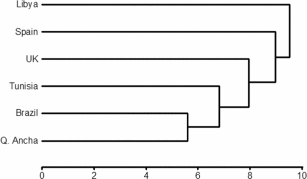
In this context, the miospores of the Los Espejos Formation, in Quebrada Ancha, display a moderate diversity with 29 trilete spores and 14 cryptospores. The diversity of coeval Ludlow–Pridoli palynological assemblages (considering only those that are relatively continuous and well-dated sections) was compared with the Precordillera assemblage and quantitatively analysed using cluster analysis (Fig. 9). In Libya, northern Gondwana, the miospore assemblage is richer than that of the Quebrada Ancha assemblage, having 71 species of trilete spores and 13 species of cryptospores (Rubinstein & Steemans, Reference Rubinstein, Steemans, Steemans, Servais and Streel2002). In Tunisia, also in northern Gondwana and close to the former locality, Spina & Vecoli (Reference Spina and Vecoli2009) recorded a less diverse miospore assemblage, with 32 species of trilete spores and 13 species of cryptospores. In northern Spain, located in the Peri-Gondwanan terrane of Armorica, Richardson, Rodríguez & Sutherland (Reference Richardson, Rodríguez and Sutherland2001) recorded 41 species of trilete spores and 12 species of cryptospores. The northern Brazilian basin is palaeogeographically closest to the Precordillera Basin. It contains 29 species of trilete spores and 8 species of cryptospores (Steemans, Rubinstein & Melo, Reference Steemans, Rubinstein and Melo2008) displaying similar diversity to that of the Precordillera (Fig. 9). It is also noteworthy that the composition of both microfloras has 70% of species in common.
Dendrogram (cluster analysis) comparing Upper Silurian miospore assemblages of Brazil (Steemans, Rubinstein & Melo, Reference Steemans, Rubinstein and Melo2008), Libya (Rubinstein & Steemans, Reference Rubinstein, Steemans, Steemans, Servais and Streel2002), Spain (Richardson, Rodríguez & Sutherland, Reference Richardson, Rodríguez and Sutherland2001), Tunisia (Spina & Vecoli, Reference Spina and Vecoli2009), the UK (Richardson & Lister, Reference Richardson and Lister1969; Richardson & McGregor, Reference Richardson and McGregor1986; Burgess & Richardson, Reference Burgess and Richardson1995) and Quebrada Ancha (present study). x-axis represents the Euclidean distance.

Even though the available distribution information is incomplete, because of the few and widely dispersed assemblages, including the total lack of information from some palaeoplates, it is possible to propose some preliminary palaeobiogeographical and palaeoecological conclusions.
The cryptospores are quite comparable in all the assemblages and are usually less represented (in abundance and diversity) in relation to trilete spores. Even if the diversity of trilete spores is variable, for example comparing those from Libyan and Precordilleran assemblages, cryptospores are more uniform in occurrence. Thus, cryptospores seem to be more constant in abundance and diversity, probably owing to the cosmopolitism of cryptospore producing plants, which are apparently more tolerant to climatic variations, as suggested by Steemans, Wellman & Filatoff (Reference Steemans, Wellman and Filatoff2007).
Conversely, trilete spore-producing plants inhabited a wider variety of biotopes but were more sensitive to climatic variations (Steemans, Wellman & Filatoff, Reference Steemans, Wellman and Filatoff2007). Therefore, palaeoplate setting and resulting palaeolatitudinal position or climatic differences, although significant, might not be the only determinant factor for the distribution of primitive vascular floras. The relatively high degree of similarity between northern Brazil and Argentina, situated at a high palaeolatitude (c. 70°S) and Tunisia, located at a mid-latitude (c. 40°S) supports this interpretation. Contrary to this, pronounced differences between Tunisian and Libyan assemblages can be easily observed on the dendrogram (Fig. 9). No palaeogeographic observation can easily explain this degree of difference. Consequently, spore-producing plants seem to have been especially sensitive to local sedimentology and/or ecology, and thus peculiarities of the local conditions were probably a major factor in controlling their distribution and evolution that overrode palaeogeographic influences. The hydrologic systems, which transport sediment to depositional basins, are important factors controlling sedimentation of miospore assemblages. Transport distance, sediment load and hydrodynamics of drainage basins are all factors that affect transport and resedimentation of miospores and the admixing of assemblages from different local floras. If long-shore currents are insignificant, there would be little mixing of sediments from different river catchments even if estuaries/deltas are relatively near.
Nevertheless, the strong relationship between Brazilian and Argentinean basins must be noted, both of them located at high palaeolatitudes under colder climatic conditions, and very similar in diversity as well as common species.
The miospore assemblage of the Los Espejos Formation contains a striking number of species with coarse ornamentation such as those included in the Chelinospora verrucata var. verrucata morphon, Hispanediscus verrucatus and Synorisporites verrucatus. Those species and other miospores such as Amicosporites spp., several Coronaspora spp. and Chelinospora spp. are frequently difficult to differentiate from each other. However, the presence of this coarse ornamentation could be an adaptation to local environmental conditions.
Interestingly, there is a degree of similarity between miospore assemblage compositions from Spain (Armorica), Libya (Gondwana) and the UK (Avalonia), which is thought to be a result of the palaeogeographic position of the Armorican/Iberic plate (Spain) situated between Baltica and Gondwana. Miospore assemblages from Spain and Brittany contain species characteristic of Baltica like S. newportensis and E. micrornatus (Steemans; Reference Steemans1989; Richardson, Rodríguez & Sutherland, Reference Richardson, Rodríguez and Sutherland2001). Those species are unknown from Gondwana, except for rare specimens from Tunisia (Loboziak et al. Reference Loboziak, Steemans, Streel and Vachard1992). On the other hand, E. splendens, well known from North Africa and Saudi Arabia and recorded from Spain (Richardson, Rodríguez & Sutherland, Reference Richardson, Rodríguez and Sutherland2001), is absent from Baltica.
6. Conclusions
The recovery of miospores in a palynological assemblage, rich in marine acritarchs and chlorophytes, demonstrates the presence of emerged land close to the Quabrada Ancha locality, San Juan Province, Argentina.
Correlations of the Quabrada Ancha assemblages with the miospore biozones from Spain allow recognition of a Ludlow age in agreement with the age based on acritarchs; however, a Pridoli age cannot be excluded for the youngest samples.
The miospore diversity is relatively low, in comparison to localities from the UK, Spain and North Africa, but similar to that from north Brazil. The Argentinean and Brazilian localities are both from relatively high palaeolatitudes (i.e. 70°S); this may explain the strong similarities between both miospore assemblages. The lower diversity of these assemblages may also be a function of their palaeolatitudinal positions, especially when compared to those assemblages from land masses present in lower palaeolatitudes. The comparison between those miospore assemblages also shows that assemblages from Spain are influenced by the vegetation from Baltica and Gondwana, reflecting Spain's intermediate position between these two palaeocontinents and its role as bridge between them.
Acknowledgements
This paper is the result of scientific collaboration between the Ministry of Science, Technology and Productive Innovation of Argentina (MINCYT) and the National Funds for Scientific Research of Belgium (NFSR). (International Cooperation Project MINCYT/NFSR, BE/09/08). Financial support for this study was also provided by the CONICET (PIP 2010-11220090100921) and FONCYT (PICT 2006-1272 and PICT 2005-33630). R. Astini (CONICET, National University of Córdoba) is acknowledged for his collaboration on the fieldwork. We thank M. Giraldo (Liège) for palynological laboratory processing and Rafael Bottero for drafting the figures. We thank Florencia Carotti for helping us to improve the English. We are very grateful to Merrell Miller for the thorough review and linguistic improvement of the manuscript before submission. We also thank Jenny Morris and Charles Wellman for critically reviewing, and improving the manuscript, and Kat Piper for the final review.