INTRODUCTION
Demodex canis (Acari, Demodicidae), a component of normal canine skin, can cause disease in immunosuppressed dogs. Skin lesions include localized or diffuse alopecia, erythema and comedones associated with papular and pustular dermatitis (Shipstone, Reference Shipstone2000). Canine demodicosis occurs as a localized (LD) or a generalized (GD) form. Although the disease is associated with immunosuppressive disorders (Jones et al. Reference Jones, Hunt and King1997), the predisposing factors for the development of generalized demodicosis are not yet fully understood. Some of the most important factors are known to include the immune status of the animal, the breed and breeding line and the age (Ghubash, Reference Ghubash2006), while genetics, nutritional status and oxidative stress (Dimri et al. Reference Dimri, Ranjan, Kumar, Sharma, Swarup, Sharma and Kataria2008), length of hair coat, stage of oestrus cycle, parturition, endoparasitism and debilitating diseases are also implicated. Of these, the immune status is thought to be the most significant (De Bosschere et al. Reference De Bosschere, Casaer, Neukermans, Baert, Ceulemans, Tavernier and Roels2007). It has been postulated that the cell-mediated immune response plays an important role in the pathogenesis of Demodex mite infestation (Caswell et al. Reference Caswell, Yager, Parker and Moore1997). The lack of pathological changes at the site of mite residence in healthy individuals might be due to local immunosuppression stimulated by the mite, which allows them to survive in the host skin (Akilov et al. Reference Akilov, Butov and Mumcuoglu2005). In dogs, it appears that puppies genetically predisposed to the disease have a selective defect of cutaneous T cell-mediated immunity that permits massive proliferation of the parasite (Scott et al. Reference Scott, Schultz and Baker1976). Generalized demodicosis has been induced using immunosuppressive drugs in adult dogs in which genetic factors are unlikely. A natural decline in non-specific immunity in older dogs can trigger the emergence of GD.
Apart from the investigation of non-specific antigen immunosuppression as a secondary feature of canine demodicosis, the character of the immune response occurring in this disease has received little attention. In dogs with either generalized or localized demodicosis, the cellular immune response has not yet been fully characterized. The detection of cellular immunity in canine demodicosis would facilitate a better understanding of its pathogenesis. The determination of CD4+ and CD8+ T cell subpopulations in the peripheral blood by flow cytometry (FC) has been proposed as a simple and fast method to evaluate the immune response. The aim of the present study was to determine the ratio of CD4+/CD8+ T cells in the peripheral blood of dogs with demodicosis.
MATERIALS AND METHODS
Animal selection criteria and design of the study
All animals included in the study were presented to the Referral Veterinary Polyclinic for clinical and dermatological examination. Skin scrapings, hair plucks and exudative swabs were obtained. Dogs positive for Demodex canis, with skin lesions distributed over more than 50% of the body surface and involvement of 2 or more feet, were classified as cases with generalized demodicosis. Dogs with small alopecic, erythematous, scaly and hyperpigmented skin lesions over the face and forelegs covering less than 50% of the total body area were classified as cases with localized demodicosis (Gortel, Reference Gortel2006). Exudative skin swabs were smeared on a clean glass slide and kept for microscopical examination for bacteria. All dogs enrolled in the study were between 1 and 10 years of age and of various breeds. The animals had received no treatment. The dogs were allocated into 2 groups, of 8 animals each. Group II consisted of dogs with LD, while Group III of dogs with GD. Eight healthy age-matched dogs (Group I) were used as controls. The means±s.d. of age for the dogs with LD, GD and control groups were 4·13±3·02, 4·06±3·09 and 4·19±2·99 years respectively and no statistically significant differences were present among the 3 groups. Approximately 3 ml of blood were obtained from each dog in EDTA-containing tubes.
Flow cytometry
FITC-labelled anti-canine CD3, RPE-labelled anti-canine CD4 and RPE-A647-labelled anti-canine CD8 monoclonal antibodies (Serotec) were used to determine the absolute numbers of CD4+ and CD8+ T cells in the peripheral blood of the dogs by use of a BD FACS Calibur flow cytometer. Following the manufacturer's instructions, aliquots of 100 μl of whole blood were added into appropriate tubes, each containing 20 μl of the combined monoclonal antibodies CD3:FITC/CD4:RPE-l and CD3:FITC/CD8:RPEA647 at a dilution of 1:100, along with isotype controls (mouse IgG1:FITC, rat IgG2a:RPE and rat IgG1:RPE-A647) (Serotec). After incubation for 15–20 min at 25°C in a dark room, red blood cells were lysed with 1 ml of 0·1X NH4Cl RBC lysing solution by incubating the samples for 7–10 min at 25°C. The samples were then centrifuged at 200 g, at 4°C for 5 min and the lysing solution was discarded. Measurement of CD3+CD4+ and CD3+CD8+ T cells was performed by gating lymphocyte areas on dot plots. Ten thousand cells were analysed, and positive fluorescence was defined to exclude 98% of the cells in the negative control. Analysis was performed in the flow cytometer using the Cell Quest Pro-software (BD, Bioscience, USA).
Statistical analysis
Comparisons between the groups were performed using the ANOVA and post-hoc Tukey's tests. The level of statistical significance for all the comparisons made was established at P⩽0·05. All data were analysed by means of the statistical package SPSS 15 (SPSS Sciences, Chicago, USA).
RESULTS
Microscopical examination of skin scrapings of dogs with GD revealed higher numbers of eggs, developing stages and adult mites per microscopic field in comparison with the dogs with LD. Partial anorexia and poor body condition were noted in dogs with GD. Three dogs with GD were found to be positive for Staphylococcus sp.
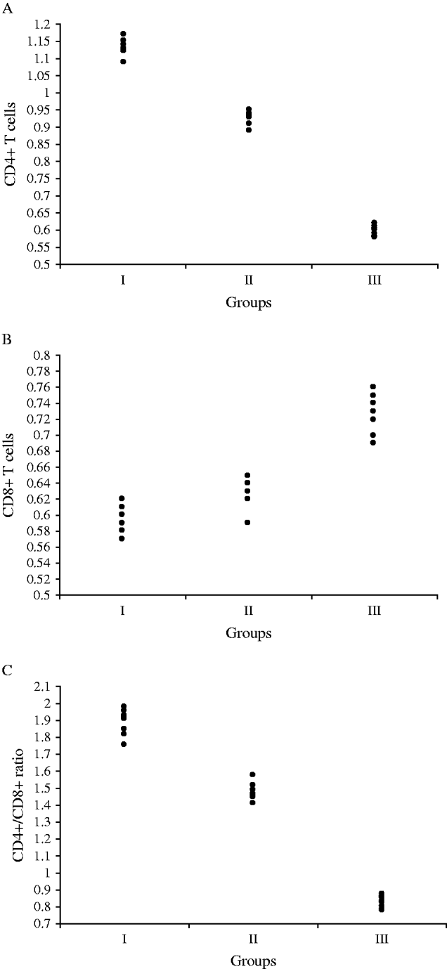
The absolute numbers of CD4+ and CD8+ T cells along with the CD4+/CD8+ ratio of diseased and healthy dogs are presented in a dot plot (Fig. 1A–C). Dogs with GD (Group III) were found to have a significantly elevated number (P<0·01) of CD8+ T cells in comparison with controls (Group I) and dogs with LD (Group II). A significantly higher number of CD8+ T cells (P<0·01) was also found in dogs with LD in comparison with the controls. On the other hand, dogs with GD had a significantly lower number (P<0·01) of CD4+ T cells in comparison with dogs with LD and healthy controls. A remarkable reduction in the number of CD4+ cells (P<0·05) was also found in dogs with LD in comparison with the healthy controls. The ratio of CD4+/ CD8+ was found to be significantly lower (P<0·01) in dogs with GD in comparison with dogs with LD and healthy controls. The CD4+/CD8+ ratio was also found to be significantly lower (P<0·01) in dogs with LD compared to healthy controls.
The absolute number (×109/L) of (A) CD4+ and (B) CD8+ T cells and the ratio of (C) CD4+/CD8+ T cells in peripheral blood of individual dogs of each group.

DISCUSSION
The increased number of adult mites along with their immature stages found during microscopical examination of skin scrapings of dogs with GD indicates that the mites are actively multiplying in these animals and might be the cause of skin lesions. Dogs with GD may be unable to restrict the multiplication of the mites. An overgrowth of secondary invaders like Malassezia sp. and Staphylococcus sp. has been reported in canine demodicosis, probably due to the immunosuppressive effect of the disease on skin microenvironment of affected dogs (Tarallo et al. Reference Tarallo, Lia, Sasanelli, Cafarchia and Otranto2009).
The increased number of CD8+ T cells in the peripheral blood of dogs with demodicosis indicates that these cells seem to play a role in the immunity of the dog against the parasite. Both immunodeficiency and immune-mediated injury to the skin may play a role in demodicosis (Frost, Reference Frost1999). Naive CD8+ T cells undergo an intense process of differentiation and proliferation after recognizing antigenic peptides derived from viruses, bacteria, or parasites (Wong and Am, Reference Wong and Am2003). Our findings regarding CD8+ cells are in agreement with those of other similar studies (Caswell et al. Reference Caswell, Yager, Parker and Moore1997; Fukata et al. Reference Fukata, Fuoki, Yoshikawa, Kambayashi, Kito and Kitagawa2005). The protective role of CD8+ lymphocytes in human demodicosis has been demonstrated. The lower density of the Demodex mites and smaller skin lesions has been found in human patients with a normal number and function of CD8+ lymphocytes (Mumcuoglu and Akilov, Reference Mumcuoglu and Akilov2005).
In this study, we also found that dogs with GD had a significantly lower number of CD4+ T cells than dogs with LD and healthy controls. Additionally, dogs with LD also had a lower number of CD4+ T cells as compared to healthy controls. This phenomenon may be due to a specific down-regulating activity of Demodex canis mites on CD4+ T cells or an increased rate of apoptosis or immunological exhaustion of these cells. A reduced number of CD4+ T cells has also been found in human demodicosis (Akilov and Mumcuoglu, Reference Akilov and Mumcuoglu2004). The lack of pathological changes at the site of mite residence in healthy individuals might be due to local immunosuppression stimulated by the parasites, which allows them to survive in the host skin (Akilov et al. Reference Akilov, Butov and Mumcuoglu2005). It has been found that other parasitic infections, like Leishmania infantum, result in reduction of CD4+ T cells and the CD4+/CD8+ ratio in symptomatic dogs (Papadogiannakis et al. Reference Papadogiannakis, Andritsos, Kontos, Spanakos, Koutis and Velonakis2009).
Effective cell-mediated immunity, essential for protection against chronic intracellular infections, cancer and parasites, depends on the cooperative action of CD8+ and CD4+ T cells to combat these conditions (Mosmann and Sad, Reference Mosmann and Sad1996). Juvenile-onset localized demodicosis resolves spontaneously in 90% of cases, whereas in some patients administration of topical or oral antibiotics is needed in order to control the secondary bacterial skin infections (Ghubash, Reference Ghubash2006). It is likely that self recovery in LD might be due to cooperative actions of CD8+ and CD4+ T cells against the mites. Conversely, generalized demodicosis is considered to be a severe disease requiring prolonged treatment, which is often difficult and potentially life-threatening (Fourie et al. Reference Fourie, Kok, Du Plessis and Rugg2007). Compromised CD4+ T cells may not be complementary to CD8+ T cells, thus self recovery in GD may not be encountered. The altered CD4+/ CD8+ ratio might be the reason for prolonged pharmacological treatment necessary in cases of GD. In conclusion, severe alteration of the CD4+/CD8+ ratio may be involved in the pathogenesis of the generalized demodicosis. Additional, large-scale studies are needed in order for these assumptions to be confirmed.