INTRODUCTION
Strongyloidiasis is a widespread infection that affects 30–100 million people across 70 countries, mainly in tropical and subtropical regions of the world (Genta, Reference Genta1989; Puthiyakunnon et al. Reference Puthiyakunnon, Boddu, Li, Zhou, Wang, Li and Chen2014). Strongyloides venezuelensis naturally infects rodents, and has been used as an experimental intestinal parasite model (Rodrigues et al. Reference Rodrigues, Cardoso, Gonçalves, Silva, Massa, Alves, Ueta, Silva and Costa-Cruz2013; Yasuda et al. Reference Yasuda, Matsumoto and Nakanishi2014; Corral et al. Reference Corral, Paula, Gottardi, Meisel, Chieffi and Gryschek2015). In experimental infections, infective-stage of S. venezuelensis larvae migrates to the lungs before implanting in the duodenal mucosa. In this way, migration in the rodent host is similar to that of Strongyloides stercoralis in humans (Negrão-Corrêa et al. Reference Negrão-Corrêa, Silveira, Borges, Souza and Teixeira2003; Ferreira et al. Reference Ferreira, Pereira, Amaral, de Souza, Coelho, Souza, Negrão-Corrêa and Teixeira2009). Thus parasite has been used as a convenient tool for development and validation of more sensitive procedures for human strongyloidiasis diagnosis (Gonçalves et al. Reference Gonçalves, Rodrigues, Silva, Gonçalves, Cardoso, Beletti, Ueta, Silva and Costa-Cruz2008, Reference Gonçalves, Silva, Ueta and Costa-Cruz2010).
Immunological assays, such as enzyme-linked immunosorbent assay (ELISA), indirect fluorescent antibody test and western blotting are useful for evaluating the host immune response and diagnosing asymptomatic cases and for epidemiological surveys (Paula and Costa-Cruz, Reference Paula and Costa-Cruz2011; Levenhagen and Costa-Cruz, Reference Levenhagen and Costa-Cruz2014; Gottardi et al. Reference Gottardi, Paula, Corral, Meisel, Costa, Abdala, Pierrotti, Yamashiro, Chieffi and Gryschek2015). The principal limitation in establishing such specific methods is the difficulty of obtaining infective S. stercoralis larvae. Parasite antigen detection in fecal samples by immunological assays may contribute to earlier diagnosis, which is particularly an important consideration for patients with increased risk of strongyloidiasis, such as immunocompromised individuals (Bailey, Reference Bailey1989; Gonçalves et al. Reference Gonçalves, Rocha, Gonzaga, Gonçalves-Pires, Ueta and Costa-Cruz2012a ).
DNA technology has greatly impacted many areas of parasitology, including infection diagnosis (Wang and Gao, Reference Wang and Gao2014). Polymerase chain reaction (PCR) has broad applicability, mainly because its high sensitivity allows the amplification of DNA fragments from minute amounts of parasite material (Gasser, Reference Gasser1999; Saugar et al. Reference Saugar, Merino, Martín-Rabadán, Fernández-Soto, Ortega, Gárate and Rodríguez2015). Several reports have shown high PCR sensitivity for detection of parasite DNA in various biological sample types (i.e. feces, tissues) and from different host species (Esteban-Redondo et al. Reference Esteban-Redondo, Maley, Thomson, Nicoll, Wright, Buxton and Innes1999; Wongratanacheewin et al. Reference Wongratanacheewin, Pumidonming, Sermswan and Maleewong2001; Sandoval et al. Reference Sandoval, Siles-Lucas, Lopez Alban, Pérez-Arellano, Gárate and Muro2006; Marra et al. Reference Marra, Chiuso-Minicucci, Machado, Zorzella-Pezavento, França, Ishikawa, Amarante, Sartori and Amarante2010).
The aim of this study was to compare the diagnostic value of three diagnostic methods: the eggs per gram of feces (EPG) technique, the detection of coproantigens by ELISA and DNA amplification/detection by conventional PCR. We tested these methods on feces samples of both immunocompetent and immunosuppressed rats that were experimentally infected with S. venezuelensis.
MATERIALS AND METHODS
Animals
Male Rattus norvegicus (Wistar) weighing 100–120 g were maintained in the Centro de Experimentação e Utilização de Animais, UFU provisioned with laboratory chow and water available ad libitum.
Parasites
Strongyloides venezuelensis third-stage infective larvae (L3) were obtained from charcoal cultures of infected rat feces. The cultures were stored at 28 °C for 48 h, and the infective larvae were collected and concentrated using the Rugai method (Rugai et al. Reference Rugai, Mattos and Brisola1954). For infection, each rat was subcutaneously inoculated with 1500 S. venezuelensis L3 larvae.
Experimental groups
The rats were divided into four groups: immunocompetent infected rats (n = 30), immunosuppressed infected rats (n = 30), control immunocompetent (n = 6) and control immunosuppressed (n = 6). Prior to infection, immunosuppressed groups received 5 µg mL−1 of dexamethasone disodium phosphate in water for 5 days, as previously described by Romand et al. (Reference Romand, Thulliez and Dubey1998). Animals from the infected groups were then inoculated subcutaneously with 1500 L3 S. venezuelensis larvae. Rats were examined in groups of 6 at 0 and 5, 8, 13, 21 and 39 days post-infection (dpi). Each fecal sample was divided into three subsamples. The first subsample was submitted to the Cornell-MacMaster quantitative method (Gordon and Whitlock, Reference Gordon and Whitlock1939) the others were stored at −70 °C for later coproantigen detection assay (ELISA) and PCR. Fecal samples used for the ELISA were prepared prior to assay by adding 0·15 m phosphate buffer pH 7·4 (PBS, containing 0·3% Tween 20) at a ratio of 1:1.
EPG
Experimentally infected rats were placed individually on clean absorbent paper moistened with distilled water, and allowed to defecate over an 18 h period at the following time points: 5, 8, 13, 21 and 39 dpi. The feces were then collected, diluted in saturated saline and homogenized. The supernatant was placed in a McMaster chamber, and the number of EPG was estimated using the Cornell-McMaster method (Gordon and Whitlock, Reference Gordon and Whitlock1939).
Production of immune serum and secondary antibodies
For the production of immune serum, two rabbits were immunized with crude larval somatic antigen as described by Gonçalves et al. (Reference Gonçalves, Rodrigues, Silva, Gonçalves, Cardoso, Beletti, Ueta, Silva and Costa-Cruz2008). Briefly, the rabbits were inoculated with 500 µg mL−1 of alkaline extract of S. venezuelensis L3 larvae emulsified in Freund's complete adjuvant in the first immunization. Inoculations were made at intervals of 2 weeks after the first immunization, using Freund's incomplete adjuvant. Blood samples were collected by auricular puncture, at intervals of 2 weeks, for evaluation of the antibody titers. Three months following immunization, when a high antibody titer (15 000) was detected, the animals were submitted to a cardiac puncture and blood was collected and an Immunoglobulin G (IgG) fraction was prepared using a Protein A Sepharose CL-4B column. After testing IgG activity against the specific antigen, a portion of the IgG fraction was stored at −20 °C to be used as capture antibody in ELISA. The other portion was conjugated with horseradish peroxidase (Sigma) according to Wilson and Nakane, (Reference Wilson, Nakane, Knapp, Holuban and Wick1978).
Coproantigen detection by ELISA
ELISA was performed according to Gonçalves et al. (Reference Gonçalves, Silva, Ueta and Costa-Cruz2010). Briefly, high-binding microtiter plates (Corning-Costar; Laboratory Sciences Company, New York, NY) were coated with 40 µg mL−1 of anti-S. venezuelensis IgG in 0·06 m carbonate bicarbonate buffer (pH 9·6) and stored overnight at 4 °C. Plates were washed three times for 5 min each with PBS containing 0·05% Tween 20 (PBS-T). After washing, plates were incubated with fecal supernatant (1:8 dilution) for 45 min at 37 °C. The secondary antibody consisting of peroxidase-labelled rabbit anti-S. venezuelensis IgG at the optimal dilution of 1:40 was added, and the plates were incubated again for 45 min at 37 °C. The reaction was revealed by adding the enzyme substrate (0·03% H2O2 and o-phenylenediamine [OPD] in 0·1 m citrate–phosphate buffer, pH 5·0) with a final incubation for 15 min at room temperature. The reaction was stopped by adding 2 N H2SO4 and the optical density (OD) was determined at 492 nm using a plate reader (Titertek Multiskan; Flow Laboratories, McLean VA). Results were arbitrarily expressed as ELISA index (EI), according to the following formula: EI = OD sample/cut-off, where the cut-off was established as the mean OD of three negative control sera plus two standard deviations and EI > 1·0 was considered positive.
DNA extraction and PCR
Genus-specific primer pairs were utilized to amplify the small subunit ribosomal RNA gene, as described by Dorris et al. (Reference Dorris, Viney and Blaxter2002) to analyze several species within the genus Strongyloides (Forward 5′-AAAGATTAAGCCATGCATG-3′; Reverse 5′- GCCTGCTGCCTTCCTTGGA-3′). The QIAamp DNA stool mini kit (QIAGEN, Hilden, Germany) was used to extract DNA from feces pool samples following the manufacturer's instructions. DNA from S. venezuelensis L3 larvae was used as positive control in all PCR reactions. DNA from feces of non-infected rats and adult Syphacia muris specimens (nematodes commonly recovered from rat DNA) was used as controls to identify similar and dissimilar bands amplified by the primers. A blank tube (PCR mix without DNA) was used as negative control.
PCR was performed in a Master Cycler ep Gradient S thermocycler (Eppendorf, Hamburg, Germany). Samples containing 10 mm dNTPs, 0·4 mm each primer, 1·5 mm MgCl2, 50 mm KCl, pH 8·3, 0·1 U of Taq polymerase® (5 U µL−1, Invitrogen, Life Technologies) and 100 ng of genomic DNA for a final reaction volume of 10 µL. PCR program (cycle specifications) were chosen according to Marra et al. (Reference Marra, Chiuso-Minicucci, Machado, Zorzella-Pezavento, França, Ishikawa, Amarante, Sartori and Amarante2010). PCR products were run on 5% polyacrylamide gels in 1× Tris-Borate-EDTA (TBE) buffer (Freitas et al. Reference Freitas, Vianna, Martins, Silva, Pesquero and Gomes2004). Direct sequencing of PCR products were performed using an automatic sequencer (ABI 3500 genetic analyzer DNA Sequencer, Applied Biosystems, Thermo Fisher Brand, Foster City, CA) and the BigDye Terminator v3.1 Cycle Sequencing kit (Applied Biosystems, Thermo Fisher Brand, Foster City, CA) according to the manufacturer's instructions. The sequences obtained were aligned using the BioEdit program (Biological Sequence Alignment Editor) (http://www.mbio.ncsu.edu/bioedit/page2.html) and compared with previously reported sequences from GenBank (http://www.ncbi.nlm.nih.gov/GenBank/tbl2asn2), using Basic Local Alignment Search Tool.
Statistical analysis
Eggs were counted and expressed as mean ± standard error of the mean (s.e.m.) percentages and were calculated using Excel. Each experiment was performed in duplicate. Data were analysed using one-way analysis of variance (ANOVA), followed by a Bonferroni test. The level of statistical significance was set at P < 0·05.
RESULTS
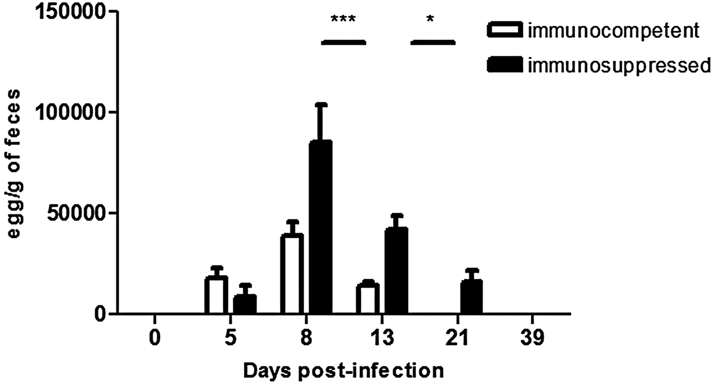
The initial time point for egg shedding was 5 dpi for both immunocompetent and immunosuppressed rats, reaching a peak at 8 dpi and decreasing at 13 dpi in both groups. Immunosuppressed rats presented higher egg counts at day 8 (P < 0·001) and 13 (P < 0·05) compared with immunocompetent rats. Strongyloides venezuelensis eggs were not detected in the feces of immunocompetent rats beyond 21 dpi, or in immunosuppressed rats beyond 39 dpi (Fig. 1).
Total number of eggs per gram in feces of immunocompetent and immunosuppressed rats infected with Strongyloides venezuelensis on 0 and 5, 8, 13, 21 and 39 dpi *P < 0·05 and ***P < 0·001. Abbreviation: dpi, days post-infection.

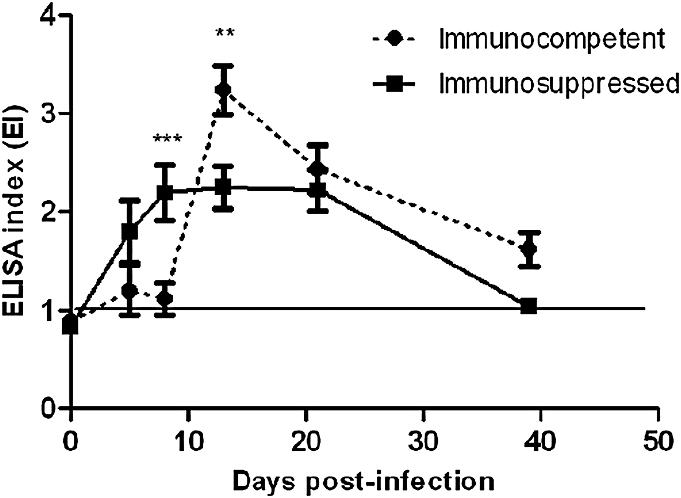
The antigen detection of immunocompetent and immunosuppressed rats was compared. In immunocompetent rats, coproantigen detection was positive during the entire kinetics (Fig. 2) and antigen was best detected on 13 dpi (P < 0·01); at 21 and 39 dpi, a lower EI was observed probably due to the reduction of antigen in feces as a result of elimination of the infection. In immunosuppressed animals, similar results were observed on 8–21 dpi and a lower EI was observed on 39 dpi.
Detection of coproantigen, with antigen levels expressed as ELISA-index in fecal samples from immunocompetent and immunosuppressed rats infected with Strongyloides venezuelensis on 0 and 5, 8, 13, 21 and 39 dpi **P < 0·01 and ***P < 0·001. Fecal supernatants were diluted 1:8 and incubated with anti-L3 polyclonal antibody. Abbreviations: dpi, days post-infection; ELISA, enzyme-linked immunosorbent assay.

In the PCR assay, a DNA fragment of approximately 405 base pairs corresponding to the positive control (L3 larvae of S. venezuelensis) was amplified. The primers amplified bands that were not associated with a specific parasite, although they were distinguishable from the positive control. PCR products (amplified bands from pooled fecal samples) from both immunocompetent and immunosuppressed rats are shown in Fig. 3. PCR amplification produced sufficient bands throughout the post-infection period in both groups.
Electrophoretic profiles of PCR products from fecal samples of rats infected with Strongyloides venezuelensis. (A) Immunocompetent and (B) immunosuppressed groups. MW lane: 100 base pairs ladder (Life Technologies); Lane 1: ~405 base pairs (positive control, S. venezuelensis L3 DNA); Lane 2: ~440 base pairs (corresponding to the Syphacia muris); Lane 3: ~480 base pairs (non-infected Rattus norvegicus fecal sample day 0); Lanes (4–8): ~405 base pairs. (pooled DNA from fecal samples collected 5, 8, 13, 21 and 39 dpi. Lane 9: negative control (without DNA). Abbreviations: dpi, days post-infection; PCR, polymerase chain reaction; MW, molecular weight.

DNA sequences obtained from filariform S. venezuelensis larvae and feces samples in both experimental groups showed 100% similarity with S. venezuelensis gene for 18S small subunit ribosomal RNA (AB923887.1) in GenBank (Fig. 4).
Alignment of products amplification from Strongyloides venezuelensis L3 larvae and feces samples from experimental groups with gene sequence of the subunit 18S rRNA of S. venezuelensis deposited in NCBI database. Abbreviations: NCBI, National Center for Biotechnology Information.

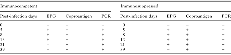
The comparison of the three diagnostic methods shows that for immunocompetent individuals, egg shedding was highest at 8 dpi, coproantigen detection by ELISA was highest at 13 dpi, and PCR amplification produced sufficient bands throughout the post-infection period. In the immunosuppressed group, egg shedding was also highest at 8 dpi, coproantigen detection by ELISA was highest at 8–21 dpi, and PCR amplification also produced sufficient bands throughout the post-infection period. Table 1 summarizes the comparison between parasitological, immunological and molecular methods for fecal samples from experimentally infected immunocompetent and immunosuppressed rats.
Comparison of eggs per gram of feces (EPG), coproantigens detected by enzyme-linked immunosorbent assay (ELISA), and polymerase chain reaction (PCR) amplified bands using fecal samples from immunocompetent and immunosuppressed rats infected with Strongyloides venezuelensis

+, positive result; −, negative result.
DISCUSSION
The detection of S. venezuelensis antigens in stool samples by ELISA may contribute to accuracy diagnostic, an important consideration for immunocompromised groups due to higher potential risk for severe forms of strongyloidiasis (Gonçalves et al. Reference Gonçalves, Silva, Ueta and Costa-Cruz2010). The coproantigen technique is more sensitive than direct observation of eggs because the antigen was detected in feces up to 39 dpi in both groups. In immunosuppressed rats, the timing of egg elimination was affected by dexamethasone disodium phosphate treatment, in which infection was more severe and long lasting. Peak egg shedding occurred at 8 dpi in both groups, with a decrease in egg counts at the end of the post-infection period. Similar results were shown by Nakai and Amarante (Reference Nakai and Amarante2001), Gonçalves et al. (Reference Gonçalves, Silva, Ueta and Costa-Cruz2010) and Paula et al. (Reference Paula, Sitta, Malta, Gottardi, Corral, Gryschek and Chieffi2013).
Coproantigen detection had higher positive detection of S. venezuelensis infection compared with the EPG method, particularly in immunosuppressed animals, in concordance with results obtained by Gonçalves et al. (Reference Gonçalves, Silva, Ueta and Costa-Cruz2010). Diagnosis using polyclonal antibodies has also been investigated by different authors, and the results revealed potential for the development of a financially viable kit for rapid coproantigen detection (Nageswaran et al. Reference Nageswaran, Craig and Devaney1994; Gonçalves et al. Reference Gonçalves, Silva, Ueta and Costa-Cruz2010; Sykes and McCarthy, Reference Sykes and McCarthy2011).
Molecular analysis of the subunit 18S ribosomal RNA gene region has been used as a target for phylogenetic analysis, especially for intestinal parasites (Dorris et al. Reference Dorris, Viney and Blaxter2002). In the present study, analysis revealed that the same region amplified a 405 base pairs fragment in fecal samples from both immunocompetent and immunosuppressed rats throughout the duration of infection. The fragment was larger than described by Marra et al. (Reference Marra, Chiuso-Minicucci, Machado, Zorzella-Pezavento, França, Ishikawa, Amarante, Sartori and Amarante2010) and Paula et al. (Reference Paula, Sitta, Malta, Gottardi, Corral, Gryschek and Chieffi2013), despite using the same set of primers; this may be due to greater separation sensitivity of the polyacrylamide gel, which is usually 10 times higher than agarose gel method (Freitas et al. Reference Freitas, Vianna, Martins, Silva, Pesquero and Gomes2004). The amplification products from S. venezuelensis L3 larvae and feces samples from both experimental groups showed high sequence similarity to ribosomal RNA genes of Strongyloides species.
PCR using genus-specific primers may function as sensitive methods for detection of S. venezuelensis genetic material in feces of rats with low parasite load (Nakai and Amarante, Reference Nakai and Amarante2001, Marra et al. Reference Marra, Chiuso-Minicucci, Machado, Zorzella-Pezavento, França, Ishikawa, Amarante, Sartori and Amarante2010; Paula et al. Reference Paula, Sitta, Malta, Gottardi, Corral, Gryschek and Chieffi2013) and it was demonstrated that the detection of S. venezuelensis in fecal samples showed higher sensitivity in the PCR assay than the parasitological methods. In this study, specific amplification occurred until 39 dpi in both, immunocompetent and immunosuppressed rat groups.
Coproantigen detection and PCR amplification showed similar results in the immunosuppressed group, perhaps a reflection of greater susceptibility to S. venezuelensis infection. According to Wallen et al. (Reference Wallen, Kita, Weiler and Gleich1991) and Gonçalves et al. (Reference Gonçalves, Ribeiro, Silva, Ueta and Costa-Cruz2012b ) the glucocorticoids inhibit the activation, proliferation and survival of inflammatory cells such as eosinophils and mast cells, and block the release of IL-4, IL-5 and IL-13 cytokines involved in response to helminthic infections.
The comparative results of the EPG method and the coproantigen detection by ELISA showed to be similar to the results of Gonçalves et al. (Reference Gonçalves, Silva, Ueta and Costa-Cruz2010), for diagnosis of S. venezuelensis, and also to the results of Gordon et al. (Reference Gordon, Zadoks, Stevenson, Sargison and Skuce2012) and Brockwell et al. (Reference Brockwell, Spithill, Anderson, Grillo and Sangster2013), for diagnosis of Fasciola hepatica. The high sensitivity of PCR for diagnosis of S. venezuelensis infection, when compared with the EPG method, was previously demonstrated by Marra et al. (Reference Marra, Chiuso-Minicucci, Machado, Zorzella-Pezavento, França, Ishikawa, Amarante, Sartori and Amarante2010).
Results from the current study demonstrate the reliability and sensitivity of the coproantigen detection by ELISA, and of the PCR assay for diagnosis of experimental strongyloidiasis. These results could help to improve the diagnosis of human strongyloidiasis as alternatives for applying in epidemiological research, considering the importance of early diagnosis to prevent the possibility of chronicity and hyperinfection establishment in endemic areas.
FINANCIAL SUPPORT
The authors are grateful to the research funding agencies, Fundação de Amparo à Pesquisa de Minas Gerais (C.C.J.M., FAPEMIG -CBB-PPM-00396-13) and Conselho Nacional de Desenvolvimento Científico e Tecnológico (C.C.J.M., CNPq- 302426/2012-4), for the financial support.
ETHICAL STANDARDS
Authors assert that all procedures contributing to this study comply with the ethical standards of laws of Brazil on the care and use of laboratory animals, in accordance with the ethical principles upon approval of the Comitê de Ética na Utilização de Animais of Universidade Federal de Uberlândia (CEUA/UFU no 004/08).