Introduction
Carcinoma of the uterine cervix is a malignancy that demonstrates stark contrast between the developing and the developed world. Worldwide it is considered to be the seventh most common cancer overall and the fourth most common in females. In India, it is the second most malignancy in females, affecting more than 20 per 100,000 females.Reference Ferlay, Soerjomataram and Dikshit1 Most patients present with locally advanced diseases and for such patients, concurrent chemoradiotherapy is the management of choice.Reference Nandakumar, Anantha and Venugopal2 Conventionally radiotherapy (RT) has been delivered through a 2-field (AP-PA) or 4-field (AP-PA and two lateral) design to cover the whole pelvis for nodal disease and parametrial spread, apart from the gross disease. After 45–50 Gy has been delivered through external beam radiotherapy (EBRT), the gross disease is given a brachytherapy boost.Reference Peters, Liu and Barrett3 This treatment is associated with significant morbidity, especially haematological and gastrointestinal, which increases with concurrent chemotherapy. Efforts have been made to reduce the dose delivered to normal structures through intensity-modulated conformal techniques since the turn of the century, with results from Mundt et al.Reference Mundt, Mell and Roeske4 and Mell et al.Reference Mell, Tiryaki and Ahn5 suggesting that these can reduce clinically observed toxicities. Various other studies, clinical and dosimetric, have suggested that intensity-modulated radiotherapy (IMRT) can spare the OARs while delivering the same dose to target volumes with clinically discernible benefit.Reference Teoh, Clark and Wood6 However, a lot of this work has included a mixed population of patients, and a significant number of patients are post-operativeReference Lukovic, Patil and D’souza7 where the uterus has been removed. The later alters clinical target volume (CTV) significantly, and one cannot extrapolate the same data to patients with an intact genital tract.
VMAT is a form of IMRT that delivers treatment in arcs by delivering dose throughout the rotation of the gantry and modulating the intensity of individual beamlets therein. VMAT is an extended form of IMRT with variable dose rate, gantry speed and dynamic multileaf collimator (MLC) movement with multiple beam entry angle options.Reference Otto8 VMAT plans with faster delivery time, fewer monitor unit (MU) and superior dose distribution than conventional step-and-shoot IMRT have been reported.Reference Palma, Vollans and James9 VMAT is rapidly becoming the treatment modality of choice when IMRT is employed, given the advantages it provides over ‘conventional’ IMRT—treatment time (by as much as 5 times shorter)Reference Richetti, Fogliata and Clivio10,Reference Cozzi, Dinshaw and Shrivastava11 and lower integral dose by almost 12% as compared to IMRT.Reference Richetti, Fogliata and Clivio10 It came into clinical practice less than a decade ago, but has rapidly been adopted for various sites due to its advantages of shorter treatment time and in many cases, improved OAR sparing. Its ability to deliver the same treatment as IMRT in lesser time needing fewer MUs is very favourable for gynaecological malignancies.Reference Teoh, Clark and Wood12 However, 3-dimensional conformal radiotherapy (3D-CRT) already provides that benefit,Reference Yang, Zhu and Cheng13 but the dosimetric advantages specifically in these cases of VMAT over 3D-CRT are not established in a head-on comparison for a uniform group of patients. Therefore, we propose to conduct a dosimetric comparison of these two techniques in patients with LACC.
Materials and Methods
This was a single institutional, prospective, observational study on patients of LACC in a tertiary cancer centre conducted from August 2015 to August 2016. The study was approved by the Institutional Ethics Committee. Patients aged 18–80, Karnofsky performance score (KPS) ≥60 with histologically proven squamous cell carcinoma of the cervix, the International Federation of Gynecology and Obstetrics (FIGO) 2009 Stage IIB–IIIBReference Pecorelli, Zigliani and Odicino14 and inoperable cases belonging to earlier stages, wherein ‘inoperable’ shall be defined as ≥1 of inoperable disease as opined by surgical oncologist and patient unfit and/or unwilling for surgery, with normal and bone marrow reserve (Haemoglobin >10 gm%, White blood cell >4000/cc, Platelet >100,000/cc), normal renal (normal KFTs) and liver (normal LFTs). Patients having Stage IV disease, positive para-aortic node, history of any other malignancy within the last 5 years, adenocarcinoma of the cervix, patient suffering from active HIV, Hepatitis B or C and pregnant and lactating women were excluded from the study. CT data of patients who fit the inclusion criteria was used for this study.
Planning was initially done by 3D-CRT technique (which was used for actual treatment), followed by VMAT planning after EBRT completion and appropriate dosimetric comparisons were done. A minimum accrual of thirty patients was planned at the initiation of the study. After a detailed history and examination, all routine blood investigations and chest X-rays were performed. Investigations like USG/CT/MRI of the abdomen and pelvis, cystoscopy, proctoscopy, bone scan and DTPA/DMSA scans for renal status were done when indicated.
Steps of RT planning
Patients were simulated in supine position with hands above the head using a 16-slice CT simulator (Siemens Somatom Sensation open, Siemens Healthineers, Forcheim, Germany). Planning CT scans were obtained from L1 (or higher if indicated) to lesser femoral trochanter with 3 mm slice acquisition and no inter-slice gap after injection of 70–100 mL of non-iodinated contrast (Siemens Somatom Sensation Open 16 slice). A bladder protocol was followed. Patients were asked to void urine followed by drinking 500 mL water over 15 minutes. Images were taken 30 minutes thereafter. No rectal protocol was specified. Patients were asked to evacuate bowel before the scan. The lowermost extent of disease was marked by a vaginal marker placed per vaginally or by placing a radiotherapy planning (RTP) marker at the skin tag between the vulva and anal verge (this along with per vaginal findings served as a surrogate for the lowermost extent of disease). Three RTP markers were placed, two lateral and one central, before simulation. These were then tattooed after the simulation to facilitate patient positioning during treatment. Images, thus, obtained were transferred to our treatment planning system (XiO Release 5·10/Monaco v3·0, Elekta AB, Stockholm, Sweden) followed by contouring.
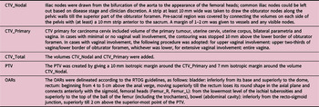
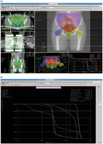
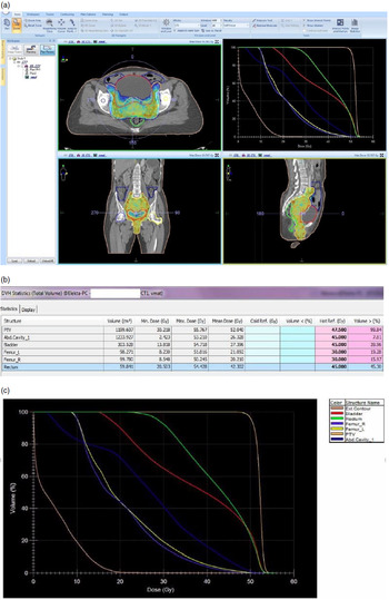
Treatment volumes were drawn according to ICRU 50, 62 and 83 definitions.15–17 OARs were drawn as per the Radiotherapy Oncology Group (RTOG) guidelines for organ delineation in pelvic RT. The volumes for CTV and OARs were delineated as per the details mentioned in Table 1.Reference Taylor, Rockall and Reznek18–Reference Small, Mell and Anderson21 Following this, planning was performed on XiO v5·10 (for 3D-CRT) and subsequently on Monaco v3·0 (for VMAT). The total dose prescribed was 45–50·4 Gy @ 1·8–2 Gy/#, 5# a week over 4·5–5·5 weeks. Appropriate dosimetric data were recorded for both planning modalities and then compared. For the PTV, homogeneity and conformity indices were also calculated (HI and CI). All patients received radical chemoradiotherapy. Treatment was performed using 3D-CRT plans. The plan evaluation criteria for both the modalities were as follows: 95% of the PTV should receive more than or equal to 95% of the prescribed dose (Figures 1a and 1b). No part of the PTV was to receive >110% of the prescribed dose for 3D-CRT. For VMAT, no volume beyond 15 mm outside the PTV was to receive >100% of the prescribed dose. It was also ensured that no volume within PTV received <93% of the prescribed dose in VMAT (Figures 2a and 2b). The dose limit to OARs was as follows: bowel (small and large): volume of abdominal cavity receiving 45 Gy to be less than 195 cc, rectum: volume of rectum receiving 45 Gy (V45) had to be less than 50%, bladder: volume of bladder receiving 45 Gy (V45) had to be less than 50%, femoral heads: volume receiving 30 Gy (V30) not to exceed 15%. Planning was assessed slice-by-slice and also via dose–volume histograms (DVHs). For the purpose of comparisons: the dose to 95 (V95) and 100% (V100) of the PTV was recorded. The dose to 95, 98 and 100% (D95, D98, D100) of the PTV volume and the maximum isodose in the PTV (Imax) were obtained. Homogeneity and conformity indices were calculated (HI and CI).Reference Feuvret, Noël and Mazeron22 HI = Imax/RI (where Imax is the maximum isodose in the target and RI is reference isodose). CI = VRI/TV (where VRI is volume covered by reference isodose and TV is target volume). To quantify the dose distribution of OAR, percentage volumes receiving 10, 20, 30, 40, 45 and 50 Gy (V10, V20, V30, V40, V45 and V50) and maximum dose (Dmax) were recorded for all OARs. Statistical analysis of data obtained from each arm was done by comparing the corresponding dose–volume parameters of the 3D-CRT and VMAT plans with the paired t-test (SPSS v21). The level of significance was set at 0·05.
Table describing contouring guidelines that were followed

(a) Image above shows a typical 3D-CRT plan. A 4-field box was employed with shielding of normal structures by MLCs. The 4-field arrangement and dose distribution (95%) can be seen. (b) The DVH of the above plan. Dose was 50 Gy/25#, with 95% volume covered by 97% isodose.

(a) The VMAT dose distribution for the same patient as above. The 95% isodose line can be seen as hugging to the PTV boundaries far more closely than the 3D-CRT plan. (b) The VMAT dose parameters for this patient. Constraints were met for the bladder and abdominal cavity but neither femur nor rectal constraints could be achieved. (c) DVH of the VMAT plan: OAR curves have shifted to the left compared to the 3D-CRT plan.

Results
Thirty-nine patients were enrolled in this study. The median age at RT registration was 57 years (range 35–78) with most patients belonging to the 50–60 years age group. Stage IIB formed the largest group of included patients (58·97%), with their disease being mostly moderately or well-differentiated SCC (58·97%). The differentiation could not be ascertained in seven patients (17·95%).
PTV coverage
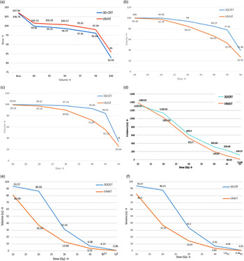
At the planning objective level of V95, VMAT provided a higher coverage of the PTV (99·44% versus 98·75%, p = 0·002) while having a narrower range (SD 0·72 versus 1·00). The isodose line covering 98% PTV volume was also recorded: 96·04 versus 97·99% for 3D-CRT versus VMAT, respectively (p < 0·0001). For VMAT, 17 out of the 39 cases (43·59%) were having 98% PTV coverage for 98% isodose line. Therefore, VMAT plans consistently delivered a higher dose to the PTV (Figure 3a). For PTV coverage, VMAT significantly improved D95, D98, D100 (p < 0·0001 for all parameters), V95 (p = 0·002) and V100 (p < 0·0001). The CI was better with VMAT (p = 0·03), whereas the 3D-CRT plans had a comparatively better HI (p = 0·003). The mean MUs delivered for 3D-CRT and VMAT were 250 + 19·01 and 776·22 + 58·42, respectively (p < 0·0001).
(a) PTV coverage, (b) dose–volume metrics for bladder: as recorded in terms of volume covering V10–V50, (c) dose–volume metrics for bladder: as recorded in terms of volume covering V10–V50, (d) dose–volume metrics for abdominal cavity: as recorded in terms of volume receiving 10–50 Gy, (e) dose–volume metrics for right femur: as recorded in terms of volume receiving 10–50 Gy, (f) dose–volume metrics for left femur: recorded in terms of volume receiving 10–50 Gy.

Bladder dose
The doses to the urinary bladder were lower in VMAT as compared with 3D-CRT for V20, V30, V40, V45 and V50 (Figure 3b). The reduction in V10 was not statistically significant. Dmax tends to be higher in VMAT but was statistically insignificant. For VMAT, dose received by 40, 45 and 50% of volume was more than 1·4 times less as compared to 3D-CRT. Dose constraint (V45 < 50%) could be met in only 1 of the 3D-CRT plans; in fact, only 4 patients had bladder doses V45 Gy< 60%. The remaining 35 patients all had higher volumes receiving 45 Gy. In comparison, for VMAT plans, the planning objective was met in 17 of the final plans, that is, 43·6% of all patients. To summarise, the bladder doses were lower with VMAT than with 3D-CRT for nearly all recorded parameters except the V10 and the Dmax. This suggests that though it struggles somewhat at the extremes, protecting the bladder through IMAT is a reasonable objective when irradiating the pelvis.
Rectum dose
The doses to the rectum were significantly reduced with VMAT compared to 3D-CRT plans for all parameters (V10, V20, V30, V40, V45 and V50) except Dmax which was significantly higher (Figure 3c). VMAT plans reduced the rectal dose (by up to 1·51 times) for all but one of the recorded parameters with statistically significant reductions. The constraint set for VMAT plans (V45 < 50%) could be met in only one of the 3D-CRT plans; two other plans had rectal doses V45 < 60%. The remaining 36 3D-CRT plans had higher volumes receiving 45 Gy. For VMAT plans, the planning objective was met in 14 of the final plans, that is, 35·90% of all patients. Also, with bladder relative reduction in doses was higher for the higher volumes than the lower volumes, that is, greater reductions in volume were seen for doses 40 Gy or higher than lower doses. To summarise, the rectal doses were lower with VMAT than with 3D-CRT for nearly all recorded parameters except the V10 and the Dmax. Both the rectum and bladder doses followed very similar patterns, which was somewhat expected given the similar anatomic relationship of the PTV to these structures.
Abdominal cavity
V10 actually saw an increase of 3·8% with VMAT plans, a difference that reached statistical significance (p = 0·038). For all subsequent dose points, VMAT plans irradiated small volumes than 3D-CRT plans. The magnitude of this reduction progressively increased from 1·1 factorial reduction for V10 to 10·05 times for volume receiving the full treatment dose, that is, 50 Gy. Thus the absolute reduction in doses to the abdominal cavity saw statistically significant reductions with VMAT for all recorded parameters except for the V10 (Figure 3 d). The VMAT plans achieved the specified constraints in 35 out of the 39 patients (89·74%); whereas, it was met in only 12 out of the 39 3D-CRT plans (30·77%). So it can be safely said that VMAT protected the abdominal cavity in over twice the number of patients.
Femur dose
The volume of femoral heads was not related to the volumes irradiated to these doses. The curves for the mean doses with both femurs (Figures 3e and 3f) separated out early to be wide apart at the femoral dose constraint of 30 Gy with VMAT producing sharp falls in volumes irradiated to 20 Gy or more. With 3D-CRT the fall came with volumes irradiated to 30 Gy, but despite this fall the dose constraint was met in only 6/39 patients (15·38%) for both femora separately, they were met for bilateral femur in four of these six patients (10·26%). The femoral doses showed that they can be protected with VMAT (by over 2 times, statistically significant dose reduction for bilateral heads of the femur).
The number of OARs for which constraints were met was higher with VMAT plans than with 3D-CRT plans, with at least four out of the five OARs protected in 46·1 versus 5·1% and all constraints were achieved in 15·4% versus none, respectively. The low dose (2 and 5 Gy) volume outside the PTV was significantly higher with VMAT plans than with 3D-CRT plans. Consequently, the integral dose was on the higher side for VMAT planning as compared to 3D-CRT plan. With VMAT, none of the constraints were met in 1 patient while the corresponding number for 3D-CRT plans were 19 (2·56 versus 48·72%) (Tables 2 and 3). With 3D-CRT, none of the plans were able to meet the dose constraints for all OARs versus 6 for VMAT. At least one constraint was met in just over half of all patients (51·3%) versus 38 (97·4%) for VMAT, with the median number of OARs receiving doses as per constraints being 3 for VMAT and 1 for 3D-CRT. Overall VMAT plans were better at sparing normal structures. Compared to 3D-CRT, they improved upon most dose–volume metrics and met the specified dose constraints often.
Dose constraints achieved in VMAT and 3D-CRT plans

Percentages of patients for whom dose constraints were achieved

Discussion
One of the major reasons why IMRT for gynaecological malignancies hasn’t been taken up as enthusiastically as in other sites is the wide range of organ motion. The uterus and cervix aren’t fixed organs within the pelvis. Lee et al.Reference Lee, Han and Huh23 working with orthogonal X-rays and fiducial markers reported inter-fraction of the cervix to a maximum of 36 mm. They used gold seeds or a uterine sleeve as fiducials. Beadle et al.Reference Beadle, Jhingran and Salehpour24 in 16 patients performed CT-based weekly assessment of cervical points of interest and found 2·3 and 1·3 cm superior and inferior; 1·7 and 1·8 cm anterior–posterior and 0·76 and 0·94 cm in the right and left lateral directions. Buchali et al.Reference Buchali, Koswig and Dinges25 in 29 cervix cancer patients took 2 CT scans, with empty and full bladder rectum, and found the median superior movement of cervix and uterus to be 4 and 7 mm, respectively. Lee et al.Reference Lee, Shrieve and Gaffney26 on comparing weekly CTs with planning CT, found uterus motion to range up to 45 mm in supero-inferior direction and 28 mm in antero–posterior direction. Chan et al.Reference Chan, Dinniwell and Haider27 using the weekly cine MRIs found uterine inter-fraction motion up to 40 mm. Taylor and Powell et al.Reference Taylor and Powell28 performed MRI scans on 33 gynaecological cancer patients for 2 consecutive days and found mean displacement of uterine body point of interests (POIs) to be 7 mm antero-posterior and supero-inferior. The tendency of IMRT plans to deliver a low dose to large volumes has been described in the literature.Reference Teoh, Clark and Wood6 While this was not a study objective, we assessed it by recording the volume outside the PTV irradiated to 2 and 5 Gy, and found both to be significantly increased with VMAT by 580 cc (2%) and 1055 cc (4%). This tendency of VMAT to deliver low dose to large volumes was also seen while noting the V10 for abdominal cavity (mean volume of which was 1717 cc) which was higher with VMAT. Additionally, the maximum dose delivered to all structures (including PTV) was higher with VMAT plans (significantly higher for PTV, rectum; trended higher without statistical significance for bladder). Thus it delivered high doses to small (point) volumes while irradiating large volumes with low doses, suggesting that VMAT struggles at the extremes.
The doses delivered to various OARs were significantly lower for nearly all recorded parameters with VMAT. The volumes receiving 10 Gy (V10) were similar for the bladder, but significantly lower for the rectum and femoral heads and significantly higher for the abdominal cavity in VMAT. V20, V30, V40 and V45 were all significantly lower with VMAT. V50 was also reduced for bladder and abdominal cavity, and similarly, for rectum and femoral heads. This was possibly due to a bias being introduced by the dose constraint introduced at that level—as this was the parameter the planning system actively attempted to reduce, and hence, it was likely to be lower. OAR sparing was most consistent in the mid-doses, that is, 20–45 Gy range, with variations at the extremes, that is, 10 and 50 Gy.
Our setup comprised of Elekta Infinity LINAC (Crawley, UK) with a maximum field size 40 x 40 cm and MLC leaf width of 1 cm at isocentre. Apart from the abdominal cavity, we found it difficult to achieve OAR constraints. The abdominal cavity was the most spared OAR with both techniques: 12 (30·77%) in 3D-CRT and 35 (89·74%) in VMAT. The bladder and rectum were most difficult to spare. This was likely due to the proximity of the OARs (bladder, rectum and abdominal cavity) to the PTV, resulting in large areas of overlapping volumes. This made it difficult to achieve all constraints while maintaining coverage of the PTV. With a smaller MLC leaf width, better sparing of OARs might have been possible, especially the bladder and rectum. The results of this study show that VMAT can significantly reduce the OAR doses while adequately covering the PTV. In fact, the PTV coverage was both higher while having a a superior CI. However, the homogeneity was lower.
The strength of this work was a homogenous study group with a small range of disease stages, so that the PTV volumes was not altered due to extent of the disease. Also, the sample size of 39 patients is a large one considering the published literature which has generally reported dosimetric studies with much fewer patients. The limitations of this study include not having delineated an ITV, so that the isotropic 1 cm margin to the CTV might be inadequate to cover internal organ motion. Also, this is a dosimetric study and correlating with clinical toxicity and disease outcomes should be done only after a clinical trial on a research-based protocol.
One of the possible limitations is the algorithm being used to calculate the dose.Reference Chen, Xiao and Li29,Reference Rana30 The two plans were generated using two different treatment planning systems—3D-CRT with XIO and VMAT with Monaco. XiO uses a superposition/convolution based algorithm while Monaco uses the Monte Carlo algorithm for dose calculation. The fundamental difference between these algorithms is in the way they incorporate the role of inhomogeneities in the path of the photon/electrons. Depending on the specific algorithm used, dose distribution differences have been demonstrated, ranging from very similar to overestimation by as much as 40%. While both these algorithms are standardised and in routine use, the inherent differences in their calculations might be responsible for some of the differences seen (or missed). The strengths of this study include a homogenous population of cervical cancer patients. While the protocol allowed for early-stage disease, all included patients belonged to the International Federation of Gynecology and Obstetrics (FIGO) Stage IIB–IIIB and those with para-aortic adenopathy were excluded. This enabled us to have uniform CTVs that did not vary significantly with the stage. We also excluded post-operative patients. The number of patients enrolled for similar previous dosimetric studies has typically ranged from about 20 to less than 10; this enabled us to detect small differences that might have otherwise been missed.
To summarise, there are limitations that preclude the widespread implementation of VMAT. Patient immobilisation is essential for daily reproducibility. Variations in daily bladder filling and bowel distention throughout the treatment are important and reconciling these variations with CTVs and patient setup remains a challenge. Studies investigating the effects of internal organ motion, both CTV and OAR, will be required to help generate consensus on the delineation of VMAT volumes, so that the use of this technique can be maximised while keeping the long-term effects of low doses in mind. The treatment of carcinoma cervix by RT entails significant toxicity. We conclude that compared to 3D-CRT, VMAT as an attempt to reduce this morbidity is a feasible approach. Large-scale clinical studies with a higher number of cases are needed to validate this approach both in terms of outcomes and toxicities to incorporate this approach into common clinical practice.
Conclusion
We conclude that in dosimetric terms, VMAT is superior to 3D-CRT for EBRT of locally advanced cervical cancers.
Acknowledgements
None.
Funding
None.
Disclosure
The authors declare no conflict of interest.