INTRODUCTION
Vancomycin-resistant Enterococcus faecium (VRE) infections are an endemic problem in many intensive care, hematology/oncology, and stem cell transplant units. VRE bloodstream infections (BSI) have been associated with increased morbidity, expense,Reference Butler, Olsen and Merz1 and mortality.Reference Vydra, Shanley and George2
Patients with acute leukemia are at high risk for VRE BSIReference Worth, Thursky, Seymour and Slavin3, Reference Bossaer, Hall and Garrett-Mayer4 owing to prolonged neutropenia, immunosuppressive medications, disruption of mucosal barriers, and indwelling central venous catheters. The purposes of this study are to describe the frequency of, risk factors for, and outcomes associated with VRE colonization and BSI in patients admitted with newly diagnosed acute leukemia.
METHODS
Patients
Two hundred fourteen consecutive patients with newly diagnosed acute leukemia who were admitted to the LDS Hospital Acute Leukemia Program between October 25, 2006 and December 31, 2012 were identified. Patients were housed in individual high-efficiency particulate air–filtered laminar air flow rooms on a hospital unit dedicated to management of patients with hematologic malignancies and stem cell transplant recipients. All patients had central venous catheters inserted. Patients were classified as having acute lymphoblastic leukemia (ALL) or acute myelogenous leukemia (AML). Patients with biphenotypic leukemia were included in one of the 2 groups depending on treatment regimen.
Stool cultures for VRE were routinely obtained on admission and weekly thereafter as part of our infection control measures. Patients colonized and/or infected with VRE were placed into contact isolation (gloves and gowns). Afebrile and noninfected neutropenic patients were given levofloxacin, penicillin, and antifungal prophylaxis. Blood cultures were performed with the occurrence of fever or other suspicion of infection and the antibiotic regimen was typically changed to include a carbapenem. Patient data were obtained from our Acute Leukemia Database, the Intermountain Healthcare Enterprise Data Warehouse, and the hospital case mix database. Total patient costs (direct and indirect) are determined quarterly by relating total hospital costs to charge codes. Contact isolation costs are not billed to individual patients. The study was approved by the Intermountain Healthcare Institutional Review Board.
Definitions
Hospital-acquired gastrointestinal colonization was defined as a positive stool culture result following one or more negative culture results. Patients with a positive initial stool culture result were considered to have community-acquired colonization if the stool was obtained within 48 hours after admission and indeterminate if more than 48 hours. A VRE BSI was defined as a VRE isolate from one or more blood cultures in the presence of signs of infection. Daily VRE colonization pressure was calculated as the number of patients on the unit who were colonized or infected with VRE each day. Severe neutropenia was defined as less than 100 neutrophils/μL.
VRE Screening and Clinical Cultures
Stools submitted for VRE screening cultures were plated onto bile esculin agar plates and BBL Campylobacter agar plates (BD). Vancomycin resistance was confirmed using MicroScan (Siemens). Blood cultures were processed using the Phoenix (BD) automated system to identify E. faecium and determine antimicrobial susceptibility. VRE isolates from surveillance and clinical cultures were cryopreserved at −80°C in skim milk broth. Molecular typing was performed using repetitive element PCR utilizing the semi-automated DiversiLab System (bioMérieux).
Statistical Analysis
Prism 6 (GraphPad) was used to construct actuarial rates curves and to calculate log-rank hazard ratios and significance determinations. For multivariable risk determinations and permutation tests on medians, the survival package from “R” statistical software was used. Variables were determined for each day of the patient’s hospitalization and hazard ratios and significance determinations were calculated using Cox proportional hazards models with time varying covariates. Variables were selected by literature review and clinician experience. Variables with a P value less than 0.1 on univariate analysis were selected for multivariable models. Statistical significance was defined as a P value less than 0.05.
RESULTS
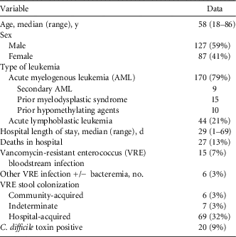
Patient characteristics are summarized in Table 1. Patient age, sex, and AML predominance are typical for this unselected population. One hundred seventy-five patients had an adequate performance status to begin induction chemotherapy. Patients with acute promyelocytic leukemia were treated with all-trans retinoic acid/chemotherapy; other patients with AML received a standard-dose anthracycline/cytosine arabinoside regimen (“3+7”). Treatment of ALL was age determined with younger patients receiving a pediatric type regimen.
Characteristics of 214 Consecutive Patients with Newly Diagnosed Acute Leukemia

Note. Data are no. (%) of patients unless otherwise indicated.
Of the 214 patients, 82 (38%) were identified as VRE colonized during the study period; 69 of these cases (32%) were hospital acquired (Table 1). Fifteen patients (7%) developed a VRE BSI. Three patients with VRE BSI had an associated VRE urinary tract infection, one of whom also had positive results for sterile fluid cultures (gall bladder, pleural fluid). One patient had a VRE urinary tract infection and 2 had VRE soft-tissue infections without VRE BSI. Hospital-acquired gastrointestinal colonization rates were proportional to length of stay (Fig. 1a). The median time to VRE colonization was 19 days (range, 6–41 days).
A: Actuarial rates of vancomycin-resistant enterococcus (VRE) colonization in 201 patients with acute leukemia and initially negative VRE stool culture results; B: Actuarial rates of VRE bloodstream infection (BSI) comparing gastrointestinal colonized patients with acute myelogenous leukemia (AML) to a combination of noncolonized AML and all acute lymphoblastic leukemia (ALL) patients; C: Overall survival comparison between patients with VRE bacteremia and patients with similar acute leukemia risk factors but without VRE bacteremia (controls); D: actuarial rates of VRE gastrointestinal colonization by type of acute leukemia (ALL vs. AML).
Molecular Characterization of VRE
VRE isolates from 14 of the 15 patients with BSI were available for molecular typing. Twelve isolates (86%) yielded unique fingerprint patterns distributed across 7 related groups (Fig. 2). Seven (58%) of the 12 isolates clustered in clonal group 3, which was detected over several years. Two patients had infection related to clonal group 1, and the remainder was unrelated isolates. Of 9 available stool-blood pairs, 7 (78%) had identical patterns and 8 (89%) were in related groups.
Dendrogram of molecular typing of vancomycin-resistant Enterococcus faecium isolates. Sample ID numbers indicate unique individuals and the letters indicate blood (A) or stool (B) isolates. Patterns (P) indicate unique strains and groups (G) designate genetically related strains. Isolates with the same pattern have more than 97% similarity with no band difference by visual inspection of electropherogram. Those with the same group designation have more than 95% similarity with up to 2 band differences on visual inspection. Bloodstream isolates collected from 14 patients were available for typing. Of these, 9 patients (Sample ID 2, 4, 7, 9, 10, 12, 29, 41, 153) had both blood and stool isolates available for typing. Five patients (Sample ID 19, 37, 47, 48, 51) had only bloodstream isolate available. One subject (Sample ID 31) had only a stool isolate available for typing.
Frequency and Risk Factors for VRE BSI
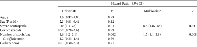
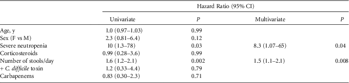
Eighty percent of patients with VRE BSI also had positive stool culture results. VRE BSI preceded detection of gastrointestinal colonization in 2 patients. In the remainder, VRE BSI occurred a median of 8.5 days (range, 3–28 days) following the first VRE-positive stool culture result. Actuarial rates of VRE BSI by type of leukemia and VRE colonization status are shown in Fig. 1b. Compared with the other groups, colonized patients with AML were at higher risk (hazard ratio, 4.9 [95% CI, 1.8–15]). Factors associated with VRE BSI include an increased number of stools/day and severe neutropenia (Table 2).
Potential Risk Factors for Vancomycin-Resistant Enterococcus Bloodstream Infection
Costs and Outcomes Associated With VRE BSI
We also assessed the impact of VRE BSI on patient mortality and hospital costs. For the former, we matched each patient with VRE BSI in a 1:3 ratio with other patients of similar leukemia type, age, admitting Karnofsky performance status, and initial treatment regimen. Overall survival curves suggested a trend towards early mortality for the VRE BSI group (hazard ratio, 1.9 [95% CI, 0.87–5.1]) (Fig. 1c) with an estimated 60-day mortality of 33% vs. 18%. Total median hospital costs were significantly higher for patients with VRE BSI ($172,000 vs. $86,000, P=0.0003), largely driven by an increased median length of stay (42 vs. 29 days, P=0.0005).
Risk Factors for VRE Gastrointestinal Colonization
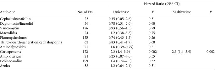
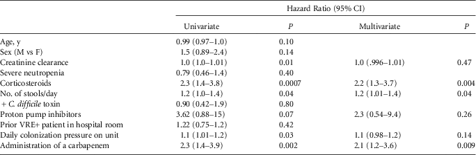
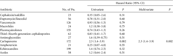
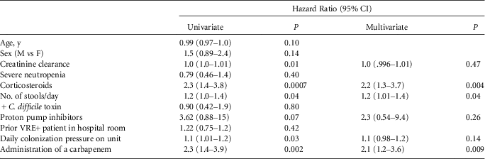
The impact of antibiotic/antifungal agents on the risk of gastrointestinal colonization is shown in Table 3. Antibiotics with primarily a gram-positive spectrum demonstrated the lowest hazard ratios, and those targeting aerobic gram-negative organisms showed minimal risk. Only exposure to carbapenems significantly increased the risk of VRE colonization. Patients with ALL had a higher rate of gastrointestinal colonization (hazard ratio, 1.9 [1.2–2.4]) (Fig. 1d) and a lower median length of stay (26 vs. 31 days, P=0.008). Other predictors of colonization include increased stools/day and exposure to high-dose corticosteroids (Table 4).
Risk of Antibiotics/Antifungals for Vancomycin-Resistant Enterococcus Stool Colonization
Potential Risk Factors for Vancomycin-Resistant Enterococcus (VRE) Stool Colonization
DISCUSSION
Our study focused on patients with acute leukemia in order to minimize the potential confounding that could result from combinations with other patient populations. Our data show that the risk of VRE acquisition is directly proportional to hospital length of stay with 50% of patients residing on our unit for 6 weeks becoming colonized (Figure 1a). Similar rates have been reported by others.Reference Colderwood, Mauer and Tolentino5, Reference Almyroudis, Lesse and Hahn6
Studies in intensive care unit patients and those with hematologic malignancies have demonstrated that administration of third-generation cephalosporins, vancomycin, and carbapenems may increase the risk of hospital-acquired VRE colonizationReference Worth, Thursky, Seymour and Slavin3, Reference Bonten, Slaughter and Ambergen7–Reference Yoon, Lee, Lee and Kin10 and BSI.Reference McKinnell, Kunz and Chamot11–Reference Ghanem, Hachem, Jiang, Chemaly and Raad13 In our cohort, only carbapenem use increased colonization. Such therapy would be most disruptive to the microbiome, leading to VRE intestinal domination.Reference Donskey, Chowdhry and Hecker14, Reference Taur, Xavier and Lipuma15 Hence, decreasing routine empirical use of carbapenems may be a strategy to limit VRE acquisition. Conversely, our prophylactic regimen (levofloxacin+penicillin) did not increase VRE colonization or infection.
We found that colonized patients with AML have a higher risk of VRE BSI. This is likely largely explained by differences in the duration of severe neutropenia. This was present during 52% and 22% of the inpatient days of patients with AML and ALL, respectively (P<0.0001). Prolonged neutropenia has also been identified as a risk factor for VRE BSI by others.Reference Worth, Thursky, Seymour and Slavin3, Reference Bossaer, Hall and Garrett-Mayer4 Our observations suggest that a VRE-colonized patient with AML should be considered for empirical VRE coverage with onset of febrile neutropenia. Conversely, colonized patients with ALL have no higher risk of BSI than those reported for colonized general medical and surgical patients.Reference Olivier, Blake, Steed and Salgado16
We found that VRE BSI has a significant negative impact on outcomes. VRE BSI was associated with a trend to early mortality and a prolonged length of stay that greatly increased costs. Similar outcomes have been reported in patients with trauma, patients with neutropenia, and stem cell transplant recipients.Reference Vydra, Shanley and George2, Reference Lodise, McKinnon, Tam and Rybak17–Reference Zirakzadeh, Gastineau, Mandrekar, Burke, Johnston and Patel19
Acquisition of gastrointestinal colonization is a critical event prior to the onset of clinical infection with VRE, as nearly all of the concomitant blood and stool isolates were genetically related. Since other risk factors for BSI in patients with AML (neutropenia, mucositis) are difficult to modify, preventing colonization is an important strategy. We found that, in addition to administration of a carbapenem and diarrhea, use of high-dose corticosteroids was associated with VRE colonization. This may explain the increased colonization rates in patients with ALL. Ninety percent of patients with ALL received high-dose corticosteroids as part of their treatment regimen. Conversely, only 16% of AML patients received these (median total dose equivalent of 920 mg prednisone) as treatment for all-trans retinoic acid syndrome and various inflammatory conditions. The mechanism(s) by which steroids might increase the risk of colonization is unclear.
In intensive care unit patients both a VRE-colonized prior room occupantReference Dress, Snydman and Schmid20 and a higher colonization pressureReference Bonten, Slaughter and Ambergen7 have been associated with an increased risk of colonization. These studies suggest that VRE can be acquired vertically or horizontally from other patients. However, in our cohort, neither variable contributed to colonization risk. These results suggest that on units such as ours where VRE is endemic, exposure to VRE is common and patient and treatment factors become more significant determinants of colonization.
Examination of our BSI strains reveals that there is strain diversity. Nevertheless, over half appeared to cluster within a related clonal group with small genetic differences. Whether this represents colonization and infection with a dominant VRE lineage or polyclonal transmission with BSI from strains with increased virulence is unclear because we limited molecular typing to BSI and stool isolates associated with BSI. Additional molecular studies with more detailed characterization of stool isolates not associated with BSI are needed to better understand the implications for infection control.
Other limitations of our study include a retrospective design, institutional practice patterns, and our molecular typing methodology. Because our clinicians prefer to use empirical carbapenems with onset of fever, we were unable to evaluate other commonly utilized agents such as beta-lactam/beta-lactamase inhibitors. Also, the molecular typing method we used is not as discriminatory as some other methods (pulsed-field gel electrophoresis, whole genome sequencing).
We have shown that VRE infections, particularly BSI, occur primarily in colonized patients with AML and are a multifactorial process, associated with increased costs to healthcare facilities. We found that VRE BSIs in patients with acute leukemia are increased in patients with prolonged neutropenia and diarrhea. Our clinical and molecular data suggest that colonization is an important factor in BSI as well, and that the use of carbapenems and high-dose corticosteroids is associated with an increased incidence of VRE gastrointestinal colonization.
Acknowledgments
Financial support. This study was supported in part by a grant from the Intermountain Healthcare Medical and Research Foundation, Salt Lake City, UT.
Potential conflicts of interest. B.K.L. reports that he has received research support from Cubist in support of a project unrelated to this study. K.K.D. reports that she has received research support from Pfizer in support of a project unrelated to this study. All other authors report no conflicts of interest relevant to this article. We thank the physicians, nurses, and other hospital staff who cared for the patients, Jo Lynn Lee and Braden Rowley for help with data abstraction, and Esar Checkrie and Jana Coombs who assisted with molecular typing.