Introduction
It is rare to see huge, lobulated juvenile nasopharyngeal angiofibromas causing extensive erosion of the skull base with intracranial spread. Complete removal of these tumours is a surgical challenge. Very few papers have reported the relevance of dura mater imaging changes to the choice of appropriate surgical management.
Our department has treated 19 cases of juvenile nasopharyngeal angiofibroma with wide extrapharyngeal extension. In four of these cases, the sphenoid sinus, sella turcica and clivus were extensively eroded, and the tumour had spread deep into the cranial fossa. Based on the intra-operative and imaging findings in these four cases, we emphasise the importance of pre-operative imaging for evaluating the: size and location of the tumour; involvement of the optic nerve, internal carotid artery and cavernous sinus; and presence of an intact dura mater. Assessment of these factors is very important when selecting suitable surgical management. Generally, if the dura mater is intact then the optic nerve, internal carotid artery and cavernous sinus are simply compressed or partially encircled by the tumour. Therefore, an extracranial approach is adequate for complete removal of the tumour. However, intracranial exploration should be available to deal with emerging complications.
Methods and materials
With the approval of the Institutional Review Board, the clinical and imaging material for four huge nasopharyngeal angiofibromas was analysed and is reported here.
Results
Case one
A 13-year-old boy presented suffering from right-sided, relapsing epistaxis and nasal obstruction for two years and from progressive swelling of the cheek and loss of vision for about six months.
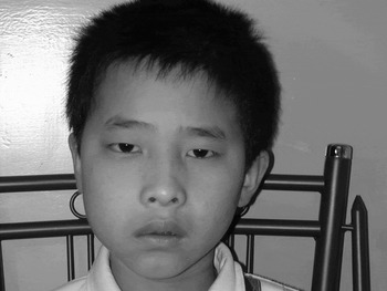
The patient was hospitalised in April 2004. Physical examination found a right-sided proptosis and a swollen cheek (Figure 1), with a mass in the nasopharynx and right nasal cavity.
Case 1: right cheek swelling and proptosis. (Photograph published with permission.)

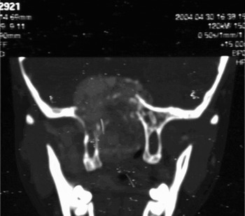
Computed tomography (CT) and magnetic resonance imaging (MRI) demonstrated a lobulated mass in the nasopharynx, right nasal cavity, infratemporal fossa and sphenoid sinus. The bone of the sella turcica and orbit was extensively eroded (Figure 2). The cavernous sinus and internal carotid artery were compressed and displaced upward. A 3D-MRA showed that the right maxillary artery was enlarged, with varicosis of the superior ophthalmic vein.
Case 1: coronal computed tomography showing the juvenile nasopharyngeal angiofibroma extending into the cranial fossa and compressing the brain; the dura mater cannot be seen.

On 20 May 2004, a transtemporal–intradural exploration was performed. At surgery, the dura mater was found to be firm, congested and haemorrhagic in the right superior orbital fissure and cavernous sinus. No perforations of the dura mater or intradural tumour lobules were found. Subsequently, a transantral approach via a midfacial degloving incision was used to remove the tumour.
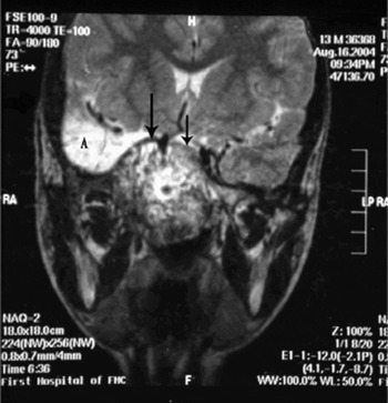
Three months later, MRI demonstrated that the tumour had recurred and spread into the cranial fossa. In addition, the right temporal lobe showed encephalomalacia due to the previous transtemporal–intradural exploration. Since the dura mater was intact (Figure 3), a transantral approach via a midfacial degloving incision was used to remove the tumour again.
Case 1: coronal T2-weighted magnetic resonance image showing recurrent tumour extending into the cranial fossa, and also the intact dura mater with low signal intensity (arrows). Note encephalomalacia (A) in the right temporal lobe due to previous transtemporal–intradural exploration.

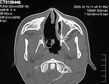
Fourteen months later, CT and clinical examination showed no tumour recurrence in the skull base (Figure 4).
Case 1: axial computed tomography, 14 months after the second operation, showing no tumour recurrence.

Case two
A 16-year-boy presented suffering from right-sided, relapsing epistaxis and progressive nasal obstruction for two years, with right-sided, severe headache and visual loss for two weeks.
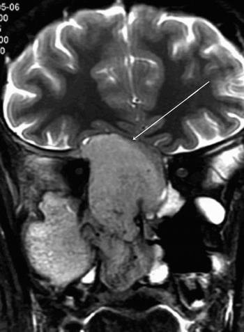
The patient was hospitalised in April 2005. A CT and MRI showed a huge, lobulated tumour in the nasopharyngeal region, with extensive extrapharyngeal spread and skull base erosion (Figure 5).
Case 2: coronal T2-weighted magnetic resonance image showing the tumour compressing and displacing the intact dura mater (arrow) upward.

The bone of the sphenoid sinus, sella turcica and clivus had been extensively destroyed. The optic nerve, optic chiasm, pituitary gland, cavernous sinus and internal carotid artery were compressed away from their original sites. Three days after embolisation of the tumour-feeding arteries, a transantral approach via a midfacial degloving incision was used to remove the tumour completely. At surgery, the tumour was found to have extended into the pterygopalatine and infratemporal fossae, eroded about 2×3 cm of the inner wall of the right orbit, destroyed much of the sella turcica and clivus, and exposed and adhered to about 3×3 cm of the dura mater. The tumour measured 6×7×8 cm (Figure 6).
Case 2: juvenile nasopharyngeal angiofibroma measuring 6×7×8 cm was removed completely.

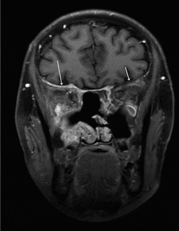
One year post-operatively, an MRI showed a thickened dura mater (red arrows) and no tumour recurrence (Figure 7).
Case 2: coronal T1-weighted+fat saturation magnetic resonance image taken one year post-operatively. The dura mater is thickened (arrows) and no tumour recurrence is observed.

Case three
A 21-year-old man was hospitalised in our department in January 1997. He had suffered from juvenile nasopharyngeal angiofibroma for the previous five years and had been managed with three previous operations, including one lateral facial approach and two transantral approaches via a Weber incision.
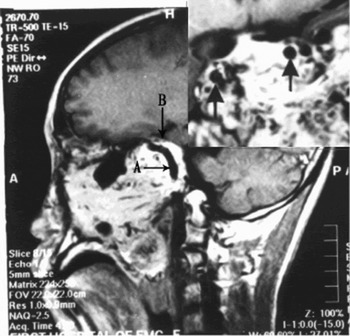
Physical examination revealed the tumour in the left nasal cavity, nasopharynx and cheek region. The left eyeball was displaced outward and upward by the tumour and the optic nerve was severely damaged, resulting in blindness. A CT and MRI showed that: the tumour involved the left maxillary sinus and orbital apex; the sphenoid sinus, sella turcica and clivus had been extensively destroyed; and the bilateral temporal lobes, cavernous sinus and internal carotid artery were compressed and displaced (Figure 8).
Case 3: sagittal T1-weighted magnetic resonance image. Arrow A indicates the internal carotid artery; arrow B indicates the intact dura mater with low signal intensity. The insert shows the internal carotid artery bilaterally (arrows) and many vascular flow voids in the tumour.

A transtemporal–extradural exploration was performed. No perforation of the dura mater was found, although the dura mater in the superior orbital fissure was congested, haemorrhagic and solid. A transantral approach via a Weber incision was then used to remove the tumour. Owing to serious intra-operative bleeding, the tumour was removed incompletely. The patient was referred for radiation therapy.
Case four
A 16-year-old boy had suffered from extensive juvenile nasopharyngeal angiofibroma for five years. Three lateral rhinotomy procedures had been performed, either elsewhere or in our department.
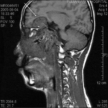
In August 2005, the tumour recurred. Physical examination revealed right-sided eye proptosis and blindness, a swollen cheek, and a mass in the nasopharynx and right nasal cavity. A CT and MRI showed that the tumour had eroded the right inner orbital wall, optic canal, sella turcica and clivus. In addition, the tumour extended deep into the anterior and middle cranial fossae. However, the dura mater was intact and the brain showed no abnormal imaging signals (Figure 9).
Case 4: sagittal magnetic resonance image, taken in 2005, showing the tumour extending deep into the anterior and middle cranial fossae, as well as an intact dura mater (arrow).

The patient declined further surgery and was referred for radiological therapy.
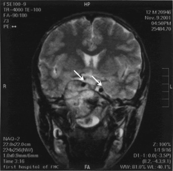
Reviewing our operative records of 2001 for this case, a transtemporal–extradural approach had been performed initially, and then a lateral rhinotomy had been used to remove the extracranial tumour. Reviewing the coronal MRI from 2001, the dura mater had been intact (Figure 10). In retrospect, we had failed to recognise the importance of the intact dura mater at that time, and we thus performed an unnecessary transcranial exploration.
Case 4: coronal magnetic resonance image, taken in 2001, showing the tumour extending into the cranial fossa and compressing the right temporal lobe and bilateral internal carotid arteries (arrows); the dura mater is intact with low signal intensity.

Discussion
Our previous research identified nasopharyngeal angiofibroma as a vascular hamartoma.Reference Liang, Yi and Liang1 As the tumour grows, it does not infiltrate the surrounding anatomical structures but induces erosion of the skull and then compresses and displaces the soft tissues, such as the dura mater, eyeball, optic nerve, optic chiasm, pituitary gland, cavernous sinus and internal carotid artery. Since the dura mater is a strong, thick layer of dense connective tissue with elastic fibresReference Eroschenko2, Reference Pick and Howden3 it has the ability to self-repair, and it can tolerate the pressure of a growing juvenile nasopharyngeal angiofibroma to a degree.
If the dura mater is not perforated, a transantral approach via a midfacial degloving incision may be able to remove a huge, lobulated nasopharyngeal angiofibroma with intracranial extension. If the dura mater has been perforated by the intracranial tumour lobule, a transtemporal–intradural approach or other suitable approach is indicated. Moreover, distal control of the cavernous sinus and internal carotid artery may be necessary.
Lloyd et al.Reference Lloyd, Howard, Phelps and Cheesman4, Reference Lloyd, Howard, Lund and Savy5 stated that good bone imaging on CT is essential to show invasion of the cancellous bone of the sphenoid. This is the main predictor of recurrence; the deeper the extension, the greater the risk that remnant tumour will be left following surgery. Our experience in the cases of 1, 3 and 4 supports these statements.
Although remnant tumour in the sphenoid sinus is the chief site of recurrence, we believe that serious intra-operative bleeding and using the finger for blind dissection may be the main reasons for residual tumour in the sphenoid sinus or elsewhere.Reference Yi, Li, Lin and Ye6
The surgical approach to juvenile nasopharyngeal angiofibroma should be determined individually based on the tumour's size and location. Specific cases may require the following approaches: endoscopic transnasal, transpalatal and combined transnasal–transpalatal; transantral via midfacial degloving;Reference Yi, Li, Lin and Ye6 and the Denker operation.Reference Hardillo, Vander Velden and Knegt7 We agree with Cansiz et al.Reference Cansiz, Guvenc and Sekercioglu8 that modified midfacial degloving is a reasonable choice for almost all advanced lesions. This approach is very useful in terms of surgical exposure, duration of surgery, cosmetic outcome and morbidity. It can be combined with other approaches or a craniotomy if necessary.
In our experience, the advantages of the transantral approach via midfacial degloving are determined by the specific anatomical location of the tumour. This approach allows ideal access to the maxillary and sphenoid sinuses, infratemporal fossa, orbit and cheek, via selective removal of the medial, external (facial), superior (orbital) and posterior (zygomatic or infratemporal) surfaces of the antrum. The tumour can then be delicately removed under direct vision. This may prevent the need for using a finger for rough and blind dissection, and the risk of severe intra-operative bleeding. Over the past decade, our department has stopped using lateral rhinotomy and the Weber incision, except in patients who have already undergone these procedures previously.
Donald et al.Reference Donald, Enepikedes and Boggan9 reported five patients with giant angiofibromas with intracranial penetration. Three of these cases involved the cavernous sinus, with angiographic evidence of a major blood supply to the tumour. In four of five patients, these authors removed the entire tumour via a skull base procedure, such as the infratemporal fossa/middle fossa approach or an anterior craniofacial approach.
We assert that intracranial exploratory management is indicated only in cases with penetration of the dura mater. Reviewing our cases one, three and four, the transtemporal–extradural or transtemporal–intradural procedure might have been avoided if pre-operative radiological evaluation had confirmed that the dura mater was not penetrated.
• Huge, lobulated juvenile nasopharyngeal angiofibromas causing extensive erosion of the skull base with intracranial spread are rare
• This study investigated the relevance of dura mater imaging changes to the choice of appropriate surgical management
• If the dura mater is not perforated, a transantral approach via a midfacial degloving incision may be able to remove a huge, lobulated nasopharyngeal angiofibroma with intracranial extension
Pre-operative super-selective embolisation of the juvenile nasopharyngeal angiofibroma feeding arteries will reduce the risk of brisk bleeding during surgical tumour removal. In case two, no blood transfusion was needed during the operation because pre-operative embolisation of the feeding arteries had been successful.
In short, radiological techniques are available for evaluating the fine involvement of the skull base, dura mater, cavernous sinus and internal carotid artery in cases of juvenile nasopharyngeal angiofibroma with intracranial extension. Hence, surgeons must consider the results of imaging when selecting the best surgical procedure for complete tumour removal.