Introduction
Human zygomycosis is a spectrum of diseases caused by fungi of the zygomycetes class.Reference Vianna, de Lacerda and de Moraes1 This class is subdivided into two orders: mucorales and entomophthorales.Reference Vianna, de Lacerda and de Moraes1 Diseases caused by mucorales are called mucormycoses, and those caused by entomophthorales are called entomophthoromycoses. Infection by entomophthorales causes a deep mycosis, and is encountered in tropical and subtropical regions such as south India.Reference Krishnan, Sentamilselvi, Kamalam, Das and Janaki2
Zygomycosis is the term accepted to refer to all disease processes caused by members of the class zygomycetes.Reference Dawood, Al-Ahmed and Hussainy3 The fungi that cause zygomycosis are ubiquitous, being commonly found in the environment.Reference Dawood, Al-Ahmed and Hussainy3 Infections by zygomycetes are usually observed in immunocompromised individuals.Reference Dawood, Al-Ahmed and Hussainy3 However, they have also been reported in immunocompetent patients.
We encountered a case of entomophthoromycosis in a 13-year-old girl, causing mastoiditis, which prompted us to report this case.
Case report
A 13-year-old girl presented in our out-patients department in December 2008 with a left post-aural abscess. The patient had had a mildly painful post-aural swelling for 15 days, and also reported mucoid left ear discharge over the previous month, which was not foul-smelling or blood-stained. There was no history of fever, headache, vomiting, decreased hearing, dizziness or tinnitus.
On examination, a 2 × 3 cm, cystic, fluctuant, tender swelling was present in the left post-aural region. Another similar but smaller swelling was also noted on the superior wall of the left external auditory canal, obscuring the tympanic membrane.
Blood investigation results were as follows: haemoglobin 11.5 g per cent; white cell count 9300/mm3; and blood differential, neutrophils 57 per cent, lymphocytes 38 per cent, eosinophils 4 per cent, monophils 1 per cent and basophils 0 per cent. Biochemical investigations (including random blood glucose concentration, liver function tests and renal function tests) were within normal limits.
Tests for human immunodeficiency viruses one and two and hepatitis B surface antigen (HBsAg) antibody tests were non-reactive.
Radiography of the mastoid revealed a sclerotic mastoid on the left and a pneumatic mastoid on the right. Chest radiography was normal.
Pure tone audiography revealed a mild conductive hearing loss in the left ear.
Incision and drainage of the post-aural and external auditory canal abscesses was performed. The pus and necrotic material removed were sent for histopathological examination.
Histopathological analysis was suggestive of a granulomatous lesion with microabscesses, most probably of zygomycotic fungal aetiology.
In view of the histopathology report, high resolution computed tomography (CT) of the temporal bone was performed to investigate the extent of the disease (Figure 1). Scans suggested the presence of a soft tissue density in the middle-ear cavity, epitympanum, aditus ad antrum and mastoid antrum. There was destruction of the mastoid septae and coalescence of the mastoid air cells, with a soft tissue density. Erosion of the lateral as well as medial cortex of the mastoid was noted.
Pre-operative, axial, high resolution computed tomography scan of the temporal bone with the arrows showing eroded mastoid cortex at two sites and soft tissue in the mastoid air cells

In view of the high resolution CT and histopathological findings, it was decided to undertake total surgical debridement of the disease.
Surgery was performed via a post-aural approach. The necrotic tissue, pus, granulation tissue and eroded mastoid bone were removed and sent for fungal culture and histopathological analysis. Granulations and osteitic bone were traced and removed in toto (i.e. cortical mastoidectomy). The intra-operative extent of disease corresponded to that seen on high resolution CT. The middle ear and the ossicles were normal.
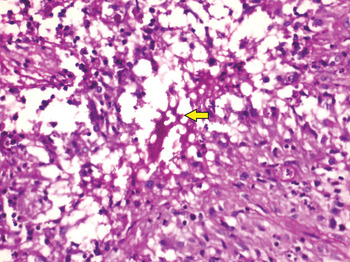
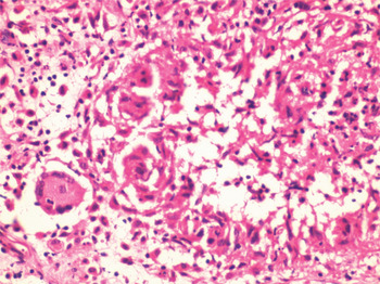
Histopathological examination showed multiple non-caseating granulomas with non-septate fungal hyphae covered with eosinophilic material (i.e. a ‘splendore hoeppli’ body) in the centre, surrounded by abundant neutrophils and eosinophils and rimmed peripherally by histiocytes. A few foreign body type giant cells and chronic inflammatory infiltrate were observed in the vicinity. The use of special stains, such as periodic acid Schiff and Gomori silver methanamine, revealed non-septate, broad, thin-walled hyphae, confirming the presence of fungal bodies (Figures 2 and 3).
Photomicrograph showing granulomatous inflammation with eosinophilic inflammatory cells and splendore hoeppli phenomenon (arrow). (Periodic acid Schiff; ×400)

Photomicrograph showing non-caseating granuloma with giant cell reaction. (H&E; ×400)

Based on these findings, a diagnosis of entomophthoromycosis (Basidiobolus ranarum type) was made.
The patient was commenced on injected amphotericin B, after renal function, liver function, serum electrolyte and blood profile tests were found to be normal. The patient was initially given a test dose comprising 0.1 ml amphotericin B in 100 ml 5 per cent dextrose over 20 minutes. No drug reaction was seen (i.e. fever, rigors or rash), and the patient was thus given 0.5 mg/kg amphotericin B.
On the third day of this treatment, a repeated blood profile test showed reductions in haemoglobin concentration (from 11.5 to 9.8 g per cent) and white blood cell count (from 9300 to 5100/mm3). The platelet count, renal function and liver function test results were normal.
In view of these findings, amphotericin B was stopped and investigations were repeated three days later. These tests showed an increased white blood cell count (to 7300/mm3) but an unchanged haemoglobin concentration.
Amphotericin B was restarted at the same dose. After three days, blood investigation results were found to be within the normal range, so the dosage was increased to 0.75 mg/kg/day and then to 1 mg/kg/day. For 21 days, the patient was continued at the same dosage, with blood investigations being performed every third day.
The treatment was stopped after four weeks as all signs of infection had subsided. At this stage, the left tympanic membrane appeared normal and the patient's post-aural scar had healed completely. Pure tone audiometry of both ears showed no hearing loss. High resolution CT scans of the temporal bone were performed at the end of therapy was normal.
The patient was discharged and followed up regularly. High resolution CT scans of the temporal bone performed three months and one year post-therapy were normal (Figure 4).
Post-operative, axial, high resolution computed tomography scan of the Temporal bone with arrow showing complete resolution of the disease in the operated cortical mastoid cavity seen after 1 year of stopping treatment

Discussion
Pathogenic zygomycosis is caused by two orders of fungi: mucorales and entomophthorales.
Infections with organisms of the order mucorales are characterised by a rapidly progressive course, and are usually fatal.Reference Vianna, de Lacerda and de Moraes1
In contrast, infections by entomophthorales organisms usually follow a benign course and are more common in immunocompromised individuals, although infections in immunocompetent individuals have also been reported.Reference Vianna, de Lacerda and de Moraes1
Entomophthorales infection is commonly reported in tropical and subtropical countries, and is the second most common deep mycosis encountered in south India.Reference Krishnan, Sentamilselvi, Kamalam, Das and Janaki2 Entomophthorales organisms have been reported to cause subcutaneous, pulmonary and rhinofacial infections. However, a careful search of the literature revealed no previous report of entomophthorales causing mastoid infection with erosion. We therefore believe that the presented patient represents the first report of such a case.
In the presented patient, histological examination with haematoxylin and eosin staining showed multiple non-caseating granulomas. The splendour hoeppli phenomenon was observed in multiple areas, with a thick eosinophilic infiltrate, indicating basidiobolus infection.
• Entomophthoromycoses are fungal infections caused by organisms of the order entomophthorales, which cause deep mycosis
• This paper reports the case of a young girl who presented with mastoid abscess
• Entomophthoromycosis was diagnosed on histopathological examination
• The patient received surgical and medical treatment, resulting in complete resolution of disease
There were no predisposing factors in this patient. A previous Indian study of entomophthoromycosis suggested that trauma may lead to implantation of the infective organism at the site of infection, but this may not be true in all cases.Reference Krishnan, Sentamilselvi, Kamalam, Das and Janaki2
Infection by entomophthoromycosis can be mistaken for abscesses or benign or malignant tumours, and patients may be subjected to surgery with no successful result.Reference Vianna, de Lacerda and de Moraes1 Our patient presented with post-aural abscess and was initially diagnosed with suppurative otitis media with mastoid abscess. The possibility of tuberculous abscess was also considered, due to its high prevalence in India and the patient's general clinical picture.
A Nigerian study of entomophthoromycosis described potassium iodide as the treatment drug of choice.Reference Martinson4 The study group included patients with rhinofacial entomophthoromycosis, treated in this way.Reference Vianna, de Lacerda and de Moraes1 Patients who do not respond to potassium iodide have been successfully treated with other drugs, including injected amphotericin B, ketoconazole, miconazole and sulphamethoxazole, in various combinations.Reference Vianna, de Lacerda and de Moraes1, Reference Dawood, Al-Ahmed and Hussainy3, Reference Martinson4
A previously reported case of a retroperitoneal mass diagnosed histopathologically as Basidiobolus ranarum infection showed good results after surgical excision together with antifungal drug treatment.Reference Vianna, de Lacerda and de Moraes1
In the present case, the patient underwent surgical debridement and was commenced on amphotericin B treatment post-operatively, as the initial histopathological report was suggestive of zycomycosis (which includes both mucormycoses and entomophthoromycoses). The clinical response of the patient to the drug was closely monitored. As the patient responded well to amphotericin B, it was continued as the drug of choice. On completion of treatment, the patient appeared to be completely cured of disease, as observed clinically and on high resolution CT temporal bone images.
Therefore, in the current patient early presentation and early detection of infection contributed to a good prognosis.
Conclusion
It is difficult to diagnose entomophthoromycoses clinically in the initial stages of infection. Therefore, the possibility of such a rare infection should be kept in mind, and relevant investigations (e.g. pus culture and histopathological examination) should be conducted routinely so as not to miss the diagnosis.