




No CrossRef data available.
Article contents
Genetic variability of glutamate-gated chloride channel genes in ivermectin-susceptible and -resistant strains of Cooperia oncophora
Published online by Cambridge University Press: 18 November 2004
Abstract
The glutamate-gated chloride channels (GluCls) are members of the ligand-gated ion channel superfamily that are thought to be involved in the mode of action of ivermectin and mechanism of resistance. Using reverse-transcriptase PCR techniques, 2 full-length GluCl cDNAs, encoding GluClα3 and GluClβ subunits, were cloned from Cooperia oncophora, a nematode parasite of cattle. The two sequences show a high degree of identity to similar subunits from other nematodes. The C. oncophora GluClα3 subunit is most closely related to the Haemonchus contortus GluClα3B subunit, while C. oncophora GluClβ subunit shares high sequence identity with the H. contortus GluClβ subunit. Using single-strand conformation polymorphism, the genetic variability of these two genes was analysed in an ivermectin-susceptible isolate and an ivermectin-resistant field isolate of C. oncophora. Statistical analysis suggested an association between the C. oncophora GluClα3 gene and ivermectin resistance. No such association was seen with the GluClβ gene.
Keywords
Information
- Type
- Research Article
- Information
- Copyright
- © 2004 Cambridge University Press
INTRODUCTION
Gastrointestinal helminth infections can severely limit the productivity of domestic ruminants, causing serious economic losses even at subclinical infection levels (Le Jambre, 1993). Control relies heavily on the use of anthelmintics, among them ivermectin. Unfortunately, the development of resistance threatens the usefulness of these drugs. While resistance is primarily a problem in sheep and goats, reports of its occurrence in cattle, though not as common, are emerging (McKenna, 1996; Williams, 1997; Coles, 2002a,b). Because they are genetically diverse, parasitic helminths are able to respond to selective pressure (Blouin et al. 1992; Grant, 1994; Otsen et al. 2001). Parasites carrying a resistance gene or genes are able to survive drug treatment and pass on their resistance genes to their offspring (Prichard, 1990; Jackson, 1993; Jackson & Coop, 2000). Over time, with ongoing therapeutic pressure, the frequency of resistant individuals in the population can increase, resulting in treatment failure.
Ivermectin resistance involving Cooperia species has been reported by several authors (Vermunt, West & Pomroy, 1995, 1996; Coles, Stafford & MacKay, 1998; Coles, Watson & Anziani, 2001; Familton, Mason & Coles, 2001; Anziani et al. 2001; Fiel et al. 2001), highlighting the need for research into the mechanisms of resistance with a view to monitoring and limiting its spread. Studies on the mode of action of ivermectin have focused largely on the free-living Caenorhabditis elegans, where the GluCls have been identified as major targets (Arena et al. 1992, 1995; Cully et al. 1994; Dent, Davis & Avery, 1997; Dent et al. 2000). These channels have also been identified in parasitic nematodes, including Haemonchus contortus (Delany, Laughton & Wolstenholme, 1998; Forrester et al. 1999; Jagannathan et al. 1999) and Ascaris suum (Martin, 1996; Jagannathan et al. 1999). As members of the ligand-gated ion channel (LGIC) superfamily, GluCls are thought to be hetero-pentameric in structure, with the 5 membrane-spanning subunits assembled around a central pore (Barnard, 1996). Binding of ivermectin opens the channel, which is permeable to chloride ions, leading to inhibitory action in the nervous system (Arena et al. 1995; Martin, 1996; Dent et al. 1997). Ivermectin also exerts its effects on other members of the LGIC superfamily, including GABA-gated chloride channels (Holden-Dye & Walker, 1990; Feng et al. 2002) and nicotinic acetylcholine receptors (Krause et al. 1998). In addition, ivermectin is a substrate for P-glycoproteins (P-gps), transmembrane proteins involved in transporting compounds across membranes (Schinkel et al. 1994; Pouliot et al. 1997).
Ivermectin resistance is thought to involve multiple mechanisms (Gill et al. 1998; Prichard, 2001). Genetic variability studies suggest a role for P-gp in ivermectin resistance in H. contortus (Blackhall et al. 1998a; Sangster et al. 1999). Xu et al. (1998) have also shown that P-gp is over-expressed in ivermectin-selected strains of H. contortus. As well, selection at an α subunit GluCl gene is associated with ivermectin resistance in H. contortus (Blackhall et al. 1998b). In C. elegans, simultaneous mutations in 3 α-subunit GluCl genes results in high-level resistance (Dent et al. 2000). Whether the GluCls also play a role in ivermectin resistance in C. oncophora in the field is not known. To determine this, we have cloned 2 GluCl subunit cDNAs (Co GluClα3 and Co GluClβ) from this nematode, and examined their genetic variability in an ivermectin-susceptible (IVS) isolate and an ivermectin-resistant field isolate (IVR) of this worm. The results suggest a role for Co GluClα3, but not GluClβ, in ivermectin resistance.
MATERIALS AND METHODS
Parasites
IVS and IVR C. oncophora isolates were provided by Dr Coles, University of Bristol, UK. The IVS isolate was passaged through calves without drug treatment at the Weybridge Experimental Station, UK, and IVR is an ivermectin-resistant field isolate (Coles et al. 1998) also from the UK. The parasites were maintained at the Macdonald Campus (McGill University) farm by passaging through male Holstein calves every 4 months without treatment. After 4 years of passage, the susceptible/resistant status of the two isolates was confirmed in a limited controlled study, in which 2 worm-free calves, for each strain, were infected with 10000 IVS and IVR L3. After the infections were patent, the calves were treated with ivermectin. Ivermectin at the recommended dose was 100% effective at eliminating the IVS isolate, with no eggs found in faeces, and no adult worms found in the small intestine of the treated IVS-infected calf. For the IVR isolate, ivermectin reduced the faecal egg count and adult worm burden by 77% and 70%, respectively, confirming the findings of Coles et al. (1998) that the IVR isolate was moderately resistant to ivermectin.
For RNA and DNA extraction, adult C. oncophora were collected live at necropsy from the small intestine, and stored in liquid nitrogen.
Isolation of two full-length C. oncophora GluCl cDNAs using RT-PCR
Using an oligo-dT primer (GibcoBRL) and murine Moloney leukaemia virus reverse transcriptase (GibcoBRL), first strand cDNA was synthesized from total RNA isolated from bulk adult worms. The cDNA served as template (~2 ng) for the initial amplification of a GluCl fragment using degenerate primers and the Advantage 2 PCR kit (ClonTech). For a nested PCR approach, 4 degenerate primers were designed based on an alignment of known GluCl sequences from H. contortus and C. elegans. In all reactions, primers were used at a concentration of 0·4 μM. In the first-round PCR reaction, the primers used were: outer sense primer 5′ TGGATGCCNGAYACNTT 3′ and the outer antisense primer 5′ CCARTANRRDATRTTRA 3′. From this reaction, 2 μl of the PCR product was then used as template for a second amplification using the nested primers 5′ ATHGAYAARCCNAAYGT 3′ (sense) and 5′ GCNCCRAADATRAANGC 3′ (antisense). For both rounds, amplification conditions were 94 °C for 30 s to denature, and 30 cycles of 94 °C for 20 s, 50 °C for 30 s, and 72 °C for 1 min, followed by a final extension at 72 °C for 5 min. PCR products from the second round were examined on a 1% agarose gel stained with 0·5 μg/ml ethidium bromide, and the fragment of the expected size (~550 bp) was purified using the Nucleospin Gel Extraction Kit (ClonTech). The purified PCR products were cloned into a TA vector (Invitrogen) and sequenced. Using the sequence information of this fragment, gene-specific primers were designed for 5′ and 3′ RACE reactions.
For the 5′ RACE reaction, the Advantage 2 cDNA kit (ClonTech) was used. Two 5′ RACE primers, 5′ RACE1 and 5′ RACE2 (5′ CGCCGATCCACACGTCCACCGCCTTTAT 3′ and 5′ CGAGCCAGAATGAAACCCAAGAGACGAC 3′, respectively) were designed and used in a semi-nested PCR reaction with the nematode splice leader sequence SL1 (5′ GGTTTAATTACCCAAGTTTGAG 3′). Amplification conditions were 94 °C for 1 min, followed by 30 cycles of 94 °C for 30 s, 54 °C for 30 s, and 72 °C for 1 min, followed by a final extension at 72 °C for 5 min. The fragment obtained after the second round of PCR was purified, ligated into a TA cloning vector (Invitrogen) and subsequently sequenced from both directions with M13 forward and reverse vector primers.
To isolate the 3′ end of the C. oncophora GluCl cDNA, the Marathon cDNA Amplification kit (ClonTech) was used. Adaptor-ligated double-stranded cDNA was prepared according to the manufacturer's recommendations and used as template. Two gene-specific sense primers 3′ RACE1 (5′ CTGGTGTTGTCCTGCCCGATGTCGTTGG 3′) and 3′ RACE2 (5′ CCAGCAAAACCAATACGGGAGAATACAG 3′) were used in a nested PCR reaction with the antisense adaptor primers AP1 and AP2, respectively. A touchdown PCR was employed as recommended by the manufacturer. The resulting product was purified, cloned and sequenced from both directions.
A second full-length GluCl cDNA, encoding a β subunit, was also isolated from C. oncophora using the same procedures outlined above, except for the primers. The degenerate primers used for the initial isolation were designed based upon an alignment of the amino acid sequences of GluClβ subunits from H. contortus (Delany et al. 1998) and C. elegans (Cully et al. 1994). Four degenerate primers were used in a nested PCR approach. In the first round of PCR, the sense and antisense primers used were 5′ GTYCGKGTYAAYATYATGAT 3′ and 5′ GACRAAYGCGTATTCSAGMA 3′, respectively. The first-round reaction was then used as template for further amplification using the nested primers 5′ GAYGTMGTYAAYATGGARTA 3′ (sense) and 5′ TTGRCASGCTCCRAGCCASA 3′ (antisense). The 736-bp product from the second round was cloned and sequenced, and gene-specific primers were then designed for 5′ and 3′ RACE reactions.
To amplify the 5′ end of the GluClβ cDNA, 2 antisense primers, 5′ RACE1 (5′ GGTACAGTGAGAAACTTTGGAGGG 3′) and 5′ RACE2 (5′ CATATGCCAGACGCGAATCGAGCC 3′), were used in a semi-nested PCR reaction with SL1 and Advantage 2 cDNA polymerase (ClonTech). For the 3′ RACE reactions, two sense primers, 3′ RACE1 (5′ AGCAGCCGAATTAGTATCACGAGC 3′) and 3′ RACE2 (5′ CCAGTGCAGCTCAAACCGGGCGTC 3′) were used with the antisense adaptor primers AP1 and AP2 in a nested PCR reaction using the Marathon cDNA amplification kit (ClonTech).
PCR amplification of genomic DNA from individual male worms for single-strand conformation polymorphism (SSCP) analysis
Genomic DNA was isolated from individual adult male worms as described by Beech, Prichard & Scott (1994). To ensure that the genomic DNA sample obtained represented one worm, only male worms were used. For the first GluCl subunit, 150 genomic DNA samples were examined (75 from each group), while 160 samples (80 from each group) were tested for GluClβ subunit. The DNA samples were amplified in a standard PCR reaction using Taq polymerase (GibcoBRL) with the gene specific primers listed in Table 1. All primers were exonic, spanning a predicted intron in both genes (Table 1). The primers were designed using the OLIGO® primer analysis software (Version 4.06). The intron position was predicted based on the C. elegans GluCl cosmid sequence (Accession number U97196). All PCR amplifications were carried out in a 25 μl reaction volume using 2 μl of genomic DNA from individual worms as template, 3 mM MgCl2, 0·2 mM dNTPs, 0·5 U Taq polymerase and 0·4 μM of each primer. Negative (no DNA) controls were included. PCR conditions were optimized individually to each region to ensure specificity. For the first GluCl subunit gene, the thermal cycling conditions included an initial incubation at 94 °C for 4 min, followed by 35 cycles of 94 °C for 30 sec, 48 °C for 30 sec, and 72 °C for 1 min, with a final extension at 72 °C for 5 min. The same conditions were used to amplify a fragment of the C. oncophora GluClβ gene from individual worms, except that the annealing temperature was 53 °C. Prior to SSCP, a 5 μl aliquot of each PCR product was checked by agarose gel electrophoresis to confirm the size and specificity of the products.
Table 1. Primers used to amplify fragments of Co GluClα3 and Co GluClβ genes from individual male worms for SSCP (The primers were exonic, and the position of the intron was predicted based on a C. elegans GluCl cosmid sequence (Accesssion number U97196).)

SSCP analysis
For SSCP screening of the first GluCl subunit gene samples, 1 μl of each PCR product was mixed with 15 μl of loading buffer containing 95% formamide, 10 mM NaOH, 0·25% bromophenol blue, and 0·25% xylene cyanol. The samples were then denatured at 95 °C for 5 min, and immediately placed on ice to stabilize the single strands of DNA. Subsequently, 10 μl of each sample were loaded onto a 15% non-denaturing polyacrylamide gel and electrophoresed in 1×TBE for 18 h at room temperature and 110 V. The conditions for SSCP electrophoresis were standardized for the optimal resolution of bands. This involved testing gels of different polyacrylamide concentrations (8%, 12%, 15%, 20%), as well as voltage settings, product:dye ratios, and gel running times. For the GluClβ gene, the PCR samples were mixed with the loading dye in the same product: dye ratio of 1[ratio ]15, and electrophoresed on a 12% polyacrylamide gel for 24 h at 90 V and room temperature. Following electrophoresis, the gels were stained with ethidium bromide and visualized using the Bio-Rad Molecular Imager FX with its corresponding Quantity One (Version 4.2.1) software. Alleles were identified based on their unique banding patterns. A Chi-square analysis was performed to test for differences in allele frequencies between the two groups, IVS and IVR. In addition, genotypic frequencies for both groups were analysed for Hardy–Weinberg equilibrium using a Chi-square test.
Sequence analysis
All alleles were investigated by direct sequencing. PCR products amplified from genomic DNA of individual worms were purified using the Nucleospin Gel Extraction kit (ClonTech) and sequenced using PCR primers. Nucleic acid sequences were analysed using MacVector 7.0 software (Oxford Molecular Groups, England). The exact size and position of the intron was determined by comparative alignment of the alleles with the corresponding full-length C. oncophora GluCl cDNA sequences.
C. oncophora GluCl predicted proteins were aligned with other GluCl sequences using CLUSTALW on the SDSC Biology Workbench (Version 3.2). Phylogenetic analyses were then carried out using MEGA Version 2.1 (Kumar et al. 2001). The strength of the tree nodes was assessed by bootstrap resampling.
RESULTS
Using degenerate primers and RT-PCR, 2 partial cDNA fragments encoding different GluCls were obtained from C. oncophora. 5′ and 3′ RACE techniques were then used to amplify the full-length sequences. The spliced leader sequence SL1, which is found on many nematode mRNAs, facilitated isolation of the 5′ end. The two cDNAs were 1818 and 1480 nucleotides long, with short 5′ untranslated regions of 97 and 12 nucleotides, respectively. The predicted protein sequences were 438 and 432 amino acids (aa) long, respectively. A database search showed the 438-aa sequence to have highest identity to the H. contortus and C. elegans GluClα3B subunits (88% and 82% identity, respectively), while the 432-aa long predicted protein sequence showed 90% and 76% identity to H. contortus and C. elegans GluClβ subunits (Table 2). Because of the high homology to GluClα3 and GluClβ subunit classes, the two cDNAs cloned from C. oncophora were assigned to these groups, and are referred to as C. oncophora (Co) GluClα3 and Co GluClβ. Nucleotide sequence data are available in the GenBank™, EMBL and DDBJ databases under the Accession numbers AY372756 and AY372757 for Co GluClα3 and Co GluClβ, respectively. These two sequences shared 54% identity at the amino acid level. A phylogenetic tree constructed by the Neighbor-Joining method placed Co GluClα3 predicted protein with GluClα3B sequences from H. contortus and C. elegans, while Co GluClβ grouped with H. contortus and C. elegans GluClβ sequences (Fig. 1). These groupings were supported by bootstrap analysis.

Fig. 1. Neighbor-joining tree showing relationships of the Cooperia oncophora GluCl subunits with full-length GluCls from Haemonchus contortus and Caenorhabditis elegans. The two C. oncophora sequences are highlighted in bold. The Co GluClα3 sequence is most closely related to GluClα3B sequences from H. contortus and C. elegans (100% bootstrap value). The Co GluClβ subunit groups with the two β subunits from H. contortus and C. elegans, and this grouping is also highly supported by bootstrap analysis.
Table 2. Amino acid sequence identities of the Co GluClα3 and GluClβ subunits with other GluCl sequences reported in GenBank (Partial sequences are indicated by an asterisk. Highest sequence identity to a different full-length sequence is shown in bold type.)

An alignment of the C. oncophora GluClα3 polypeptide with GluClα3B sequences from H. contortus and C. elegans is shown in Fig. 2A, while an alignment of GluClβ polypeptide sequences from C. oncophora, H. contortus and C. elegans is shown in Fig. 2B. The C. oncophora GluClα3 and GluClβ predicted protein sequences contain features common to ligand-gated ion channel subunits – a long N-terminal extracellular domain containing 4 cysteine residues, 4 predicted membrane-spanning domains (TM1–4), a long intracellular loop between TM3 and TM4, and a very short C-terminal extracellular domain. The estimated molecular weights of the Co GluClα3 and GluClβ predicted proteins were 50667 and 49651 Daltons respectively. Putative signal peptides were identified at the amino termini of both sequences, and these were predicted to be cleaved between amino acids 30 and 31 for Co GluClα3 (Fig. 2A), and 16 and 17 for Co GluClβ (Fig. 2B). Co GluClα3 contains one predicted N-linked glycosylation site at amino-acid residue 57, as well as consensus sites for phosphorylation by casein kinase II and protein kinase C in the intracellular loop between TMs 3 and 4 (Fig. 2A). These are conserved in H. contortus and C. elegans GluClα3B. The Co GluClβ sequence contains two conserved glycosylation sites at asparagines residues 52 and 209, and phosphorylation sites for protein kinase C and casein kinase II at amino acid residues 337 and 392 respectively (Fig. 2B).

Fig. 2. Alignment of GluClα3 (A) and GluClβ (B) deduced amino acid sequences of Cooperia oncophora, Haemonchus contortus and Caenorhabditis elegans. Completely conserved residues are shaded grey. Transmembrane domains are indicated (TM1–TM4). Predicted phosphorylation (protein kinase C ([bull ]) and casein kinase 2 (○)) and N-linked glycosylation sites ([loz ]) are shown. The glycosylation and phosphorylation sites are conserved among similar subunits. The putative signal peptide cleavage sites are indicated by ([dtrif ]). The four extracellular cysteine residues are indicated by ([lozf ]). The first two cysteines, separated by 13 residues, are common to all members of the ligand-gated ion channel superfamily, and this region forms the neurotransmitter signature sequence. The second cysteine pair, separated by 10 residues, is unique to glutamate-, glycine- and histamine-gated chloride channel subunits.
To examine genetic variation of the Co GluCla3 and GluClβ genes, genomic DNA was prepared from individual IVS and IVR male worms. Co GluCla3 and GluClβ gene fragments, respectively 228 bp and 278 bp, were amplified separately and subjected to agarose gel electrophoresis. While no size variations were detected on agarose gels, SSCP revealed differences among the samples (Fig. 3). For the Co GluCla3 gene, 75 IVS and 75 IVR individual male worms were analysed. A total of 9 different alleles (A–J) were identified (Fig. 4). Allele A was the most common allele in the IVS group, with a frequency of 0·406. The frequency of allele A in the IVR group was 0·21. Allele E was the most common allele in the IVR group, with a frequency of 0·27. This allele was also found in the IVS group, but at a lower frequency of 0·18. As with allele A, alleles C and G were found at a higher frequency in the IVS group, while alleles D, F and H were found at higher frequencies in the IVR group. Allele J was found only in the IVR group. Chi-square analysis revealed a significant difference in allele frequencies between IVS and IVR groups (P<0·001). Genotypic frequencies did not differ significantly from Hardy–Weinberg equilibrium.

Fig. 3. Single-strand conformation polymorphism analysis of Co GluClβ PCR products. (A) Agarose gel showing the quality of the PCR products derived from 10 individual male worms. M, molecular weight marker; C, no-DNA control. (B). SSCP patterns of 11 individual IVS worms at the Co GluClβ gene. Worms shown here are homozygous BB (2 bands) or heterozygous AB (3 bands).

Fig. 4. Co GluClα3 allele frequencies in IVS and IVR worms.
For the Co GluClβ gene, 160 worms (80 IVS and 80 IVR) were examined. Only two alleles, A and B, were detected by SSCP (Figs 3 and 5). Allele A had frequencies of 0·137 and 0·1 in the IVS and IVR groups, respectively, while allele B had frequencies of 0·863 and 0·9 in the same respective groups. Allele frequencies were not significantly different between IVS and IVR groups, and genotypic frequencies did not differ significantly from Hardy–Weinberg equilibrium.

Fig. 5. Co GluClβ allele frequencies in IVS and IVR worms.
Following the SSCP evidence that there were significant differences in genetic polymorphism in Co GluClα3, but not in Co GluClβ, the nucleotide sequences of the different alleles were determined. Amplicons representing all Co GluClα3 and GluClβ alleles were subjected to direct sequencing. For the Co GluClα3 gene, 228 bp of sequence was obtained for each allele, with a 93-bp intron flanked by exons. No length polymorphisms were detected. Alignment of the alleles revealed 18 polymorphic sites, distributed across the length of the sequence (Fig. 6A). Of these, 13 were singletons, while 5 were found twice or more among the 9 allele sequences. All were single base substitutions, with 14 (77·8%) being transitions (A[harr ]G, n=9; C[harr ]T, n=5) and 4 (22·2%) being transversions (C[harr ]G, n=2; A[harr ]T, n=1; A[harr ]C, n=1). Thirteen of the polymorphic sites were found in the intron region. Of the 5 nucleotide changes found in the exon, 4 were at the third codon position, and one was at the first codon position. All exon variants were silent mutations, consisting of single nucleotide substitutions with no amino acid changes.

Fig. 6. Positions of polymorphic sites in the region of the Co GluClα3 and Co GluClβ genes analysed. Polymorphic sites are highlighted as vertical bars. (A) Eighteen positions were found to be polymorphic for the Co GluCla3 gene. Five of these were in the exon, and are marked by arrows. All were silent mutations. (B). Only one polymorphic site was identified in the Co GluClβ gene in an exon region. The nucleotide substitution at this position did not result in amino acid change.
For the Co GluClβ gene, 278 bp of sequence was obtained for both alleles A and B. The intron was 68 bp long. Fig. 6B shows that only one polymorphic site was identified following alignment of the alleles. This nucleotide difference, a C[harr ]T transition, was found in an exon region, and represented a silent mutation at the third codon position.
DISCUSSION
GluCls are members of the ‘cys-loop’ class of LGICs that are found only in invertebrates (Cleland, 1996). Among nematodes, full-length GluCl cDNAs have been cloned from C. elegans (Cully et al. 1994, 1996; Dent et al. 1997; Vassilatis et al. 1997; Dent et al. 2000; Horoszok et al. 2001), and H. contortus (Delany et al. 1998; Forrester et al. 1999; Jagannathan et al. 1999). Partial GluCl cDNA sequences have also been obtained from A. suum (Jagannathan et al. 1999), Dirofilaria immitis and Onchocerca volvulus (Cully et al. 1996). In the present study, 2 full-length GluCl cDNAs, encoding GluClα3 and GluClβ subunits, were cloned from C. oncophora. The Co GluClα3 predicted protein sequence showed high identity to GluClα3 sequences from H. contortus (88%), O. volvulus (88%), C. elegans (82%), A. suum (76%) and D. immitis (85%) (Table 2). The conservation of sequence among GluClα3 subunits from different nematodes, which was previously highlighted by Jagannathan et al. (1999), suggests that these subunits may play an important role in the function of nematode nerve cells.
The Co GluClα3 and GluClβ subunits exhibit features that are characteristic of cys-loop LGICs – a long hydrophobic N-terminal extracellular domain containing a pair of cysteine residues separated by 13 amino acids, and 4 transmembrane domains. A second pair of cysteine residues, separated by 10 amino acids, is also present in the N-terminal extracellular region. This is conserved in GluCls, vertebrate glycine-gated chloride channels (Vassilatis et al. 1997), and the recently characterized histamine-gated chloride channels (Zheng et al. 2002).
Studies of C. elegans GluCls have shown that these receptors are targets of the avermectin class of endectocides (Arena et al. 1992, 1995; Cully et al. 1994, 1996; Vassilatis et al. 1997; Dent et al. 1997, 2000; Horoszok et al. 2001). When expressed in Xenopus oocytes, the C. elegans GluClα subunit forms homomeric channels that are activated by ivermectin (Cully et al. 1994). Other C. elegans GluCls that are sensitive to ivermectin include AVR-15/GluClα2 (Dent et al. 1997), AVR-14B/gbr-2B/GluClα3B (Dent et al. 2000) and glc-3 (Horoszok et al. 2001). While ivermectin sensitivity appears to be a feature of α-like GluCl subunits, the C. elegans AVR-14A subunit, which is a splice variant of the avr-14/GluClα3 gene, does not respond to ivermectin (Dent et al. 2000). As well, the C. elegans GluClβ subunit is sensitive to glutamate, but not ivermectin (Cully et al. 1994).
GluCls have also been implicated in the mechanism of resistance to ivermectin. In C. elegans, simultaneous mutation of 3 GluCl α-subunit genes, glc-1, avr-15 and avr-14, confers high-level ivermectin resistance (Dent et al. 2000). In H. contortus, changes in allele frequencies as a result of ivermectin selection have been observed in a GluCl gene encoding a putative α-subunit (Blackhall et al. 1998b). No such selection occurred at the GluClβ gene (Blackhall et al. 1998b). Ivermectin gates the GluCl channel by binding to the α-subunit, and not the β-subunit (Cully et al. 1994). In the present study, genetic variability of 2 C. oncophora GluCl genes, Co GluClα3 and Co GluClβ, was analysed in IVS and IVR worms using SSCP in combination with direct sequencing. Amplicons of Co GluClα3 and Co GluClβ genes analysed were ~228 and 278 bp in length, respectively, well within the high sensitivity range of 100–350 bp for SSCP (Benkwitz, Oberdorf-Maass & Neyses, 1999). A significant difference in allele frequencies was observed between IVS and IVR worms at the Co GluClα3 gene locus, suggesting that this gene may be involved in ivermectin resistance. Two alleles, E and H, were found at higher frequencies in the IVR group, while allele A had a higher frequency in the IVS group. Differences in allele frequencies between the two groups in the Co GluClβ gene locus were not significant and, consistent with the information in C. elegans and H. contortus, this gene appeared not to be involved in ivermectin resistance. In addition to this ligand-gated ion channel subunit gene, similar analyses were performed on the genes encoding actin and beta-tubulins, in the same group of worms as additional controls. All 32 IVS and 32 IVR worms examined were heterozygous for 2 actin alleles, A and B. At the variable beta-tubulin isotypes 1 and 2 loci, where 10 alleles were identified, allele frequencies were not significantly different between the two groups (Njue & Prichard, 2003). These findings suggest that the differences seen at the Co GluClα3 locus were most likely due to ivermectin selection, and not population bottlenecking, which would have affected the other polymorphic genes.
While a significant difference in Co GluClα3 allele frequencies was evident, there was no reduction in genetic variability in the IVR group. Similar results were also obtained by Blackhall et al. (1998b), who found 5 alleles of a putative GluClα gene in ivermectin-selected and unselected H. contortus. With benzimidazole resistance, significant reductions in genetic variability of beta-tubulin isotype 1 and 2 genes have been reported in H. contortus, Trichostrongylus colubriformis and Teladorsagia circumcincta (Kwa et al. 1993; Beech et al. 1994; Grant & Mascord, 1996; Elard & Humbert, 1999). With no detectable loss of genetic variability, selection at the Co GluClα3 gene may be at the early stages. This is consistent with the relatively low to moderate level of resistance of the IVR field isolate.
For both Co GluClα3 and GluClβ genes, the region analysed by SSCP lay between the two dicysteine loops. Sequencing of the two GluClβ alleles revealed that they were identical at the amino acid level. As well, all nine Co GluClα3 alleles had identical amino acid sequences, suggesting that this region may not directly be involved in ivermectin resistance. For both GluClα3 and GluClβ genes, a second region, the intracytoplasmic loop, was also analysed by SSCP and sequencing of alleles, and no amino acid changes were seen among GluClα3 alleles or the GluClβ alleles identified (data not shown). SSCP cannot detect mutations located outside the region being analysed, and it is possible that mutations in other regions of the gene may be involved in ivermectin resistance.
The expression sites of the Co GluClα3 and GluClβ subunits remain to be determined. In C. elegans, the avr-14/GluClα3 subunits, which are homologous to Co GluClα3, are expressed in extrapharyngeal neurons (Dent et al. 2000). Ivermectin-induced hyperpolarization of extrapharyngeal neurons can inhibit pharyngeal pumping, an action mediated via gap junctions (Dent et al. 2000). The C. elegans GluClβ subunit is expressed in the pharynx (Laughton, Lunt & Wostenholme, 1997). In H. contortus, the GluClβ subunit is expressed in motor neuron commissures, and may therefore have some effect on body wall muscle (Delany et al. 1998; Portillo et al. 2003). The H. contortus (Hc) splice variants Hc GluClα3A and 3B are also expressed in the motor neuron commissures (Jagannathan et al. 1999; Portillo et al. 2003). In other regions, these two splice variants have unique expression patterns (Portillo et al. 2003). Hc GluClα3A is found in a pair of lateral neurons in the head of the worm, which are most likely amphidial neurons. Hc GluClα3B is expressed in nerve cords, as well as 3 cell bodies on the pharynx. A third H. contortus subunit, Hc GluCla, is expressed in motor neuron commissures (Portillo et al. 2003). Green fluorescent protein under the control of the Hc GluCla promoter was expressed in the M2 and MC neurons, in C. elegans, which innervate the terminal bulb and anterior of the isthmus of the pharynx (Liu et al. 2004). This suggests that pharyngeal receptors containing Hc GluClα3B and Hc GluCla may be involved in mediating ivermectin's inhibitory effects on pharyngeal pumping in H. contortus.
The two isolates used in this study, IVS and IVR, were originally obtained from different locations in the UK, and their geographical separation, albeit not great, may have contributed to the difference in allele frequencies at the Co GluClα3 gene. However, no significant differences in allele frequencies were seen on the Co GluClβ subunit, actin or the beta-tubulin genes. The products of these genes are not known to bind ivermectin and to be the primary site of action of this anthelmintic. In addition, the same Co GluClα3 alleles were found in the IVS and IVR groups, suggesting that the significant differences observed in Co GluClα3 allele frequencies were most likely due to ivermectin selection. In H. contortus, selection with benzimidazoles has been shown to select for the same allelic types in independently derived field isolates, suggesting that geographical separation does not preclude relevant comparison of the allele frequencies of such isolates (Kwa et al. 1993; Beech et al. 1994). However, further work is required to determine whether there are any functional differences between Co GluClα3 alleles. Indeed, expression of Co GluClα3 alleles in Xenopus oocytes results in formation of channels with different sensitivities to ivermectin and moxidectin (Njue et al. 2004).
The authors thank Dr G. Coles for providing the parasite isolates used in this study. The technical assistance of D. Eshelby and G. Bingham is greatly appreciated. This study was supported by grants from Fort Dodge Animal Health and NSERC, Canada.
References
REFERENCES
ANZIANI, O. S.,
ZIMMERMANN, G.,
GUGLIELMONE, A. A.,
VAZQUEZ, R. &
SUAREZ, V. (
2001).
Avermectin resistance in Cooperia pectinata in cattle in Argentina.
Veterinary Record
149,
58–59.CrossRefGoogle Scholar
ARENA, J. P.,
LIU, K. K.,
PARESS, P. S.,
SCHAEFFER, J. M. &
CULLY, D. F. (
1992).
Expression of a glutamate-activated chloride current in Xenopus oocytes injected with Caenorhabditis elegans RNA: evidence for modulation by avermectin.
Brain Research Molecular Brain Research
15,
339–348.CrossRefGoogle Scholar
ARENA, J. P.,
LIU, K. K.,
PARESS, P. S.,
FRAZIER, E. G.,
CULLY, D. F.,
MROZIK, H. &
SCHAEFFER, J. M. (
1995).
The mechanism of action of avermectins in Caenorhabditis elegans: correlation between activation of glutamate-sensitive chloride current, membrane binding, and biological activity.
Journal of Parasitology
81,
286–294.CrossRefGoogle Scholar
BARNARD, E. A. (
1996).
The transmitter-gated channels: a range of receptor types and structures.
Trends in Pharmacological Sciences
17,
305–309.CrossRefGoogle Scholar
BEECH, R. N.,
PRICHARD, R. K. &
SCOTT, M. E. (
1994).
Genetic variability of the beta-tubulin genes in benzimidazole-susceptible and -resistant strains of Haemonchus contortus.
Genetics
138,
103–110.Google Scholar
BENKWITZ, C.,
OBERDORF-MAASS, S. &
NEYSES, L. (
1999).
Combined SSCP and heteroduplex analysis of the human plasma membrane Ca(2+)-ATPase isoform 1 in patients with essential hypertension.
Biochemical and Biophysical Research Communications
261,
515–520.CrossRefGoogle Scholar
BLACKHALL, W. J.,
LIU, H. Y.,
XU, M.,
PRICHARD, R. K. &
BEECH, R. N. (
1998 a).
Selection at a P-glycoprotein gene in ivermectin- and moxidectin-selected strains of Haemonchus contortus.
Molecular and Biochemical Parasitology
95,
193–201.Google Scholar
BLACKHALL, W. J.,
POULIOT, J. F.,
PRICHARD, R. K. &
BEECH, R. N. (
1998 b).
Haemonchus contortus: selection at a glutamate-gated chloride channel gene in ivermectin- and moxidectin-selected strains.
Experimental Parasitology
90,
42–48.Google Scholar
BLOUIN, M. S.,
DAME, J. B.,
TARRANT, A. &
COURTNEY, C. H. (
1992).
Unusual population genetics of a parasite nematode: mtDNA variation with and among populations.
Evolution
46,
470–476.CrossRefGoogle Scholar
CLELAND, T. A. (
1996).
Inhibitory glutamate receptor channels.
Molecular Neurobiology
13,
97–136.CrossRefGoogle Scholar
COLES, G. C. (
2002 a).
Cattle nematodes resistant to anthelmintics: why so few cases?
Veterinary Research
33,
481–489.Google Scholar
COLES, G. C. (
2002 b).
Sustainable use of anthelmintics in grazing animals.
Veterinary Record
151,
165–169.Google Scholar
COLES, G. C.,
STAFFORD, K. A. &
MACKAY, P. H. (
1998).
Ivermectin-resistant Cooperia species from calves on a farm in Somerset.
Veterinary Record
142,
255–256.Google Scholar
COLES, G. C.,
WATSON, C. L. &
ANZIANI, O. S. (
2001).
Ivermectin-resistant Cooperia in cattle.
Veterinary Record
148,
283–284.Google Scholar
CULLY, D. F.,
WILKINSON, H.,
VASSILATIS, D. K.,
ETTER, A. &
ARENA, J. P. (
1996).
Molecular biology and electrophysiology of glutamate-gated chloride channels of invertebrates.
Parasitology
113,
S191–S200.Google Scholar
CULLY, D. F.,
VASSILATIS, D. K.,
LIU, K. K.,
PARESS, P. S.,
VAN DER PLOEG, L. H.,
SCHAEFFER, J. M. &
ARENA, J. P. (
1994).
Cloning of an avermectin-sensitive glutamate-gated chloride channel from Caenorhabditis elegans.
Nature, London
371,
707–711.CrossRefGoogle Scholar
DELANY, N. S.,
LAUGHTON, D. L. &
WOLSTENHOLME, A. J. (
1998).
Cloning and localization of an avermectin receptor-related subunit from Haemonchus contortus.
Molecular and Biochemical Parasitology
97,
177–187.CrossRefGoogle Scholar
DENT, J. A.,
DAVIS, M. W. &
AVERY, L. (
1997).
avr-15 encodes a chloride channel subunit that mediates inhibitory glutamatergic neurotransmission and ivermectin sensitivity in Caenorhabditis elegans.
EMBO Journal
16,
5867–5879.CrossRefGoogle Scholar
DENT, J. A.,
SMITH, M. M.,
VASSILATIS, D. K. &
AVERY, L. (
2000).
The genetics of ivermectin resistance in Caenorhabditis elegans.
Proceedings of the National Academy of Sciences, USA
97,
2674–2679.CrossRefGoogle Scholar
ELARD, L. &
HUMBERT, J. F. (
1999).
Importance of the mutation of amino acid 200 of the isotype 1 beta-tubulin gene in the benzimidazole resistance of the small-ruminant parasite Teladorsagia circumcincta.
Parasitology Research
85,
452–456.CrossRefGoogle Scholar
FAMILTON, A. S.,
MASON, P. &
COLES, G. C. (
2001).
Anthelmintic-resistant Cooperia species in cattle.
Veterinary Record
149,
719–720.Google Scholar
FENG, X. P.,
HAYASHI, J.,
BEECH, R. N. &
PRICHARD, R. K. (
2002).
Study of the nematode putative GABA type-A receptor subunits: evidence for modulation by ivermectin.
Journal of Neurochemistry
83,
870–878.CrossRefGoogle Scholar
FIEL, C. A.,
SAUMELL, C. A.,
STEFFAN, P. E. &
RODRIGUEZ, E. M. (
2001).
Resistance of Cooperia to ivermectin treatments in grazing cattle of the Humid Pampa, Argentina.
Veterinary Parasitology
97,
211–217.CrossRefGoogle Scholar
FORRESTER, S. G.,
HAMDAN, F. F.,
PRICHARD, R. K. &
BEECH, R. N. (
1999).
Cloning, sequencing, and developmental expression levels of a novel glutamate-gated chloride channel homologue in the parasitic nematode Haemonchus contortus.
Biochemical and Biophysical Research Communications
254,
529–534.CrossRefGoogle Scholar
GILL, J. H.,
KERR, C. A.,
SHOOP, W. L. &
LACEY, E. (
1998).
Evidence of multiple mechanisms of avermectin resistance in Haemonchus contortus – comparison of selection protocols.
International Journal for Parasitology
28,
783–789.CrossRefGoogle Scholar
GRANT, W. N. (
1994).
Genetic variation in parasitic nematodes and its implications.
International Journal for Parasitology
24,
821–830.CrossRefGoogle Scholar
GRANT, W. N. &
MASCORD, L. J. (
1996).
Beta-tubulin gene polymorphism and benzimidazole resistance in Trichostrongylus colubriformis.
International Journal for Parasitology
26,
71–77.CrossRefGoogle Scholar
HOLDEN-DYE, L. &
WALKER, R. J. (
1990).
Avermectin and avermectin derivatives are antagonists at the 4-aminobutyric acid (GABA) receptor on the somatic muscle cells of Ascaris; is this the site of anthelmintic action?
Parasitology
101,
265–271.Google Scholar
HOROSZOK, L.,
RAYMOND, V.,
SATTELLE, D. B. &
WOLSTENHOLME, A. J. (
2001).
GLC-3: a novel fipronil and BIDN-sensitive, but picrotoxinin-insensitive, L-glutamate-gated chloride channel subunit from Caenorhabditis elegans.
British Journal of Pharmacology
132,
1247–1254.CrossRefGoogle Scholar
JACKSON, F. (
1993).
Anthelmintic resistance – the state of play.
British Veterinary Journal
149,
123–138.CrossRefGoogle Scholar
JACKSON, F. &
COOP, R. L. (
2000).
The development of anthelmintic resistance in sheep nematodes.
Parasitology
120,
S95–S107.CrossRefGoogle Scholar
JAGANNATHAN, S.,
LAUGHTON, D. L.,
CRITTEN, C. L.,
SKINNER, T. M.,
HOROSZOK, L. &
WOLSTENHOLME, A. J. (
1999).
Ligand-gated chloride channel subunits encoded by the Haemonchus contortus and Ascaris suum orthologues of the Caenorhabditis elegans gbr-2 (avr-14) gene.
Molecular and Biochemical Parasitology
103,
129–140.CrossRefGoogle Scholar
KRAUSE, R. M.,
BUISSON, B.,
BERTRAND, S.,
CORRINGER, P. J.,
GALZI, J. L.,
CHANGEUX, J. P. &
BERTRAND, D. (
1998).
Ivermectin: a positive allosteric effector of the α7 neuronal nicotinic acetylcholine receptor.
Molecular Pharmacology
53,
283–294.CrossRefGoogle Scholar
KUMAR, S.,
TAMURA, K.,
JAKOBSEN, I. B. &
NEI, M. (
2001).
MEGA2: molecular evolutionary genetics analysis software.
Bioinformatics
17,
1244–1245.CrossRefGoogle Scholar
KWA, M. S.,
KOOYMAN, F. N. J.,
BOERSEMA, J. H. &
ROOS, M. H. (
1993).
Effect of selection for benzimidazole resistance in Haemonchus contortus in β-tubulin isotype 1 and isotype 2 genes.
Biochemical and Biophysical Research Communications
191,
413–419.CrossRefGoogle Scholar
LAUGHTON, D. L.,
LUNT, G. G. &
WOLSTENHOLME, A. J. (
1997).
Reporter gene constructs suggest that the Caenorhabditis elegans avermectin receptor beta-subunit is expressed solely in the pharynx.
Journal of Experimental Biology
200,
1509–1514.Google Scholar
LE JAMBRE, L. F. (
1993).
Molecular variation in trichostrongylid nematodes from sheep and cattle.
Acta Tropica
53,
331–343.CrossRefGoogle Scholar
LIU, J.,
DENT, J. A.,
BEECH, R. N. &
PRICHARD, R. K. (
2004).
Genomic organization of an avermectin receptor subunit from Haemonchus contortus and expression of its putative promoter region in Caenorhabditis elegans.
Molecular and Biochemical Parasitology
134,
267–274.CrossRefGoogle Scholar
MARTIN, R. J. (
1996).
An electrophysiological preparation of Ascaris suum pharyngeal muscle reveals a glutamate-gated chloride channel sensitive to the avermectin analogue, milbemycin D.
Parasitology
112,
247–252.CrossRefGoogle Scholar
McKENNA, P. B. (
1996).
Anthelmintic resistance in cattle nematodes in New Zealand: is it increasing?
New Zealand Veterinary Journal
44,
76.Google Scholar
NJUE, A. I. &
PRICHARD, R. K. (
2003).
Cloning two full-length beta-tubulin isotype cDNAs from Cooperia oncophora, and screening for benzimidazole resistance-associated mutations in two field populations.
Parasitology
127,
579–588.CrossRefGoogle Scholar
NJUE, A. I.,
HAYASHI, J.,
KINNE, L.,
FENG, X. P. &
PRICHARD, R. K. (
2004).
Mutations in the extracellular domains of glutamate-gated chloride channel α3 and β subunits from ivermectin-resistant Cooperia oncophora affect agonist sensitivity.
Journal of Neurochemistry
89,
1137–1147.CrossRefGoogle Scholar
OTSEN, M.,
HOEKSTRA, R.,
PLAS, M. E.,
BUNTJER, J. B.,
LENSTRA, J. A. &
ROOS, M. H. (
2001).
Amplified fragment length polymorphism analysis of genetic diversity of Haemonchus contortus during selection for drug resistance.
International Journal of Parasitology
31,
1138–1143.CrossRefGoogle Scholar
PORTILLO, V.,
JAGANNATHAN, S. &
WOLSTENHOLME, A. J. (
2003).
Distribution of glutamate-gated chloride channel subunits in the parasitic nematode Haemonchus contortus.
Journal of Comparative Neurology
462,
213–222.CrossRefGoogle Scholar
POULIOT, J. F.,
L'HEUREUX, F.,
LIU, Z.,
PRICHARD, R. K. &
GEORGES, E. (
1997).
Reversal of P-glycoprotein-associated multidrug resistance by ivermectin.
Biochemical Pharmacology
53,
17–25.CrossRefGoogle Scholar
PRICHARD, R. K. (
1990).
Anthelmintic resistance in nematodes: extent, recent understanding and future directions for control and research.
International Journal for Parasitology
20,
515–523.CrossRefGoogle Scholar
PRICHARD, R. (
2001).
Genetic variability following selection of Haemonchus contortus with anthelmintics.
Trends in Parasitology
17,
445–453.CrossRefGoogle Scholar
SANGSTER, N. C.,
BANNAN, S. C.,
WEISS, A. S.,
NULF, S. C.,
KLEIN, R. D. &
GEARY, T. G. (
1999).
Haemonchus contortus: sequence heterogeneity of internucleotide binding domains from P-glycoproteins.
Experimental Parasitology
91,
250–257.CrossRefGoogle Scholar
SCHINKEL, A. H.,
SMIT, J. J.,
VAN TELLINGEN, O.,
BEIJNEN, J. H.,
WAGENAAR, E.,
VAN DEEMTER, L.,
MOL, C. A.,
VAN DER VALK, M. A.,
ROBANUS-MAANDAG, E. C.,
TE RIELE, H. P.,
BERNS, A. J. &
BORST, P. (
1994).
Disruption of the mouse mdr1a P-glycoprotein gene leads to a deficiency in the blood-brain barrier and to increased sensitivity to drugs.
Cell
77,
491–502.CrossRefGoogle Scholar
VASSILATIS, D. K.,
ARENA, J. P.,
PLASTERK, R. H.,
WILKINSON, H. A.,
SCHAEFFER, J. M.,
CULLY, D. F. &
VAN DER PLOEG, L. H. (
1997).
Genetic and biochemical evidence for a novel avermectin-sensitive chloride channel in Caenorhabditis elegans.
Journal of Biological Chemistry
272,
33167–33174.CrossRefGoogle Scholar
VERMUNT, J. J.,
WEST, D. M. &
POMROY, W. E. (
1995).
Multiple resistance to ivermectin and oxfendazole in Cooperia species of cattle in New Zealand.
Veterinary Record
137,
43–45.CrossRefGoogle Scholar
VERMUNT, J. J.,
WEST, D. M. &
POMROY, W. E. (
1996).
Inefficacy of moxidectin and doramectin against ivermectin-resistant Cooperia spp. of cattle in New Zealand.
New Zealand Veterinary Journal
44,
188–193.CrossRefGoogle Scholar
WILLIAMS, J. C. (
1997).
Anthelmintic treatment strategies: current status and future.
Veterinary Parasitology
72,
461–470.CrossRefGoogle Scholar
XU, M.,
MOLENTO, M.,
BLACKHALL, W.,
RIBEIRO, P.,
BEECH, R. &
PRICHARD, R. (
1998).
Ivermectin resistance in nematodes may be caused by alteration of P-glycoprotein homolog.
Molecular and Biochemical Parasitology
91,
327–335.CrossRefGoogle Scholar
ZHENG, Y.,
HIRSCHBERG, B.,
YUAN, J.,
WANG, A. P.,
HUNT, D. C.,
LUDMERER, S. W.,
SCHMATZ, D. M. &
CULLY, D. F. (
2002).
Identification of two novel Drosophila melanogaster histamine-gated chloride channel subunits expressed in the eye.
Journal of Biological Chemistry
277,
2000–2005.CrossRefGoogle Scholar

Table 1. Primers used to amplify fragments of Co GluClα3 and Co GluClβ genes from individual male worms for SSCP
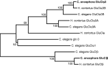
Fig. 1. Neighbor-joining tree showing relationships of the Cooperia oncophora GluCl subunits with full-length GluCls from Haemonchus contortus and Caenorhabditis elegans. The two C. oncophora sequences are highlighted in bold. The Co GluClα3 sequence is most closely related to GluClα3B sequences from H. contortus and C. elegans (100% bootstrap value). The Co GluClβ subunit groups with the two β subunits from H. contortus and C. elegans, and this grouping is also highly supported by bootstrap analysis.
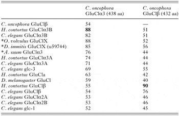
Table 2. Amino acid sequence identities of the Co GluClα3 and GluClβ subunits with other GluCl sequences reported in GenBank
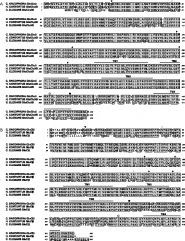
Fig. 2. Alignment of GluClα3 (A) and GluClβ (B) deduced amino acid sequences of Cooperia oncophora, Haemonchus contortus and Caenorhabditis elegans. Completely conserved residues are shaded grey. Transmembrane domains are indicated (TM1–TM4). Predicted phosphorylation (protein kinase C ([bull ]) and casein kinase 2 (○)) and N-linked glycosylation sites ([loz ]) are shown. The glycosylation and phosphorylation sites are conserved among similar subunits. The putative signal peptide cleavage sites are indicated by ([dtrif ]). The four extracellular cysteine residues are indicated by ([lozf ]). The first two cysteines, separated by 13 residues, are common to all members of the ligand-gated ion channel superfamily, and this region forms the neurotransmitter signature sequence. The second cysteine pair, separated by 10 residues, is unique to glutamate-, glycine- and histamine-gated chloride channel subunits.
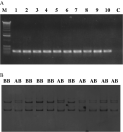
Fig. 3. Single-strand conformation polymorphism analysis of Co GluClβ PCR products. (A) Agarose gel showing the quality of the PCR products derived from 10 individual male worms. M, molecular weight marker; C, no-DNA control. (B). SSCP patterns of 11 individual IVS worms at the Co GluClβ gene. Worms shown here are homozygous BB (2 bands) or heterozygous AB (3 bands).
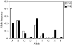
Fig. 4. Co GluClα3 allele frequencies in IVS and IVR worms.
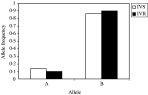
Fig. 5. Co GluClβ allele frequencies in IVS and IVR worms.

Fig. 6. Positions of polymorphic sites in the region of the Co GluClα3 and Co GluClβ genes analysed. Polymorphic sites are highlighted as vertical bars. (A) Eighteen positions were found to be polymorphic for the Co GluCla3 gene. Five of these were in the exon, and are marked by arrows. All were silent mutations. (B). Only one polymorphic site was identified in the Co GluClβ gene in an exon region. The nucleotide substitution at this position did not result in amino acid change.
 You have
Access
- 47
- Cited by