Introduction
Assisted reproductive technology had become an important means of treating infertility. Classic in vitro fertilization (IVF) treatment has been to transplant an embryo formed on the third day of in vitro fertilization and at the cleavage stage into the patient's uterus. With continuous improvement in in vitro embryo culture systems, cultured blastocysts have also been widely used. Theoretically, blastocyst transplantation had many advantages as it involves a re-screening process, therefore implantation rate may be higher after blastocyst transplantation, and the rate of live births could thus be increased. One study showed that transplantion of the embryo at the blastocyst stage could not only avoid multiple pregnancy and its related complications, but also improve the pregnancy rate of each cycle (Glujovsky et al., Reference Glujovsky, Blake, Farquhar and Bardach2012).
Blastocyst development rate is not consistent for in vitro development of human embryos, as most blastocysts form either on the fifth (D5) or sixth day (D6) after in vitro fertilization. Currently, the Gardner scoring system is the most widely used method for assessing blastocyst quality for blastocyst morphological evaluation (Gardner et al., Reference Gardner, Lane, Stevens, Schlenker and Schoolcraft2000). At this time, blastocyst development rate of blastocyst is considered an important reference factor – the faster the blastocyst develops, the higher the growth potential. In the published literature, research findings on frozen–thawed blastocyst cycles are not consistent. Certain studies have reported that the rates of clinical pregnancy, implantation and birth obtained with D5 frozen–thawed blastocysts are significantly higher than those of D6 embryos (Kovalevsky et al., Reference Kovalevsky, Carney, Morrison, Boylan, Neithardt and Feinberg2013), while other studies find that the clinical outcomes of D5 and D6 frozen–thawed blastocysts are not significantly different (Hiraoka et al., Reference Hiraoka, Hiraoka, Miyazaki, Fukunaga, Horiuchi, Kusuda, Okano, Kinutani and Kinutani2009). One study found that the implantation rate of D5 blastocysts was significantly higher than that of D6 blastocysts (Muthukumar et al., Reference Muthukumar, Kamath, Mangalaraj, Aleyamma, Chandy and George2013); but a difference in implantation rate on its own does not fully represent the developmental potential of D5 or D6 blastocysts, and blastocysts can not be selected based only on the implantation rate. Much more effective selection criteria should also include birth outcomes after the corresponding embryos are transplanted. Some studies have shown that the chance of premature delivery and of low birth weight after blastocysts transplantation was increased (Kalra et al., Reference Kalra, Ratcliffe, Barnhart and Coutifaris2012; Dar et al., Reference Dar, Joanne and Librach2013a, Reference Dar, Librach, Gunby, Bissonnette and Cowan2013b). Other studies also showed that the proportion of pregnancies involving fetal macrosomia after transplantation of frozen–thawed blastocysts increased (Mäkinen et al., Reference Mäkinen, Söderström-Anttila, Vainio, Suikkari and Tuuri2013), while others showed that if culture was extended to the blastocyst stage, there was no effect on neonatal outcomes (Fernando et al., Reference Fernando, Halliday, Breheny and Healy2012; Oron et al., Reference Oron, Sokal-Arnon, Son, Demirtas, Buckett, Zeadna, Holzer and Tulandi2014). Comparisons were made only between blastocysts and the cleavage-stage embryos, however, as no study was available that compared neonatal birth outcomes after transplantation of D5 or D6 frozen–thawed blastocysts.
In this study, birth outcomes of frozen blastocysts formed at D5 or D6 were compared. In order to avoid any effect of multiple births on results, patients were divided into either a single-birth or twins-birth group according to fetal numbers. Birth outcomes between the D5 and D6 groups were compared within the single-birth group and in the twins-birth group, respectively, aiming to provide further guidance on selecting the best blastocyst recovery day.
Materials and methods
Subjects
Data for 1109 patients that underwent recovered blastocyst transplantation at the Assisted Reproduction Center, Beijing Union Hospital for the period January 2005 to December 2012 were analyzed retrospectively. Patients with the following infertility criteria in a new cycle were included: (1) uterine tube factor; (2) endometriosis and metrial adenomyosis; (3) male factor; (4) ovulation failure (including polycystic ovary syndromes, etc.); or (5) unknown reasons. Exclusion criteria were: (1) ovary dysfunction (ova obtained <5); (2) performed D5/D6 frozen–thawed blastocyst transplantation simultaneously in one recovery cycle; (3) drug or alcohol abuse, smoking, and a history of chronic medical disease (heart diseases, hepatonephric dysfunction). Among the 1109 patients included according to the above criteria, 515 had currently given birth at the time of analysis. All blastocysts had undergone the vitrification method for freezing and recovery. Based on blastocyst formation time (D5 or D6), blastocysts frozen on the fifth day of insemination were put into the D5 group and blastocysts frozen on the sixth day of insemination were put into the D6 group. Patients were also divided by fetal number into the single-birth group or the twins-birth group. This study was conducted in accordance with the Declaration of Helsinki and conducted with the approval of the Ethics Committee of Beijing Union Hospital. Written informed consent was obtained from all participants.
Blastocyst culture and scoring
Controlled ovarian hyperstimulation was performed routinely according to the centre's regulations. Oocytes were obtained, then insemination was performed naturally or by intracytoplasmic sperm injection (ICSI) according to individual circumstances for semen, fertilization was measured within 24 h of ovulation. Morphology of the cleavage-stage embryo was observed within 72 h, and the embryo was then selected for transplantation based on its morphological score. The remaining embryos after the transplantation underwent blastocyst culture after obtaining informed consent from the patient. Blastocyst formation and morphology were assayed on the fifth or sixth day of fertilization, and the Gardner scoring system was used to score the blastocysts (Gardner et al., Reference Gardner, Lane, Stevens, Schlenker and Schoolcraft2000). Firstly, blastocysts were categorized according to the their degree of expansion: stage 1, blastocyst size was less than 50% of the embryo; stage 2, blastocyst size was more than 50% of the embryo; stage 3, the blastocyst occupied the entire embryo; stage 4, the blastocyst expanded and the zona pellucida became thinner; stage 5, the blastocyst began to hatch; stage 6, the blastocyst was fully hatched. The qualities of the inner cell mass (ICM) and the trophectoderm layer were further evaluated for stage 4 to stage 6 blastocysts. For ICM scoring: A, the cells were numerous, tightly packed; B, the cells were less, loosely arranged; C, the cells were rare. For trophectoderm (TE) scoring: A, the cells were numerous, the blastocyst was surrounded by cells, tightly packed; B, the cells were less, loosely arranged; C, the cells were rare. Because the previous observation had found that the post-freezing recovery rates of early blastocyst and already-hatched blastocysts were not ideal, we performed freezing when the blastocyst developed to stage 4. Blastocysts with ICM score A or B were used for freezing, the C blastocysts were not used, while TE quality was not considered.
Embryo freezing
The vitrification method was used for embryo freezing. Before freezing, the blastocyst was artificially shrunk, then placed into a pre-balanced solution containing 7.5% (v/v) dimethyl sulphoxide (DMSO) (Sigma, USA) and 7.5% (v/v) ethylene glycol (Beijing Chemical Plant, Beijing, China) for equilibration for 2 min, then the embryo was transferred into the vitrification solution (15% (v/v) DMSO + 15% (v/v) ethylene glycol + 0.65 mol/L sucrose (Sigma, USA) + 10 mg/ml Ficoll (Sigma, USA) for 30 s equilibration. The embryo was then transferred onto the cryoloop ring, and immersed directly into liquid nitrogen for preservation.
Embryo recovery
Recovery was performed on the same day as blastocyst transplantation. Before the blastocyst was recovered, the recovery solution was prepared: 0.33 M sucrose solution, 0.2 M sucrose solution and basic culture medium (G-MOPS Plus, VitroLife, Sweden) were added in turn to the embryo culture dish. The cryoloop ring was taken from the liquid nitrogen freezer, kept for 5 s in air, then directly immersed into the above resuscitation solution, for 2 min, 3 min or 5 min, respectively. The fully recovered blastocysts were then subjected to laser-assisted hatching, about one-quarter of the upper part of zona pellucida that was away from ICM was cut; the blastocyst was moved into the prepared blastocyst culture medium (G2, VitroLife, Sweden), and incubated at 37°C, in an atmosphere of 6% CO2 and 5% O2 for 2 h. Embryo survival was assessed before transplantation, the re-expanded blastocyst cavity could confirm the survival of embryo.
Preparation of endometrium and embryo transplantation
Preparation methods for endometrium:
-
(1) Natural cycle method: patients with a regular menstrual cycle and ovulation were injected with HCG2000IU (Eiser, Serono, Switzerland) before the ovulation. When follicular rupture was monitored, luteal support (intramuscular injection of progesterone 20–40 mg/day) was then started. The embryo was transplanted on the sixth day after ovulation.
-
(2) Artificial cycle method: the patient was given conventional ultrasound on the second to fourth day of menstruation, and orally administrated estradiol valerate (Progynova, Schering, Germany). The starting dose was set according to the patient's previous endometrial conditions (2–8 mg/day). The endometrium was monitored with ultrasound. When endometrium thickness reached 8 mm, progesterone was injected intramuscularly (20 mg × 2 days, 40 mg × 2 days, 20–40 mg × 1 days). Blastocyst recovery transplantation was performed on the sixth day. Embryo transplantation was performed under ultrasound guidance, one or two embryos were transplanted per cycle.
After transplantation, the patient was conventionally injected with progesterone intramuscularly for luteal support. Serum human chorionic gonadotrophin (HCG) was tested on the eighth to the 10th day. Ultrasound was performed on the 25th to 30th day of transplantation; appearance of the blastocyst could be used to determine clinical pregnancy.
Follow-up
All patients were given a telephone follow-up 1–3 months after childbirth. The follow-up contents included obstetric complications, neonatal birth date, delivery mode, gestational age, sex, birth weight, length, Apar score and birth defects, etc.
Statistical analysis
SPSS19.0 software was used for statistical analysis, the measured data were subjected to a t-test. Categorized variables were performed, i.e. chi-squared test; a P < 0.05 considered to indicate statistical significance.
Results
General information
In total, 1109 recovery periods were performed, and 2092 blastocysts were recovered, including 2069 blastocysts that survived after recovery (with blastocoelic expansion); the survival rate was 98.9%. The average number of transplanted blastocysts was (1.78 ± 0.41). Overall, 888 gestational sacs could be seen under ultrasound, and the transplantation rate was 44.9% (888/1978); 651 cases obtained clinical pregnancy, with the clinical pregnancy rate as 58.7% (651/1109); 40 cases were lost during the post-pregnancy follow-up, with the loss rate as 3.6% (40/1109); 515 cases gave live birth, with the live birth rate as 46.4% (515/1109); 96 cases aborted, with the abortion rate as 13.8% (90/651); six cases of ectopic pregnancy (0.5%) occurred.
Impacts of blastocyst development rate on pregnancy outcomes
The average recovery ages (33.8 ± 6.4, 33.5 ± 4.3 years old) and mean transplanted intimal thickness (10.7 ± 1.6 mm, 9.9 ± 1.7 mm) in the D5 and D6 group showed no significant difference (P > 0.05). The average number of transplanted blastocysts at D5 cycle was (1.80 ± 0.39), showing no significant difference with that at D6 cycle (1.76 ± 0.43) (P > 0.05). The biochemical pregnancy rate at D5 (3.9%) was significantly lower than D6 (6.5%), and the difference was statistically significant (P = 0.049). The implantation rate, clinical pregnancy rate and live birth rate at D5 (50.3%, 65.2% and 54.1%) were statistically significantly higher than that at D6 (38.2%, 51.9% and 38.3%) (P < 0.05). When compared with the abortion rate at D6 (16.4%), the abortion rate at D5 was slightly lower (11.9%), but the difference was not statistically significant (P = 0.095) (Table 1).
Outcomes of frozen-thawed blastocysts between the D5 and D6 groups

note: *comparison between the D5 and D6 groups showed statistical difference, P < 0.05.
Impacts of blastocyst development rate on birth outcomes
In order to avoid the effects of multiple births on the birth outcomes, we divided the patients into single-birth and twins-birth groups based on fetal numbers, and analysed their birth outcomes, respectively.
Birth outcomes of single-birth group
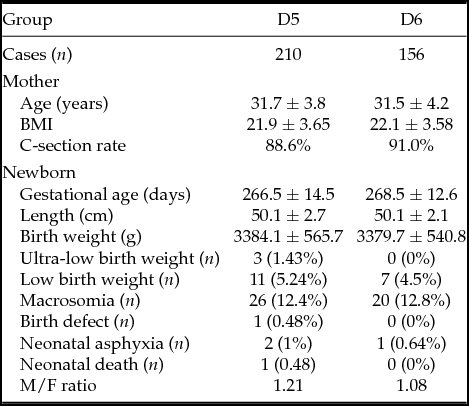
In order to exclude the effects of twins towards birth outcomes, we performed statistics on birth outcomes according to single birth or twin birth. The average age of the single-birth group was 31.7 ± 4 years old, and they delivered 366 newborns. The gestational age, birth weight and birth length were 267.3 ± 13.7 days, 3384.1 ± 565.7 g and 50.1 ± 2.5 cm, respectively. There was no statistically significant differences in age, BMI and delivery weight of the patients between the D5 and D6 groups (P > 0.05). The D5 group had 210 newborns, the gestational age, birth weight and birth length were 266.5 ± 14.5 days, 3387.4 ± 584.8 g and 50.1 ± 2.7 cm, respectively, exhibiting no significant difference from the D6 group (268.5 ± 12.6 days, 3379.7 ± 540.8 g and 50.1 ± 2.1 cm) (P > 0.05). There was no case of very low birth weight in the D6 group. The rate of low birth weight in the D5 group was slightly higher than that of the D6 group, while the rate of macrosomia in the D5 group was slightly lower than that in the D6 group, but there was no significant difference between these two groups (P > 0.05). The male/female ratio of the D5 group was 1.21, slightly higher than that in the D6 group, but the difference was not statistically significant (P > 0.05). In addition, one incident of birth defect occurred in the D5 group, which was patent ductus artery combined with hydrocephalus, and two incidences of mild neonatal asphyxia occurred (Apar score was 7 points). One child died. One incidence of neonatal asphyxia occurred in the D6 group (Table 2).
comparison of birth outcomes between the D5 and D6 single-birth groups

Birth outcomes of twins-birth group
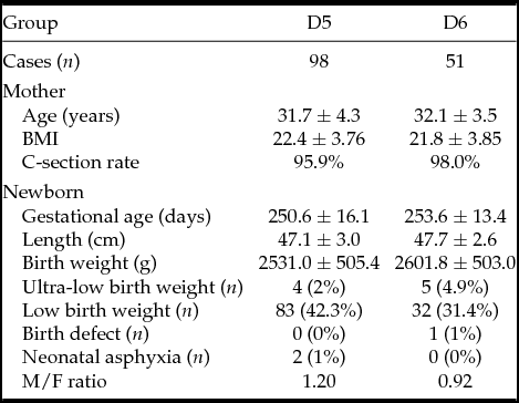
The average age of patients with twin birth was 31.8 ± 4.07 years old. In total, 149 patients delivered 298 newborns, the gestational age, birth weight and birth length were 251.6 ± 15.3 days, 2555.09 ± 504.9 g and 47.3 ± 2.9 cm, respectively. There was no statistically significant difference in age, BMI and delivery method for patients in the D5 and D6 groups (P > 0.05). The D5 group had 196 newborns, the gestational age, birth weight and birth length was not significantly different from the D6 group (P > 0.05), but the birth weight of the D5 group (2531.0 ± 505.4 g) was slightly lower than that of the D6 group (2601.8 ± 503.0) g. No case of macrosomia occurred in both groups, the incidence of low birth weight of the D5 group (42.3%) was significantly higher than the D6 group (31.4%), but the difference was not statistically significant (P > 0.05). Although the male/female ratio of the D5 group was 1.2, significantly higher than the D6 group (0.92), there was no statistically significant difference (P > 0.05). In addition, two cases of mild neonatal asphyxia occurred in the D5 group (Apar score was 7 points), while the D6 group had one case of birth defect (bilateral undescended testes) (Table 3).
Comparison of birth outcomes between the D5 and D6 twins-birth groups

Discussion
For in vitro embryonic development, at the point when the cleavage stage embryo develops to the blastocyst stage, re-selection could eliminate embryos that have poor developmental potential. Blastocyst transplantation is considered to be more like the natural conditions, and more conducive to embryo implantation, thus resulting in better clinical outcomes. Some studies have found that, in fresh cycles, the implantation rate and pregnancy rate of the blastocysts formed at D5 were significantly higher than those at D6, due to endometrial receptivity (Shapiro et al., Reference Shapiro, Daneshmand, Garner, Aguirre and Ross2008; Van Voorhis & Dokras, Reference Van Voorhis and Dokras2008). In order to avoid any endometrial receptivity effect on results, we only analysed the transplantation data from frozen–thawed blastocysts, and compared neonatal outcomes in D5 and D6 frozen–thawed blastocysts.
Hiraoka et al. (Reference Hiraoka, Hiraoka, Miyazaki, Fukunaga, Horiuchi, Kusuda, Okano, Kinutani and Kinutani2009) froze and recovered D5 and D6 blastocysts, and the survival rates were 99.5 and 97.9%, respectively. There was no statistically significant difference, similar to that found by Liebermann (Reference Liebermann2009), and consistent with our findings, so vitrification freezing of embryos formed at different stages would not affect post-recovery survival rate. However, findings for implantation rates and pregnancy rates of D5 and D6 blastocysts have been inconsistent. Hiraoka et al. (Reference Hiraoka, Hiraoka, Miyazaki, Fukunaga, Horiuchi, Kusuda, Okano, Kinutani and Kinutani2009) found no statistically significant difference in implantation rates and pregnancy rates of D5 and D6 blastocysts, while Levens et al. (Reference Levens, Whitcomb, Hennessy, James, Yauger and Larsen2008) thought the implantation rate of D5 frozen–thawed blastocysts was significantly higher than that of D6, but no significant difference was found in the clinical pregnancy rates. A recent meta-analysis found that the clinical pregnancy rate, ongoing pregnancy rate, and live birth rate of D5- frozen–thawed blastocysts were significantly higher than that of D6, but the transplantation outcomes of D5 and D6 frozen–thawed blastocysts showed no statistically significant difference when developed to the same stage (Sunkara et al., Reference Sunkara, Siozos, Bolton, Khalaf, Braude and El-Toukhy2010). In contrast, in our study, for freezing we chose blastocysts that had developed to the four stage, so the compared D5 and D6 blastocysts were almost at the same stage, and thus were more homogeneous and would not be affected by other embryonic factors. In the present study, we found that the implantation rate, clinical pregnancy rate, and live birth rate of the D5 group were significantly higher than that of the D6 group (P < 0.05), the biochemical pregnancy rate of the D5 group was significantly lower than that of the D6 group (P = 0.049). The abortion rates of the two groups showed no statistically significant difference although that of the D5 group (11.9%) was slightly lower than that of the D6 group (16.4%). This was similar to recent studies, which believed that implantation rate, clinical pregnancy rate, and live birth rate of D5 blastocysts were significantly higher than that of D6 blastocysts (Kovalevsky et al., Reference Kovalevsky, Carney, Morrison, Boylan, Neithardt and Feinberg2013; Kato et al., Reference Kato, Ueno, Yabuuchi, Uchiyama, Okuno, Kobayashi, Segawa and Teramoto2014). Similarly, Liebermann (Reference Liebermann2009) also found that the outcomes of the D5 blastocysts were better than of the D6 blastocysts, so it was considered that embryonic development rate would affect transplantation outcomes of frozen–thawed blastocysts, and the delayed blastocyst formation would mean poor embryonic qualities. One study used trophoblastic biopsy to analyse blastocyst chromosomes and found that the incidence of aneuploidy was higher in stunted blastocysts (Alfarawati et al., Reference Alfarawati, Fragouli, Colls, Stevens, Gutiérrez-Mateo, Schoolcraft, Katz-Jaffe and Wells2011). In the future, preimplantation genetic screening (PGS) might be an important means to select blastocysts with higher vitality and transplantation potentials. Transplantation of embryos with PGS-screened normal chromosome could further improve implantation rate and clinical pregnancy rate.
In the present study, in order to avoid the effects of birth numbers on the study results, the patients were divided into single-birth and twin-birth groups for analysis. We found that for the patients with a single birth, there was no statistically significant difference in the age, BMI and delivery method between the D5 and D6 groups (P > 0.05). The gestational age of the D5 group (266.5 ± 14.5 days) was slightly lower than the D6 group (268.5 ± 12.6 days), and the proportion of newborns with a low birth weight in the D5 group was slightly higher than that of the D6 group. For the twin-birth group, there was no statistically significant difference in the age, BMI and delivery method between the D5 and D6 groups (P > 0.05). Gestational age (250.6 ± 16.1 days) of the D5 group was slightly lower than that of the D6 group (253.6 ± 13.4 days). The proportion of low birth weight newborns in the D5 group was higher than that the D6 group. We found that the birth weight of the D5 group was slightly lower than that the D6 group. These differences were not statistically significant, but there was a trend that the longer the in vitro culture, the longer the gestation age after the transplantation, and the heavier the birth weight, similar to the study of Mäkinen et al. (Reference Mäkinen, Söderström-Anttila, Vainio, Suikkari and Tuuri2013), who found that OR values of D5 or D6 embryos that would cause the fetus with larger for gestational age (LGA) were significantly higher than that of D2 embryos, so it was considered that the longer the in vitro culture time, the more prone the LGA fetus. In addition, two recent Swedish studies revealed that the longer the in vitro culture time of embryos, the lower the proportion of fetuses with smaller for gestational age (SGA) (3.2 to 2.5%), although the difference was not statistically significant (Källén et al., Reference Källén, Finnström, Lindam, Nilsson, Nygren and Olausson2010; Sazonova et al., Reference Sazonova, Källen, Thurin-Kjellberg, Wennerholm and Bergh2011). In a recent large-scale study, the proportion of SGA or LGA infants was not significantly difference independent of whether embryos were cultured in vitro for 3, 5 or 6 days (Fernando et al., Reference Fernando, Halliday, Breheny and Healy2012). This was similar to findings by Hiraoka et al. (Reference Hiraoka, Hiraoka, Miyazaki, Fukunaga, Horiuchi, Kusuda, Okano, Kinutani and Kinutani2009), who compared the birth outcomes after transplantation of frozen–thawed D5, D6 or D7 blastocysts, and found no significant differences in gestational age, premature birth rate and average birth weight among these three groups. It was found that the blastocysts formed on either day had similar developmental potentials after transplantation. Fetal gestational age, birth weight and length might be affected by many factors, the specific reasons are not clear. Animal studies have found that when the blastocyst is formed, changes in TE and ICM affect fetal size, when the TE was rich, the placental volume would increase, but when the numbers of ICM were increased, the fetus would also increase (Leese et al., Reference Leese, Donnay and Thompson1998; Walker et al., Reference Walker, Hartwich and Robinson2000). Other animal studies showed that components of in vitro and in vitro culture would induce macrosomia (Young et al., Reference Young, Sinclair and Wilmut1998). A recent randomized study on human embryos discovered that neonatal birth weight could be affected by different media, but gestational age would not be affected (Young et al., Reference Young, Sinclair and Wilmut1998; Nelissen et al., Reference Nelissen, Van Montfoort, Coonen, Derhaag, Geraedts, Smits, Land, Evers and Dumoulin2012). In short, many factors can affect birth weight, so whether the birth weights of frozen–thawed D5 and D6 blastocysts after transplantation differed still needs to be studied further.
In this study, we found that in the singe-birth group, the male/female ratio at D5 (1.21) was higher than that at D6 (1.08), and the difference in the twins-birth group was more significant (D5 1.2, D6 0.92), consistent with previous studies. Previous studies in mice, cattle, pigs and other animals suggested that male embryos developed faster than female embryos (Burgoyne, Reference Burgoyne1993; Pergament et al., Reference Pergament, Fiddler, Cho, Johnson and Holmgren1994), this was also true for human embryos in in vitro culture. It was considered that the faster the rate of cell division, the more possibility the embryo with more cells had male genetic materials. Appropriately extending the in vitro culture time would make it easier to select for male embryos, because the male/female ratio at the blastocyst stage was higher than that of the embryos at the cleavage stage, and the male/female ratio transplanted after freeze–thawing was higher than that with the fresh blastocyst (Alfarawati et al., Reference Alfarawati, Fragouli, Colls, Stevens, Gutiérrez-Mateo, Schoolcraft, Katz-Jaffe and Wells2011; Bu et al., Reference Bu, Chen, Huang, Zhang, Wu, Ma, Shi, Xu, Zhang, Zhang, Zhao, Zhang, Huang, Sun, Kang, Wu, Wu, Sun and Sun2014; Maalouf et al., Reference Maalouf, Mincheva, Campbell and Hardy2014). Studies had found that, compared with the female embryos developed in the same period, the morphological score of male embryos would be better, for example 72% of D5 or D6 blastocysts were male embryos, while 60% of D3 blastocysts were female embryos (Alfarawati et al., Reference Alfarawati, Fragouli, Colls, Stevens, Gutiérrez-Mateo, Schoolcraft, Katz-Jaffe and Wells2011). Recently, Bronet et al. (Reference Bronet, Nogales, Martínez, Ariza, Rubio and García-Velasco2015) used time lapse photography to observe embryonic morphologic changes and analysed the relationship to fetal gender. They came to similar conclusions that embryonic development was affected by embryo gender, and male embryos would develop faster. When the embryos were cultured to D5, the better blastocysts with higher ICM and TE numbers, as well as the more closely arranged, were selected for cryopreservation, and selected for transplantation after freeze–thawing, therefore the D5 blastocysts had a higher proportion of males. Although the embryo score differences between male and female embryos could not reflect the activity differences among the embryos, these studies still suggested that, compared with female embryos, male embryos could reach the final blastocyst stage faster (Mittwoch, Reference Mittwoch1993). The specific mechanism is still unclear, but more and more evidence shows that certain growth factors on the Y chromosome have a role in promoting embryo development, while the second X chromosome that only exists in female embryos has a role in blocking the embryo at the cleavage stage and preventing development to the morula stage. So male and female embryos responded differently towards in vitro culture conditions (Gutiérrez-Adán et al., Reference Gutiérrez-Adán, Oter, Martínez-Madrid, Pintado and De La Fuente2000). These preliminary data still needed further study.
The results of this study suggest that implantation rate, clinical pregnancy rate and live birth rate in the D5 group were all significantly higher than that in the D6 group. In both the single-birth group and the twin birth group, the gestational age, birth weight and length of D5 and D6 blastocysts were not significantly different after transplantation following freeze–thawing. The male/female ratio of the D5 group was higher than that of the D6 group, but, because of the limited case numbers, there appeared to be no significant difference. Therefore, whether differences in the birth outcomes between the D5 and D6 frozen–thawed blastocysts exist still needs to be investigated in a further large-sample-size follow-up study.