Introduction
In order to protect aquatic ecosystems the EU water framework directive (WFD) was agreed upon in the year 2000. The fundamental objective of the WFD aims at achieving at least ’good status’ in relation to all waters by 2015 (Directive 2000/60/EC). However, a variety of abiotic and biotic parameters still threatens the health of aquatic habitats. The main factors influencing the well being of aquatic organisms such as fish and benthic macroinvertebrates are anthropogenic impacts (man-made eutrophication, chemical pollution, degradation, etc.) and pathogens (Braunbeck et al., Reference Braunbeck, Hinton and Streit1998). In recent years, an increasing body of knowledge on adverse effects of pollutants on organisms has accumulated. Some of these effects, especially alterations in biochemical processes (commonly referred to as biomarkers) in aquatic animals such as gammarids, molluscs and fish, seem to be a promising tool for assessment of environmental pollution (Segner & Braunbeck, Reference Segner, Braunbeck, Schüürmann and Markert1998). Biomarker analyses in suitable organisms allow conclusions to be drawn on the contamination of habitats with substances like polychlorinated biphenyls (PCBs), polyaromatic hydrocarbons (PAHs), metals and others. While some biomarkers show a high specificity for particular contaminants (e.g. metallothioneins as markers for metals) others are less specific and must be considered as part of a general stress response. Among these less-specific markers heat shock proteins (hsp) play an important role. They are widespread in plants, bacteria and animals and belong to a group of proteins known as chaperones, which are important for protein folding, protein transport and cell stabilization (Schröder et al., Reference Schröder, Batel, Hassanein, Lauenroth, Jenke, Simat, Steinhart and Müller1999; Werner & Hinton, Reference Werner and Hinton1999). In terms of ecotoxicological research hsp are used as biomarkers for a wide range of adverse conditions such as heat stress and metal pollution (reviewed in Sanders, Reference Sanders1993). Their induction is usually interpreted as a general sign of protein damage (Lewis et al., Reference Lewis, Handy, Cordi, Billinghurst and Depledge1999; Perceval et al., Reference Perceval, Pinel-Alloul, Méthot, Couillard, Giguère, Campbell and Hare2001; Köhler et al., Reference Köhler, Bartussek, Eckwert, Farian, Gränzer, Knigge and Kunz2001; Radlowska & Pempkowiak, Reference Radlowska and Pempkowiak2002).
Among aquatic pollutants, metals still play an important role (Scherer et al., Reference Scherer, Fuchs, Behrendt and Hillenbrand2003; Merian et al., Reference Merian, Anke, Ihnat and Stoeppler2004). One major anthropogenic source for heavy metals in freshwater systems is automobile traffic (Harrison & Wilson, Reference Harrison and Wilson1985a, Reference Harrison and Wilsonb; Ward, Reference Ward1990; Maltby et al., Reference Maltby, Forrow, Boxall, Calow and Betton1995a, Reference Maltby, Boxall, Forrow, Calow and Bettonb; Greenstein et al., Reference Greenstein, Tiefenthaler and Bay2004). Following the introduction of automobile catalytic converters for exhaust purification some 20 years ago, a new group of metals, the so-called platinum group metals platinum (Pt), palladium (Pd) and rhodium (Rh), have been increasingly emitted by cars. These metals are found in relatively high concentrations in road dust, and are transported via road runoff into aquatic habitats. Several studies show that road runoff entering aquatic habitats is usually associated with toxic effects on the respective organisms (Harrison & Wilson, Reference Harrison and Wilson1985a, Reference Harrison and Wilsonb; Ward, Reference Ward1990; Maltby et al., Reference Maltby, Forrow, Boxall, Calow and Betton1995a, Reference Maltby, Boxall, Forrow, Calow and Bettonb; Greenstein et al., Reference Greenstein, Tiefenthaler and Bay2004). With respect to hsp, evidence emerged recently that hsp can also be induced by exposure of organisms to Pd (Singer et al., Reference Singer, Zimmermann and Sures2005).
Another important factor that affects the health of organisms is infection with pathogens such as parasites. A possible influence of parasites on the expression of biomarkers is, however, seldom considered in ecotoxicological studies (Sures, Reference Sures2004, Reference Sures2006). Therefore, the present study aims at investigating the interaction of parasitism and pollution with respect to induction of biomarkers in hosts. As model organisms the amphipod Gammarus roeseli partly infected with larvae of the acanthocephalan Polymorphus minutus were used. This host–parasite system was exposed under laboratory conditions to Pd at relatively low aqueous concentrations (10 μg l− 1). After exposure, relative levels of the heat shock protein 70 (hsp70) were analysed in tissues of parasites and intermediate hosts. Simultaneously, metal accumulation in gammarids and parasites was compared.
Materials and methods
Experimental design
Unparasitized and naturally infected Gammarus roeseli harbouring cystacanths of Polymorphus minutus were collected by pond net from the brook Giessbach close to Karlsruhe, Germany, in February 2006. They were returned to the laboratory in cooled river water and thereafter divided into four groups, with 140 individuals per treatment (see table 1). Infected gammarids were provisionally separated after inspection by eye, as cystacanths of P. minutus are usually visible as oval orange structures in the haemocoel of G. roeseli. Final information on the infection status was obtained after dissecting animals at the end of the exposure period. As the water temperature of the brook was 6°C, handling of the crustaceans was performed in plastic containers on ice in order to avoid heat stress. After provisional determination of the infection status, gammarids were placed in 20-litre plastic tanks with 15 litres of aerated, de-chlorinated tap water. Amphipods were fed alder and horse chestnut leaves. The water was replaced twice weekly. Two groups of G. roeseli (CoInf and CoUninf) were used as unexposed controls, the other two groups (PdInf and PdUninf) were exposed to nominal Pd concentrations of 10 μg l− 1. Due to precipitation processes it can be assumed that only 50% of Pd is present after 24 h (Sures & Zimmermann, Reference Sures and Zimmermann2007). The resulting concentration of 5 μg l− 1 is expected to be still above environmental levels (these could not be determined due to analytical problems). Pd exposure was performed by adding 150 μl of Palladium standard solution (Pd 1000 μg ml− 1, Ultra Scientific ICP-045, North Kingstown, Rhode Island, USA) to the tank water at the beginning of the experiment and after each water replacement. Gammarids were maintained under these conditions for 24 days. Water characteristics and data on amphipods are summarized in table 1.
Experimental design and characteristics of the tank water and of Gammarus roeseli. Values for pH, water temperature and conductivity are means±SD, for eight observations made through time during the 24 days' exposure.

a No palladium (Pd) added to tank water.
d.l., detection limit.
Sample collection and processing
Water samples were taken from each tank directly before and 1 h after each water replacement during the exposure period, according to the following procedure: 10 ml tank water was filtered (cellulose nitrate filter, pore size 0.45 μm, Sartorius AG, Göttingen, Germany), acidified with 10 μl HNO3 (65 vol.%, suprapure quality, Merck, Darmstadt, Germany) and stored at − 21°C until metal analysis. After an exposure period of 24 days crustaceans were collected from each group and killed by decapitation. After killing, cystacanths were separated from host tissue. Subsequently, crustacean tissue from the same group was pooled and frozen at − 21°C in Eppendorf tubes. Cystacanths collected from all gammarids of one tank were divided in to three aliquots and frozen in Eppendorf tubes. To avoid metal contamination, all instruments used for sampling were cleaned with 0.1% EDTA solution and double-distilled water.
Metal analysis
Concentrations of Pd in water, gammarids and cystacanths were determined by electrothermal atomic absorption spectrometry (ET-AAS) as described in Zimmermann et al. (Reference Zimmermann, Messerschmidt, von Bohlen and Sures2003). The water samples were measured without any pre-treatment in duplicate by ET-AAS. For metal analysis three aliquots of the pooled gammarid tissues (approximately 150 mg wet weight) or 15 mg (wet weight) of cystacanth tissue were digested with 1.8 ml nitric acid (65%, suprapure, Merck) using a microwave digestion procedure described by Sures et al. (Reference Sures, Taraschewski and Haug1995). To determine the analytical detection limits, blanks were prepared without insertion of sample material. After the digestion was completed, the clear solutions were filled with HNO3 (65%, suprapure, Merck) up to 2 ml in graduated flasks. Metal concentrations of the digested tissues, as well as of the water samples, were determined with ET-AAS using a Perkin–Elmer model 4100ZL spectrometer (Uberlingen, Germany) equipped with a Zeeman effect background correction system. Calibration was performed by standard addition. Pd concentrations in each sample were calculated by fitting linear regression lines to the points defined by the spiked concentration values and the corresponding integrated peak areas. Correlation coefficients were always better than 0.99.
Heat shock protein analysis
The detection of hsp70 was performed in accordance with Singer et al. (Reference Singer, Zimmermann and Sures2005) and Tokalov & Gutzeit (Reference Tokalov and Gutzeit2003). For protein quantification, 0.1 g (fresh weight) of homogenized crustacean or parasite tissue was mixed with 1.5 ml phosphate-buffered saline, pH 7.2 (PBS) and 2% sodium dodecyl phosphate (SDS) and sonicated on ice for 45 s using a Sonifier HD 2070, Sonotrode MS 72 (Bandelin Sonopuls Electronic, Berlin, Germany). After the lysate was centrifuged at 19,000 g for 10 min the total protein concentration was determined in the supernatant using the Pierce BCA Protein assay Kit (Pierce Biotechnology, Rockford, Illinois, USA). To ensure constant protein concentrations, the samples were diluted with PBS+2% SDS to a protein concentration of 2 μg μl− 1 and subsequently separated by SDS-PAGE.
For SDS-PAGE, 4% stacking and 12% resolving gels were used. Samples were diluted 1:2 with sample loading buffer and denatured in a boiling water bath for 5 min. Subsequently, 25 μl sample solution was loaded in every well of the gel. Each sample was processed three times and a protein marker (Bio-Rad Perfect Stain, Bio-Rad, Munich, Germany) was used to visualize the molecular weight. SDS-PAGE was carried out in two steps (80 V for 20 min and 120 V for 100 min) in a Mini protean cell 3 (Bio-Rad).
After SDS-PAGE, blotting was performed in a semi-dry blotting cell (Bio-Rad) at 24 V for 60 min. The membranes were blocked for 90 min in a 5% non-fat dry milk solution and subsequently incubated with a monoclonal anti-hsp70 IgG1 (H5147, Sigma–Aldrich, Siegen, Germany), diluted 1:5000 with 5% non-fat dry milk in Tris-buffered saline containing 1% Tween (TBS-T; Fluka, Buchs, Switzerland) overnight at 4°C. The membranes were washed three times for 10 min in TBS-T and then incubated for 90 min with a secondary antibody (polyclonal goat anti-mouse immunoglobulins conjugated with horseradish peroxidase; P0447, Dako A/S, Denmark, diluted 1:2500 with 5% non-fat dry milk in TBS-T). After three washing steps, hsp70 bands were visualized on a high performance chemiluminescence film (ECL-Hyperfilm™, Amersham Pharmacia, Buckinghamshire, UK) using an enhanced chemiluminescence detection kit (ECL plus kit, Amersham Pharmacia). The films were scanned at 300 dpi and the density of the bands was subsequently analysed using the software Gelscan V4.0 (BioSciTec, Marburg, Germany).
Data analysis
Metal concentrations in the crustacean and cystacanth tissues were determined as μg g− 1 (fresh weight). For evaluation of the hsp70 induction, it was necessary to normalize the data of each gel to a constant reference. Therefore, a hsp70-positive sample had been run as internal standard in all gels. To calculate the relative hsp70 concentration in the gammarid samples, the following equation was used:

where rel. c sample is the relative hsp70 concentration in the gammarid sample related to the hsp70 positive control sample; d std, gel is the mean density of the triple analysis of the hsp70 internal standard on the gel; and d sample is the mean density of the triple analysis of the exposed sample.
For statistical analysis of the differences in the metal concentrations between hosts and parasites and the hsp levels the Mann–Whitney U-test was applied; significance was accepted when P ≤ 0.05.
Heat stress experiment
A follow-up experiment was performed to investigate the effects of heat stress on infected and uninfected G. roeseli. Therefore, naturally infected and uninfected gammarids were collected in the field (water temperature of 6.7°C), returned to the laboratory in cooled river water and acclimatized to a water temperature of 13°C overnight in plastic tanks. The next day half of the gammarids were transferred to water with a temperature of 29°C. After 3 h, crustaceans from the heat treatment and from the 13°C control were collected, killed by decapitation and their infection status was determined. Three subsamples of uninfected G. roeseli, infected G. roeseli and P. minutus, respectively, were collected and frozen at − 21°C from both groups (heat treatment and 13°C control).
In a second follow-up experiment, cystacanths of P. minutus were separated from freshly collected G. roeseli and divided into two groups: one group was placed in 0.8% NaCl solution at a temperature of 5°C and the other group was placed in NaCl solution at a temperature of 18°C. Three hours later cystacanths were collected from each solution and frozen at − 21°C.
Animal tissues of both follow-up experiments were analysed for their hsp70 concentration as described above.
Results
Mortality rates were high (32–54%) among both Pd exposed und unexposed gammarids (table 1). Interestingly, infection of G. roeseli with P. minutus seems to reduce mortality. Among metal-exposed G. roeseli infected amphipods showed a 10% lower mortality. A 20% lower mortality was observed for the unexposed infected G. roeseli when compared with the unexposed uninfected conspecifics. Mean infection intensity of G. roeseli with P. minutus was found to be 2 ± 1 cystacanth per gammarid, with a maximum infection intensity of 11 larvae per host. However, it remains unclear if infection intensity affects metal accumulation or hsp response, as tissues of animals with different infection intensities were pooled in order to obtain enough material for subsequent analyses.
Palladium concentration in water and animal tissues
The detection limits for palladium (3 × SD) in water, gammarids and cystacanths were determined as 4.8 μg l− 1, 0.80 μg g− 1 (wet weight) and 3.5 μg g− 1 (wet weight), respectively.
ET-AAS analyses of Pd in water revealed that the concentrations were usually lower than the detection limit. This is not only true for the unexposed controls but also for the Pd-exposed groups. Only those water samples taken 1 h after the renewal of water and Pd showed Pd concentrations around 5 μg l− 1, which is approximately half the nominal concentration applied.
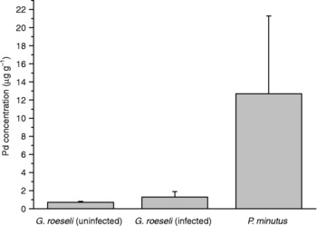
Metal accumulation in gammarids and their cystacanths is summarized in fig. 1. Pd concentrations in gammarids and parasites from the unexposed groups were all below the detection limit. Uninfected, exposed G. roeseli exhibited Pd concentrations close to the detection limit. Only the exposed host–parasite system showed Pd concentrations clearly above the detection limit. Although the mean metal concentration in infected G. roeseli was twice as high as the levels in uninfected gammarids, this difference was not found to be significant (Mann–Whitney U-test, P > 0.05). The highest significant (Mann–Whitney U-test, P < 0.05) Pd accumulation was determined for the cystacanths of P. minutus, with 10 times higher levels than the host.
Mean concentrations of palladium [μg g− 1 (wet weight)] in Gammarus roeseli and their acanthocephalan parasites, Polymorphus minutus, after exposure to Pd for 24 days (mean ± SD, n = 3).

Induction of hsp70 in Gammarus roeseli and Polymorphus minutus after Pd exposure
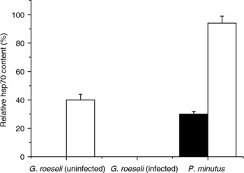
Heat shock proteins were detected only in P. minutus (from exposed and control gammarids) and in uninfected gammarids exposed to Pd (fig. 2). Both infected groups of G. roeseli failed to show any hsp70 levels, as did the unexposed amphipods. Concerning the hsp70 response of uninfected gammarids, one can conclude that exposure to Pd is a suitable stressor to induce hsp70 synthesis. Interestingly, although no hsp70 response was found for exposed, infected gammarids, their parasites inside the haemocoel showed significantly (Mann–Whitney U-test, P < 0.05) higher hsp70 levels than P. minutus collected from unexposed G. roeseli. Furthermore, the cystacanths of infected amphipods also showed significantly (Mann–Whitney U-test, P < 0.05) higher hsp70 levels than their hosts. Thus, one may assume that the presence of cystacanths in the haemocoel prevents hsp70 induction as a response to Pd exposure. In order to investigate whether this suppression of hsp70 induction is specific for exposure to Pd or a more general effect of P. minutus on its intermediate host, additional heat experiments were performed.
Levels of the heat shock protein hsp70 (mean ± SD, n = 3) in Gammarus roeseli and their acanthocephalan parasites, Polymorphus minutus, after exposure to Pd for 24 days (black bars, control; white bars, Pd exposed).

Induction of hsp70 in Gammarus roeseli and Polymorphus minutus after heat exposure
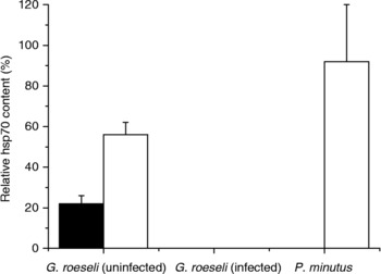
Exposure of the host–parasite system to heat reveals similar results as exposure to Pd (fig. 3). Again, parasitized gammarids showed no hsp70 response, while hsp70 levels increased significantly (Mann–Whitney U-test, P < 0.05) with heat exposure in uninfected G. roeseli. Furthermore, the uninfected amphipods showed a basal level of hsp70 which was absent in the infected conspecifics. Although the intermediate host showed no heat response, hsp70 levels in larval parasites increased with water temperature. Furthermore, an increase in hsp70 levels in P. minutus was also found after heat treatment of isolated cystacanths. The larvae maintained in NaCl solution at 18°C showed 20% higher hsp levels than their conspecifics maintained at 5°C (data not shown).
Levels of hsp70 (mean ± SD, n = 3) in Gammarus roeseli and their acanthocephalan parasites, Polymorphus minutus, after exposure to heat (black bars, 13°C; white bars, 29°C).

Discussion
The present study demonstrates that parasites may interact with pollutants in different ways: on the one hand, the acanthocephalan larvae were able to take up and accumulate Pd to a higher degree than their intermediate hosts. Secondly, infection of G. roeseli with cystacanths of P. minutus prevents synthesis of hsp70 in the host.
A couple of recent studies concentrate on heavy metals in acanthocephalans (summarized in Sures, Reference Sures2003, Reference Sures2004). From these studies it emerged that adult acanthocephalans within the intestine of their final hosts are able to accumulate metals to a far higher degree than their hosts. This enormous accumulation capacity was described for several metals and for different limnic and marine host–parasite combinations (summarized in Sures, Reference Sures2003, Reference Sures2004). In contrast to the high heavy metal concentrations recorded in adult acanthocephalans, the larval stages in their respective crustacean intermediate hosts show little tendency to accumulate metals (Brown & Pascoe, Reference Brown and Pascoe1989; Sures & Taraschewski, Reference Sures and Taraschewski1995; Siddall & Sures, Reference Siddall and Sures1998). Whereas metal concentrations in larval Acanthocephalus lucii were seven times lower for lead and 16 times lower for cadmium when compared with the intermediate host Asellus aquaticus (Sures & Taraschewski, Reference Sures and Taraschewski1995), these elements had been detected at approximately the same concentrations in cystacanths of Pomphorhynchus laevis and its naturally infected crustacean host Gammarus pulex (Brown & Pascoe, Reference Brown and Pascoe1989; Siddall & Sures, Reference Siddall and Sures1998). Studies by Sures and co-workers indicated that the location of the parasite inside the host determines the availability of metals to the parasite (Sures & Siddall, Reference Sures and Siddall1999, Reference Sures and Siddall2001; Sures et al., Reference Sures, Siddall and Taraschewski1999). For example, young adults of P. laevis were only able to bioconcentrate lead above host tissue levels if they were located within the intestine (Sures & Siddall, Reference Sures and Siddall2001). In contrast, conspecifics which passed through the intestinal wall and were located in the body cavity of the fish host showed no, or only a very poor, accumulation of lead (Sures & Siddall, Reference Sures and Siddall2001). Accordingly, the difference in metal accumulation between larval P. laevis and P. minutus might be due to small-scale differences in the location within the intermediate host. Whereas P. minutus shows an intimate contact with the alimentary tract of its intermediate host, P. laevis is completely isolated from the intestine of the gammarid (Dezfuli & Giari, Reference Dezfuli and Giari1999). This discrepancy might be the reason for the differences in metal accumulation. Also, in the intermediate host the location of the parasite appears to be the decisive factor that determines the degree of metal uptake.
Apart from the unexpectedly high metal accumulation in P. minutus, it emerged from our results that infected G. roeseli showed no hsp70 response. Following exposure to Pd only uninfected G. roeseli had an increased hsp70 content, whereas infected amphipods failed to show hsp70 at all. This was not only true for Pd-exposed animals but also for infected gammarids that were exposed to heat. In both gammarid treatments their parasites showed an increase in hsp70 levels due to heat or metal exposure of the intermediate host. Additional in vitro experiments with isolated cystacanths revealed that P. minutus is able to produce its own hsp70. Heat shock proteins are commonly used as a biomarker to indicate sublethal environmental stress due to chemicals or heat (Sanders, Reference Sanders1993; Lewis et al., Reference Lewis, Handy, Cordi, Billinghurst and Depledge1999; Köhler et al., Reference Köhler, Bartussek, Eckwert, Farian, Gränzer, Knigge and Kunz2001; Schirling et al., Reference Schirling, Jungmann, Ladewig, Ludwichowski, Nagel, Köhler and Triebskorn2006). Recently we showed that exposure to Pd results in increased hsp70 levels in mussels (Singer et al., Reference Singer, Zimmermann and Sures2005), a result that is confirmed in the present study for the amphipod G. roeseli. However, presence of cystacanths of P. minutus suppresses the hsp70 response in G. roeseli. Reasons for this suppression of hsp70 remain unclear. Köhler et al. (Reference Köhler, Bartussek, Eckwert, Farian, Gränzer, Knigge and Kunz2001) showed that hsp70 contents in fish can decrease if stressors were too severe or if organisms face a combination of strong stressors. If we follow these arguments, P. minutus must be considered a strong stressor. It is clear that acanthocephalan cystacanths cause a number of physiological changes in gammarid intermediate hosts. These include changes in host integumental pigmentation (Dezfuli et al., Reference Dezfuli, Franzoi, Trisolini and Rossi1989), reproduction (Oetinger, Reference Oetinger1987), respiration (Rumpus & Kennedy, Reference Rumpus and Kennedy1974) and behaviour (Bakker et al., Reference Bakker, Mazzi and Zala1997; Bauer et al., Reference Bauer, Trouve, Gregoire, Bollache and Cezilly2000; Tain et al., Reference Tain, Perrot-Minnot and Cezilly2006). As very fundamental processes of the host are affected by acanthocephalan larvae, it appears likely that P. minutus can also alter the stress protein response of gammarids. However, the precise mechanisms by which cystacanths can interfere with the intermediate host's stress response have yet to be elucidated.
From our results it is clear that analyses of hsp70 levels as biomarkers for pollution or heat are only reliable if uninfected gammarids are investigated. Especially in ecotoxicological studies examining whether exposure to certain chemicals affects the physiological homeostasis of a test organism, it is important to use organisms that are known to be uninfected. Otherwise, wrong conclusions can be drawn as parasites may influence the metabolism of pollutants in infected hosts. Additionally, parasites and pollutant can interact in synergistic or antagonistic ways; or parasites may induce physiological reactions in hosts which were thought to be pollutant induced. Due to these variable ways of interaction between pollutants and parasites, this topic should be elucidated in more detail in future studies.
Acknowledgements
Thanks are due to Sabrina Frank, Nadine Haus and Christoph Singer for their help during gammarid sampling and laboratory work.