Introduction
Digenean trematode species richness appears to be particularly high in the Eastern Indo-Pacific (EIP) and Central Indo-Pacific (CIP) marine ecoregions (Yamaguti, Reference Yamaguti1970; Cribb et al., Reference Cribb, Bray, Barker, Adlard and Anderson1994). Several studies have examined adult digenean trematodes of marine fishes in these ecoregions, including Australia, the South Pacific regions of French Polynesia, New Caledonia and Hawaii (see, for example, Cribb et al., Reference Cribb, Bray, Barker, Adlard and Anderson1994, Reference Cribb, Bray, Wright and Pichelin2002; Nolan & Cribb, Reference Nolan and Cribb2005). However, only a few such studies have considered digenean metacercariae (e.g. Yamaguti, Reference Yamaguti1970; Køie & Lester, Reference Køie and Lester1985; Cribb, Reference Cribb1998 and references therein), perhaps partly because metacercariae are more difficult to locate, recognize and identify than adult worms. However, knowledge of metacercariae is obviously necessary for understanding the population dynamics of both digeneans and hosts, the ecological impacts on hosts and the broader role of digeneans in ecosystems. For example, metacercariae, being trophically transmitted, require their host to be eaten by a final host and they therefore have an inherently different relationship with their hosts than do adult trematodes (which die if their host is eaten). Metacercariae can dominate the parasite community of fishes in both numbers of individuals and species (Salgado-Maldonado & Kennedy, Reference Salgado-Maldonado and Kennedy1997; Rodríguez-González & Vidal-Martínez, Reference Rodríguez-González and Vidal-Martínez2008). More generally, trophically transmitted parasites, including abundant metacercariae, had substantially greater biomass than did adult parasites in a study in three estuarine ecosystems (Kuris et al., Reference Kuris, Hechinger, Shaw, Whitney, Aguirre-Macedo, Boch, Dobson, Dunham, Fredensborg, Huspeni, Lorda, Mababa, Mancini, Mora, Pickering, Talhouk, Torchin and Lafferty2008).
Palmyra Atoll is one of the northern Line Islands, located in the East Indo-Pacific marine ecoregion (sensu Spalding et al., Reference Spalding, Fox, Allen, Davidson, Ferdaña, Finlayson, Halpern, Jorge, Lombana, Lourie, Martin, McManus, Molnar, Recchia and Robertson2007), 1680 km SSW of Hawaii (5°52′55.54′N; 162°04′59.05″W). During the Second World War, Palmyra supported a few thousand military personnel. The atoll has otherwise never had a permanent human population. All fishing has been prohibited at Palmyra since it became a US National Wildlife Refuge in 2000 (before that, its remoteness kept fishing pressure low).
As a part of a larger research project on the role of parasites in EIP atoll food webs (Lafferty et al., Reference Lafferty, Shaw and Kuris2008), a survey of the parasites of the fishes from the lagoon flats of Palmyra Atoll was conducted. A substantial diversity and abundance of digenean metacercariae infecting fish as second intermediate hosts was found. The lack of comprehensive taxonomic studies of digenean metacercariae in the EIP prompted us to provide morphological descriptions of these parasites, and to summarize their prevalence, mean intensity and host and tissue use. The primary aim of this paper is to provide a document that can facilitate ecological, taxonomic and other biological research involving these trematodes at Palmyra Atoll, and perhaps more broadly throughout the EIP.
Materials and methods
Between 13 October–10 November 2009 and 22 June–28 July 2010, fish were collected by seine, spear, and hook and line from the intertidal sand flats bordering the Palmyra Atoll lagoon (see Fig. 1 in Papastamatiou et al., Reference Papastamatiou, Lowe, Caselle and Friedlander2009). Fish were individually bagged and examined fresh within 24 h (except for four bonefish, which were frozen for 4 months at − 60°C before examination).
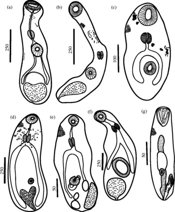
Excysted metacercariae from lagoon-flat fishes of Palmyra Atoll (all worms in ventral view). (a) Stephanostomum sp.; (b) Acanthocolpidae gen. sp.; (c) Cryptogonimidae gen. sp. 1; (d) Cryptogonimidae gen. sp. 2; (e) Stellantchasmus sp.; (f) Zoogonidae gen. sp.; (g) Bucephalus sp. 1. All scale bars are in micrometres.

Parasitological examinations were conducted using stereomicroscopes, using 0.7% saline solution as necessary. Metacercaria abundance was quantified by squashing tissues, organs and body parts between two glass plates. For most body parts with clear bilateral symmetry (i.e. body surface, muscles, pectoral and pelvic fins, eyes and gills), metacercariae were enumerated in the left half and multiplied by two to estimate the expected intensity for both sides. Brain, liver, gall bladder, spleen, kidney, intestine, stomach, heart and swim bladder were squashed in their entirety. For body parts>25 g (primarily large jack muscle fillets and shark livers), tissue interspersed throughout the body part area was excised to squash a maximum of 25 g. The observed intensities were multiplied by the appropriate weight proportion to estimate intensity in the whole body part. Both prevalence and mean intensity values were calculated using ‘expected’ data, while reported total number of individuals recovered reflect only directly ‘observed’ metacercariae.
Metacercariae generally lack diagnostic adult structures that characterize most species descriptions. Thus, it is generally not possible to identify metacercariae to the species level without previous life cycle or molecular work linking them to adults. However, metacercariae do have a suite of morphological characters. This makes it possible frequently to place them into the appropriate family and genus, and to designate them as different morphospecies. Even if morphospecies designations are somewhat provisional, they provide a clear, working hypothesis of specific identity that can immediately facilitate research. To identify metacercariae, up to 10–20 cysts of a putative species were removed from host tissues and individually isolated on a slide with a drop of saline solution. The site of infection, shape, movements, colour and number of metacercarial cysts were recorded. The metacercarial cyst dimensions were measured for those that withstood coverslip pressure. Surgical needles were used to free metacercariae from cysts that did not excyst with coverslip pressure. A coverslip was added to flatten the worm, and the corners of the coverslip were fixed to the slide with Du-Noyer sealant (1 part dehydrated lanolin and 4 parts melted resin) that was melted to the slide using an alcohol lamp. Then, a small amount of ammonium picrate fixative (1:1 saturated ammonium picrate and glycerin) was added to one side of, and drawn under, the coverslip using a Kimwipe® on the opposite side. Although metacercariae suffer some deformation by flattening, fixation with ammonium picrate works well for fieldwork because of its simplicity, rapid staining and its ability to clarify internal morphology and external sclerotized structures. This technique produces semi-permanent slides which can be preserved for 1–2 years by adding small amounts of fixative after monthly inspection (for further details see Scholz & Aguirre-Macedo, Reference Scholz and Aguirre-Macedo2000). Sketches or high-quality digital photographs were used to document morphological details. Measurements of metacercariae were based on flattened specimens fixed with ammonium picrate, unless otherwise indicated. In some cases, which are indicated, we acquired morphometrics from excysted specimens that were killed in hot water, fixed in 4% formalin, stained with carmine and mounted in Canada balsam. Images of the metacercariae were captured using a digital camera and drawn on a digitizing tablet using Adobe Illustrator software. All other helminthological methods followed were those recommended by Vidal-Martinez et al. (Reference Vidal-Martinez, Aguirre-Macedo, Scholz, González-Solís and Mendoza-Franco2001). Synonyms for each host species were obtained from FishBase (http://www.fishbase.com) and helminth records for each of the possible host synonyms were researched using Web of Science (ISI Web of KnowledgeSM; http://wok.mimas.co.uk), Google Scholar (http://scholar.google.com), and the Host–Parasite Database of the Natural History Museum, London (Gibson et al., Reference Gibson, Bray and Harris2005). In the tables, fish species are arranged by family, in taxonomic order following Randall et al. (Reference Randall, Allen and Steene1997). Voucher specimens of the metacercariae have been deposited in the United States Parasitological collection (USNPC), Beltsville, Maryland. All measurements below are in micrometres (range, mean ± standard deviation), unless otherwise stated. For each taxon, the prevalence and mean intensity were calculated following Bush et al. (Reference Bush, Lafferty, Lotz and Shostak1997). Observations of other parasite taxa and stages will be reported elsewhere.
Results
Table 1 presents the species and numbers of host fishes examined, and the prevalence and mean intensity ( ± SD) of digenean metacercariae. Up to 425 individual fish were dissected, belonging to 42 species and 21 families. Three hundred and sixty-four (86%) of the 425 dissected fish were infected by one or more digenean species. The following species were not infected by metacercariae: Carcharhinus melanopterus (Quoy & Gaimard, 1824) (number of fish examined = 4), Albula glossodonta (Forsskål, 1775) (12), Gymnothorax pictus (Ahl, 1789) (2), G. rueppelliae (McClelland, 1844) (1), Chanos chanos (Forsskål, 1775) (3), Epinephelus merra Bloch, 1793 (1), Caranx melampygus Cuvier, 1833 (2), Parapercis sp. (16), Oplopomus oplopomus (Valenciennes, 1837) (19), Istigobius rigilius (Herre, 1953) (1), Psilogobius prolatus Watson & Lachner, 1985 (4) and Asterropterix semipunctata Ruppell, 1830 (8).
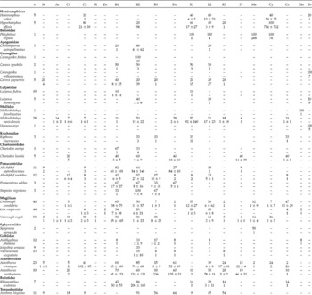
Prevalence (above, in percentage) and mean intensity (below ± SD) of digenean metacercariae of marine fishes from Palmyra Atoll. All fish species are in phylogenetic order following Randall et al. (Reference Randall, Allen and Steene1997), and digeneans following Olson et al. (Reference Olson, Cribb, Tkach, Bray and Littlewood2003). Acronyms are as follows: n, number of fish sampled; St, Stephanostomum sp., Ac, Acanthocolpidae gen. sp.; C1–C2, Cryptogonimidae gen. spp. 1–2; Sl, Stellantchasmus sp.; Zo, Zoogonidae gen. sp.; B1–B3, Bucephalus spp. 1–3; Do, Dollfustrema sp.; R1–R3, Rhipidocotyle spp. 1–3; Pr, Prosorhynchus sp.; Me, Mesostephanus sp.; Cy, Cyathocotylidae gen. sp.; Ca, Cardiocephaloides sp.; Mo, Monolicaecum-type; To, Torticaecum-type.

Of the 33,964 metacercariae counted, 19 digenean species were recognized comprising 8 families. The digenean families recovered, in decreasing order of number of species and individuals, were Bucephalidae Poche 1907 (8 species with 29,480 individuals), Cyathocotylidae Mühling 1898 (2 and 2209), Cryptogonimidae Ward, 1917 (2 and 1923), Strigeidae Railliet, 1919 (1 and 270), Didymozoidae Monticelli, 1888 (2 and 42), Acanthocolpidae Lühe, 1906 (2 and 38), Heterophyidae Leiper, 1909 (1 and 1) and Zoogonidae Odhner, 1902 (1 and 1).
In general, the most prevalent taxon was Bucephalus sp. 2 with prevalence values between 15% in Valenciennea sexguttata (Valenciennes, 1837) and 91% in Arothron hispidus (Linnaeus, 1758), followed by other bucephalids such as Rhipidocotyle spp. 1–3 (table 1). This range does not include the 100% prevalence of Bucephalus sp. 2 observed in three Stegastes nigricans (Lacepède, 1802). In this case, and others with low host sample size, there is substantial error around prevalence estimates. The species with the highest mean intensity in our samples was also Bucephalus sp. 2 with 1053 ± 1365 individuals in A. hispidus, followed by Cyathocotylidae with 714 ± 732 individuals in Hyporhamphus affinis (Günther, 1866) and Rhipidocotyle sp. 1 with 147 ± 187 in A. hispidus.
Table 1 presents 167 host records for the 19 digenean species, none of which have been recorded from Palmyra Atoll. The following section presents morphological descriptions for each of the recovered species, including host information and site(s) of infection.
Family Acanthocolpidae
Stephanostomum sp. (fig. 1a)
Description
Based on three flattened individuals, one from Arothron hispidus (USNPC No. 104843) and two from Acanthurus triostegus. Body length 810–2050 (1230 ± 710), maximum width 220–350 (264 ± 74) towards posterior end of the body, tegument densely covered with spines at anterior half. Eye-spot pigment present scattered in anterior part of the body. Oral sucker 92–210 (132 ± 68) long and 117–320 (187 ± 115) wide, with double row of 30 spines and without ventral hiatus. Acetabulum equatorial, 90–200 (134 ± 58) long and 85–190 (121 ± 60) wide. Prepharynx 132–250 (207 ± 64) long. Oesophagus short, 20–62 (44 ± 22) long. Caeca bifurcating immediately preacetabularly, extending to anterior excretory vesicle. Developing testes median, tandem. Anterior (right) testis 55–150 (102 ± 67) long, 50–125 (87 ± 53) wide. Posterior (left) testis 70 (n = 1) long, 55 (n = 1) wide. Ovary not distinguishable. Excretory vesicle V-shape, excretory pore terminal.
Hosts
Carangidae: Caranx papuensis Alleyne & MacLeay, 1877 (encysted in intestine wall); Pomacentridae: Abudefduf septemfasciatus (Cuvier, 1830) (muscle); Mugilidae: Valamugil engeli (Bleeker, 1858–59) (fins); Acanthuridae: Acanthurus triostegus (Linnaeus, 1758) (fins); Tetraodontidae: A. hispidus (fins).
Comments
Stephanostomum Looss, 1899 is a large genus comprising 118 species (Bray & Cribb, Reference Bray and Cribb2003, Reference Bray and Cribb2006, Reference Bray and Cribb2008). However, due to the number of spines around the oral sucker (30) and the lack of a ventral hiatus, only two species previously described in the Indo-Pacific region could be the potential adult stages: S. madhaviae Bray et Cribb, 2003 (30–34 spines) from Caranx ignobilis (Forsskål, 1775) from Queensland and S. rachycentronis Shen, 1990 (30–34 spines) from Rachycentron canadum (Linnaeus, 1766) from Hainan Island, China (Bray & Cribb, Reference Bray and Cribb2003).
Acanthocolpidae gen. sp. (fig. 1b)
Description
Based on two flattened individuals; one from Mulloidichthys vanicolensis (Valenciennes, 1831) (USNPC No. 104844) and one from Valamugil engeli. Body length 510–715, maximum width 75–82 at posterior part of body, tegument densely covered with fine spines, especially at anterior half. Eye-spot pigment present at prepharyx level. Oral sucker subterminal 75–82 long and 80–87 width. Acetabulum postequatorial, 67 long and 72 width. Prepharynx 267 long, pharynx 34 long and 33 wide, nearly equatorial. Oesophagus and caeca not distinguishable. Genitalia non-developed. Excretory vesicle clavate, occupying last quarter of body.
Hosts
Mullidae: Mulloidichthys vanicolensis (fins); Mugilidae: Valamugil engeli (brain, heart, muscle).
Comments
This metacercaria is likely an acanthocolpid, based on the tegument covered with fine spines, lack of circumoral spines, neck region enlarged, pharynx present and small acetabulum close to midbody. This species may be one of the several acanthocolpid genera that lack oral spines, e.g. Acanthocolpus Luhe, 1906; Lepidauchen Nicoll, 1913; Neophasis Stafford, 1904; Spinoplagioporus Skrjabin et Koval, 1958; and Ningalooia Bray et Cribb, 2007 (Bray & Cribb, Reference Bray and Cribb2007).
Family Cryptogonimidae
Cryptogonimidae gen. sp. 1 (fig. 1c)
Description
Based on two excysted specimens; one flattened (USNPC No. 104845) from Arothron hispidus and one unflattened from Valamugil engeli. Body oval, 260–350 long, 150–200 wide, tegument covered with spines. Eye spots scattered in anterior third. Oral sucker 20–82 by 35–77; acetabulum postequatorial, 25–35 in diameter. Sucker ratio 1:0.45–0.71. Prepharynx 37–45 long, pharynx 30–42 by 42; oesophagus short, 5 long; caeca bifurcation at equatorial level, caeca extending near to posterior end; excretory vesicle V-shaped, observed in fresh material but not apparent in fixed individuals.
Hosts
Mullidae: Mulloidichthys vanicolensis (eye, intestine, kidney); Chaetodontidae: Chaetodon lunula (Lacepède, 1802) (fins), Abudefduf sordidus (Forsskål, 1775) (fins); Mugilidae: Crenimugil crenilabris (Forsskål, 1775) (kidney, spleen), Valamugil engeli (intestine); Acanthuridae: Acanthurus triostegus (fins), Acanthurus xanthopterus Valenciennes, 1835 (fins); Tetraodontidae: Arothron hispidus (fins).
Comments
This metacercaria has key characteristics of the family Cryptogonimidae, such as a spiny body with eye spots, a well-developed oral sucker and pharynx and an acetabulum smaller than oral sucker (Miller & Cribb, Reference Miller and Cribb2008). Unfortunately, a V-shaped excretory vesicle was observed only in fresh material but was not apparent in fixed individuals.
Cryptogonimidae gen. sp. 2 (fig. 1d)
Description
Based on two flattened specimens; one (USNPC Nos 104846 and 104847) from Hemiramphus lutkei Valenciennes, 1847, and one from Hyporhamphus affinis. Body elongated, slightly clavate without spined tegument, maximum length 490–820, maximum width 147–155 at equatorial level. Eye-spot pigment scattered in the anterior third of the body. Oral sucker subterminal, 70–97 long and 80–82 wide. Acetabulum postequatorial, 70–80 in diameter. Sucker ratio 1:0.87–0.97. Prepharynx absent or very short, pharynx 32–35 by 22–25; oesophagus short or absent; caeca long, reaching posterior end of body; excretory vesicle Y-shaped and excretory pore subterminal.
Hosts
Hemiramphidae: Hemiramphus lutkei (liver), Hyporhamphus affinis (liver); Mullidae: Mulloidichthys vanicolensis (intestine, stomach); Pomacentridae: Abudefduf septemfasciatus (liver), Abudefduf sordidus (liver); Mugilidae: Liza vaigiensis (Quoy & Gaimard, 1825) (intestine), Valamugil engeli (liver); Acanthuridae: Acanthurus triostegus (fins, muscle); Tetraodontidae: Arothron hispidus (fins).
Comments
This metacercaria has key characteristics of the family Cryptogonimidae, such as a spiny body with eye spots, a well-developed oral sucker and pharynx, acetabulum smaller than oral sucker, and excretory vesicle Y-shaped (Miller & Cribb, Reference Miller and Cribb2008).
Family Heterophyidae
Stellantchasmus sp. (fig. 1e)
Description
Based on two flattened specimens (one sent to USNPC, No. 104848) from Valamugil engeli. Cyst 220 long and 120 wide. Thickness of cyst wall 5. Body elliptical 250–530 long and 122–217 wide. Tegument highly spined. Oral sucker rounded, 25–50 long and 35–47 wide, mouth subterminal. Ventral sucker, postequatorial, rounded to oval, 24–43 long and 34–40 wide, slightly smaller than oral sucker, without visible spines. Seminal vesicle provided with a thick muscular wall and transformed in an expulsor, 40 long and 12 wide. Prepharynx 29–57 long, pharynx 27–35 long and 20–37 wide. Intestinal bifurcation in first third of body. Caeca long, narrow, terminate blindly, unequal, reaching anterior margin of testis anlagen. Testis slightly diagonal, 26–32 long and 20–32 wide. Ovary anlagen oval, 36 long and 23 wide. Excretory vesicle saccular and intertesticular, 74 long and 30 wide.
Hosts
Mugilidae: Valamugil engeli (kidney).
Comments
This metacercaria was identified based on the presence of nearly symmetrical testes, the seminal vesicle modified as a muscular expulsor and the caeca reaching the testis anlagen. The lowest values of the range for each one of the measurements obtained are well in the range of those reported by Yamaguti (Reference Yamaguti1970) and Scholz et al. (Reference Scholz, Ditrich and Giboda1991). In contrast, the largest values for each measurement are, in general, out of ranges reported by these authors, which is possibly explained by our specimens being slightly flattened. Yamaguti (Reference Yamaguti1970) reported the metacercariae of Stellantchasmus falcatus Onji et Nishio, 1915 from Mugil cephalus Linnaeus, 1758 from Hawaii. There are several reports of Stellantchasmus for the CIP region because it is zoonotic (Rekharani & Madhavi, 1985; Hong, Reference Hong2000).
Family Zoogonidae (Odhner, 1902)
Zoogonidae gen. sp. (fig. 1f)
Description
Based on one specimen from water in the holding bag of Liza vaigiensis. Cyst oval, 328 long, 304 wide. Body pear-shaped, 550 long, 209 wide; tegument with spines extending to up to ventral sucker, and sparse posteriorly. Eye-spot pigment absent. Oral sucker small, 71 long, 70 wide; ventral sucker very large, 117 long, 111 wide. Sucker ratio 1:1.58. Prepharynx 56 long; pharynx 54 long, 37 wide; oesophagus short, 51 long; caeca short, extending to about midlevel of ventral sucker. Excretory vesicle saccular. Excretory pore terminal.
Hosts
Mugilidae: Liza vaigiensis (water in the holding bag).
Comments
The morphological characteristics of this metacercaria, such as the absence of a prepharynx, the small size of the pharynx, the short caeca and sucker ratio (1:1.58), are similar to those of the members of Zoogonidae. This is a cosmopolitan family, also widely distributed in the Indo-Pacific region (see Bray, Reference Bray1987). The metacercariae of a member of this family have been recorded encysted on plants (Bray, Reference Bray1987), which could have been eaten by the mullet and regurgitated after capture. Unfortunately, the specimen was lost during processing.
Bucephalidae
Bucephalus sp. 1 (fig. 1g)
Description
Based on ten individuals flattened: three from Cheilodipterus quinquelineatus Cuvier, 1828, three from Lutjanus fulvus, one from Abudefduf sordidus (USNPC No. 104849), and three from Liza vaigiensis. Cyst spherical, 110–180 (148 ± 17) long, 70–150 (105 ± 23) wide. Body shape ellipsoid, total length 166–285 (228 ± 41), maximum width 49–135 (85 ± 25). Width represents 26–56% (38 ± 10) of body length. Tegument with fine spines. Rhynchus sucker-like, 31–70 (50 ± 13) long, 31–70 (48 ± 13) wide, with seven tentacular appendages. Rhynchus length as % of body length: 17–25 (22 ± 3). Mouth 117–225 (174 ± 37) from anterior end, or 68–87% (76 ± 6%). Pharynx 14–31 (21 ± 6) in diameter. Intestine sac-like, 40–90 (63 ± 18) long, oriented anteriorly. Testis primordia smooth. Post-testicular distance 60–92 (77 ± 16). Post-testicular distance as % of body length: 27–38 (33 ± 5). Cirrus sac 41–88 (55 ± 15) long, 9–21 (13 ± 4) wide, opens terminally at posterior. Anterior-most reach of cirrus sac as % of body length: 15–32% (24 ± 6%). Excretory vesicle simple, elongate saccular, 132–206 (169 ± 25) long, empties terminally at posterior body.
Hosts
Apogonidae: Cheilodipterus quinquelineatus (heart); Carangidae: Caranx ignobilis (intestine), Caranx papuensis (heart, intestine); Lutjanidae: Lutjanus fulvus (Forster, 1801) (heart); Mullidae: Mulloidichthys vanicolensis (heart, eye); Chaetodontidae: Chaetodon auriga Forsskål, 1775 (heart), Chaetodon lunula (heart); Pomacentridae: Abudefduf septemfasciatus (gills, heart, kidney), Abudefduf sordidus (heart, kidney, tissue surrounding eye), Pomacentrus adelus Allen, 1991 (fins), Stegastes nigricans (heart); Mugilidae: Crenimugil crenilabris (fins, heart, kidney, spleen), Liza vaigiensis (heart, kidney), Valamugil engeli (heart, kidney); Gobiidae: Amblygobius phalaena (Valenciennes, 1837) (eye); Acanthuridae: Acanthurus triostegus (fins, muscle), Acanthurus xanthopterus (fins, heart); Balistidae: Rhinecanthus aculeatus (Linnaeus, 1758) (kidney, tissue surrounding eye).
Comments
These metacercariae were identified as belonging to the genus Bucephalus Baer, 1827 because the rhynchus was a simple sucker with tentacles around the anterodorsal rim, the mouth was between middle- and hind-body, the caecum was sac-like and variably directed from pharynx, the pars prostatica was straight (not figured), and the excretory vesicle was variable in length. This metacercaria mainly infected host hearts. It is 0.58 times smaller in length than Bucephalus sp. 2 (Student's t 0.05,21 test: 8.34, P < 0.05), which also infected the heart. The only way to distinguish these species without excystation is by cyst length, Bucephalus sp. 1 being 0.55 smaller than that of Bucephalus sp. 2 (Student's t 0.05,18 test: − 6.22, P < 0.01 for length; t 0.05,18 test: − 3.87, P = 0.001 for width). Bucephalus sp. 1 was not found as an adult in the predatory fishes examined at Palmyra (see table 1).
Although it was possible to distinguish Bucephalus spp. 1–3 as separate species, it was not possible to assign specific species names without adults. However, these metacercariae could belong to one of the species already described in the Central Indo-Pacific (CIP) or East Indo-Pacific (EIP) marine ecoregions. Bucephalus species recorded for these marine ecoregions are: B. fragilis Velasquez, Reference Velasquez1959 for Megalaspis cordyla (Linnaeus, 1758); B. leognathi Velasquez, Reference Velasquez1959 for Leognathus sp.; B. pseudovaricus Velasquez, Reference Velasquez1959 for Caranx sp. and B. paraheterotentaculatus Velasquez, Reference Velasquez1959 for Seriolina nigrofasciata (Rüppell, 1829); all of them from the Philippines (Velasquez, Reference Velasquez1959). Other species described in these ecoregions are: B. varicus Manter, 1940 from Caranx sp. from Fiji Islands (Manter, Reference Manter1963); B. sphyraenae Yamaguti, Reference Yamaguti1952 for Sphyraena sp., and B. retractilis Yamaguti, Reference Yamaguti1952 for Caranx sp. from Celebes Islands (Yamaguti, Reference Yamaguti1952); B. carangis Yamaguti, Reference Yamaguti1970 for Caranx lugubris Poey, 1860, B. carangoides Yamaguti, Reference Yamaguti1970 for Carangoides sp., B. kaku Yamaguti, Reference Yamaguti1970 for Sphyraena barracuda (Edwards, 1771), B. sextentaculatus Yamaguti, Reference Yamaguti1970 for Caranx sexfasciatus Quoy & Gaimard, 1825, B. ulua Yamaguti, Reference Yamaguti1970 for Carangoides ferdau (Forsskål, 1775), all from Hawaii (Yamaguti, Reference Yamaguti1970); and B. gorgon (Linton, 1905) Eckmann, 1932 for Seriola lalandi Valenciennes, 1833 from New South Wales, Australia (Hutson et al., Reference Hutson, Ernst and Mooney2007). It is also possible that not enough predatory fish were examined to find adult bucephalids. Adult bucephalids have been recorded previously from jacks (C. sexfasciatus and C. ignobilis) in Japan (Sakaguchi, Reference Sakaguchi1966).
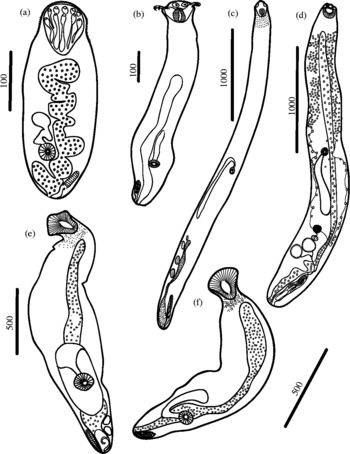
Bucephalus sp. 2 (fig. 2a)
Description
Based on 13 individuals: five from Abudefduf septemfasciatus, five from Abudefduf sordidus (one sent to USNPC, Nos 104850, 104851 and 104852) and three from Acanthurus triostegus. Cyst oval, 190–290 (215 ± 29) long, 120–200 (146 ± 25) wide. Body shape ellipsoid, total length 300–445 (385 ± 47), maximum width 90–152 (118 ± 23). Width as % of body length: 24–39% (31 ± 5%). Tegument with fine spines. Rhynchus sucker-like, 55–100 long, 47–80 width (69 ± 11 long, 68 ± 9 width) with 7–10 tentacular appendages. Rhynchus length as % of body length: 14–23 (18 ± 2%). Mouth 155–345 (271 ± 54) from anterior end. Pre-mouth distance as % of body length: 40–81% (71 ± 12%). Pharynx 20–37 (28 ± 5) in diameter. Intestine saccular, 25–208 long (98 ± 61 long), oriented anteriorly. Testis primordia smooth. Post-testicular distance 0–34 (11 ± 30). Post-testicular distance as % of body length: 0–28% (2 ± 8%). Cirrus sac 55–107 (74 ± 15) long, 10–25 (16 ± 5) width. Anterior-most reach of cirrus sac as % of body length: 15–26% (18 ± 7%). Excretory vesicle elongate, saccular, with undulating margins, 195–342 (267 ± 64 long).
Excysted metacercariae from lagoon-flat fishes of Palmyra Atoll (all worms in ventral view). (a) Bucephalus sp. 2; (b) Bucephalus sp. 3; (c) Rhipidocotyle sp. 1; (d) Rhipidocotyle sp. 2; (e) Rhipidocotyle sp. 3; (f) Prosorhynchus sp. All scale bars are in micrometres.

Hosts
Hemiramphidae: Hyporhamphus affinis (muscle); Apogonidae: Cheilodipterus quinquelineatus (heart, muscle); Carangidae: Carangoides ferdau (Forsskål, 1775) (fins), Caranx ignobilis (intestine), Caranx papuensis (heart, intestine); Lutjanidae: Lutjanus monostigma (Cuvier, 1828) (heart, fins); Mullidae: Mulloidichthys vanicolensis (heart, eye); Kyphosidae: Kyphosus cinerascens (Forsskål, 1775) (fins); Chaetodontidae: Chaetodon auriga (heart), Chaetodon lunula (heart); Pomacentridae: Abudefduf septemfasciatus (heart), Abudefduf sordidus (heart, gills), Pomacentrus adelus (heart, kidney), Stegastes nigricans (eye, fins, gills, heart, kidney); Mugilidae: Crenimugil crenilabris (heart), Liza vaigiensis (eye, fins, gills, heart, intestine, muscle, stomach), Valamugil engeli (body cavity, brain, heart, muscle, kidney); Gobiidae: Amblygobius phalaena (tissue surrounding eye), Istigobius ornatus (Rüppell, 1830) (eye, gills, muscle), Valenciennea sexgutatta (gills, muscle); Acanthuridae: Acanthurus triostegus (fins, gills, heart, kidney, liver), Acanthurus xanthopterus (fins); Balistidae: Rhinecanthus aculeatus (fins, tissue surrounding eye); Tetraodontidae: Arothron hispidus (fins, heart).
Comments
See comments for Bucephalus sp. 1.
Bucephalus sp. 3 (fig. 2b)
Description
Based on five individuals (one sent to USNPC, No. 104853) from Acanthurus triostegus. Cyst fragile, oval, 217 long, 110 wide (n = 2). Body shape ellipsoid, total length 325–680 (498 ± 176), maximum width 117–190 (145 ± 33). Width as % of body length: 17–49% (33 ± 14). Tegument with fine spines. Rhynchus funnel-shape, 62–95 (74 ± 13) long, 65–77 (70 ± 5) width, with seven tentacular appendages. Rhynchus length as % of body length: 10–21% (16 ± 5). Mouth in last fifth of body, 240–558 (446 ± 132) from anterior end. Pre-mouth distance as % of body length: 69–87% (77 ± 7). Pharynx 27–42 (34 ± 7) in diameter. Intestine saccular, 108–150 (129 ± 24) long, oriented anteriorly. Excretory vesicle elongate, sac-like, 198 long.
Hosts
Carangidae: Caranx papuensis, (stomach); Kyphosidae: Kyphosus cinerascens (heart); Pomacentridae: Abudefduf sordidus (heart, kidney), Pomacentrus adelus (eye, muscle), Stegastes nigricans (eye, kidney, liver, muscle); Mugilidae: Crenimugil crenilabris (fins, heart, kidney, muscle), Valamugil engeli (muscle); Gobiidae: Amblygobius phalaena (eye, muscle), Valenciennea sexguttata (muscle); Acanthuridae: Acanthurus triostegus (gills), Acanthurus xanthopterus (fins); Tetraodontidae: Arothron hispidus (fins).
Comments
This is the largest metacercaria of the three members of Bucephalus in the present study. However, only a few specimens were recovered. Additional comments as for Bucephalus sp. 1.
Rhipidocotyle sp. 1 (fig. 2c)
Description
Based on ten individuals; three from Hyporhamphus affinis, one from Crenimugil crenilabris (USNPC, No. 104854) and six from Liza vaigiensis. Cyst elliptical, 400–625 long, 175–273 wide. Body shape cylindrical, total length 2380–3630 (2843 ± 387), maximum width 210–510 (320 ± 83). Width as % of body length: 6–18% (11 ± 3%). Tegument with fine spines. Rhynchus funnel shaped, 90–160 long (133 ± 24), 100–170 width (130 ± 26) with a crown of five lobes. Rhynchus length as % of body length: 4–6% (5 ± 1%). Mouth opening 890–1890 (1275 ± 291) from anterior end. Pre-mouth distance as % of body length: 33–52% (45 ± 7%). Pharynx 40–80 (57 ± 12) in diameter. Intestine tubular 240–810 (571 ± 176), oriented posteriorly. Pars prostatica slightly bent. Post-testicular distance 270–530 (412 ± 85). Post-testicular distance as % of body length: 10–19% (15 ± 3%). Cirrus sac 190–370 long (283 ± 53), 40–80 width (55 ± 13). Anterior-most reach of cirrus sac as % of body length: 7–13% (10 ± 2%). Ovary pre-testicular (not measured). Vitelline glands two bands along the body sides anterior to ovary.
Hosts
Hemiramphidae: Hemiramphus lutkei (body cavity, brain, kidney, muscle, tissue surrounding brain), Hyporhamphus affinis (fins, spleen, tissue surrounding eye and intestine); Belonidae: Platybelone argalus (Lesueur, 1821) (kidney); Carangidae: Caranx ignobilis (intestine), Caranx papuensis (fins, intestine); Lutjanidae: Lutjanus fulvus (spleen, body cavity); Mullidae: Mulloidichthys vanicolensis (heart, kidney); Kyphosidae: Kyphosus cinerascens (fins, heart, kidney); Pomacentridae: Abudefduf sordidus (fins); Mugilidae: Crenimugil crenilabris (fins, gills, muscle, heart, kidney, tissue surrounding brain and eye), Liza vaigiensis (heart, kidney, tissue surrounding brain and intestine, liver, body cavity); Acanthuridae: Acanthurus xanthopterus (fins); Balistidae: Rhinecanthus aculeatus (tissue surrounding eye); Tetraodontidae: Arothron hispidus (fins).
Comments
These metacercariae belong to the genus Rhipidocotyle Diesing, 1858 because they had a rhynchus with a crown of five lobes, a pars prostatica slightly bent, and a pre-testicular ovary, following the generic definition of Overstreet & Curran (Reference Overstreet and Curran2002). This metacercaria is different than Riphidocotyle sp. 2 in being significantly larger in total body length (Student's t 0.05,16 test: 8.54, P < 0.001), by having a rhynchus with a pentagonal hood and by having a smaller rhynchus length to body length ratio (Student's t 0.05,16 test: − 3.65, P < 0.002). These metacercariae could belong to one of the species of Rhipidocotyle already described in the CIP or EIP marine ecoregions and include: Rhipidocotyle khalili Nagaty, 1937 from Sphyraena sp. from Makassar, India (Yamaguti, Reference Yamaguti1953); R. eggletoni Velasquez, Reference Velasquez1959 in Sillago sihama (Forsskål, 1775) and R. laruei Velasquez, Reference Velasquez1959 from Psettodes erumei (Bloch & Schneider, 1801) from the Philippines (Velasquez, Reference Velasquez1959); R. labrodei Jones, Grutter & Cribb, 2003 from Labroides dimidiatus (Valenciennes, 1839) from Lizard Island, Australia (Jones et al., Reference Jones, Grutter and Cribb2003); R. danai Bray & Palm, Reference Bray and Palm2009 from Thyrsitoides marleyi Fowler, 1929 and R. jayai Bray & Palm, Reference Bray and Palm2009 from Johnius macropterus (Bleeker, 1853) from Java, Indonesia (Bray & Palm, Reference Bray and Palm2009).
Rhipidocotyle sp. 2 (fig. 2d)
Description
Based on seven individuals; five from Hemiramphus lutkei, one from Crenimugil crenilabris and one from Liza vaigiensis (USNPC No. 104855). Cyst oval, 520–680 length (617 ± 85), 150–420 width (313 ± 144). Body shape cylindrical, total length 890–1860 (1410 ± 305), maximum width 170–490 (281 ± 107). Width as % of body length: 10–30% (20 ± 7%). Tegument with fine spines. Rhynchus sucker-like, 60–200 long (116 ± 51 long), 70–240 wide (124 ± 59) with 3–4 rounded lobes on the anterior end. Rhynchus length as % of body length: 5–12% (8 ± 3%). Mouth opening 410–1370 (737 ± 247) from anterior end. Pre-mouth distance as % of body length: 43–87% (57 ± 17%). Pharynx 40–120 (59 ± 28) in diameter. Intestine tubular, 150–880 long (313 ± 263), oriented posteriorly. Vitelline glands two bands along body sides anterior to ovary. Post-testicular distance 40–370 (196 ± 113). Post–testicular distance as % of body length: 2–24% (14 ± 7%). Cirrus sac 60–190 long (139 ± 45), 30–90 width (47 ± 21). Anterior-most reach of cirrus sac as % of body length: 5–14% (10 ± 4%). Excretory vesicle 1170–1350 (n = 2) length.
Hosts
Hemiramphidae: Hemiramphus lutkei (fins), Hyporhamphus affinis (gills, heart, liver); Belonidae: Platybelone argalus (kidney, water); Apogonidae: Cheilodipterus quinquelineatus (eye); Carangidae: Caranx ignobilis (intestine), Caranx papuensis (intestine, stomach); Lutjanidae: Lutjanus monostigma (fins); Mullidae: Mulloidichthys vanicolensis (heart, eye); Pomacentridae: Abudefduf septemfasciatus (gills), Abudefduf sordidus (fins, kidney, muscle); Mugilidae: Crenimugil crenilabris (fins, heart, kidney, muscle), Liza vaigiensis (muscle, heart, kidney, tissue surrounding brain, body cavity, intestine), Valamugil engeli (tissue surrounding brain, heart); Gobiidae: Amblygobius phalaena (muscle); Acanthuridae: Acanthurus triostegus (fins, gills), Acanthurus xanthopterus (fins); Balistidae: Rhinecanthus aculeatus (heart); Tetraodontidae: Arothron hispidus (fins).
Comments
See comments for Rhipidocotyle sp. 1.
Rhipidocotyle sp. 3 (fig. 2e)
Description
Based on four individuals; one from Mulloidichthys vanicolensis (USNPC No. 104856) and three from Acanthurus xanthopterus. Cyst oval, 480–600 length, 187–320 width. Body shape cylindrical, total length 610–820 (712 ± 87), maximum width 215–325 (295 ± 53). Width as % of body length: 35–47% (41 ± 5%). Tegument with fine spines. Rhynchus sucker-like, 127–182 long (152 ± 23), 132–160 wide (151 ± 13), with a long tranverse bump on anterior end. Rhynchus length as % of body length: 21–22 (21 ± 1%). Mouth 497–652 (563 ± 65) from anterior end. Pre-mouth distance as % of body length: 75–81% (79 ± 3%). Pharynx 62–77 (69 ± 7) in diameter. Intestine tubular, 277–545 (422 ± 114), oriented posteriorly. Ovary pre-testicular, 10–25 long (n = 2), 5–12 wide (n = 2). Testis smooth, 25–37 long (32 ± 7), 12–22 (15 ± 3) wide. Post-testicular distance 57–62 (60 ± 3). Post-testicular distance as % of body length: 7–9% (8 ± 1%). Cirrus sac 95–120 long (105 ± 13), 27–35 (32 ± 4) width. Anterior-most reach of cirrus sac as % of body length: 12–20 (15 ± 4%). Excretory vesicle 477–615 long (532 ± 72).
Hosts
Hemiramphidae: Hyporhamphus affinis (gills); Carangidae: Caranx papuensis (fins); Mullidae: Mulloidichthys vanicolensis (fins, heart, kidney, muscle); Mugilidae: Crenimugil crenilabris (kidney), Valamugil engeli (heart, kidney); Gobiidae: Valenciennea sexgutatta (muscle); Acanthuridae: Acanthurus triostegus (body cavity, fins, heart, kidney, muscle), Acanthurus xanthopterus (fins); Balistidae: Rhinecanthus aculeatus (muscle); Tetraodontidae: Arothron hispidus (fins, heart).
Comments
This species is smaller than Rhipidocotyle spp. 1 and 2. Additional comments as for Rhipidocotyle sp. 1.
Prosorhynchus sp. (fig. 2f)
Description
Based on four individuals from Acanthurus xanthopterus (one sent to USNPC, No. 104857). Cysts oval to elliptical, 270–500 (378 ± 105; n = 5) long, 190–200 (197 ± 6; n = 3) wide. Total body length 800–1475 (1184 ± 317), maximum width 210–300 (271 ± 42). Width as % of body length: 20–29% (24 ± 4). Tegument with fine spines. Rhynchus funnel-shaped, 100–180 (144 ± 33) long, 150–185 (174 ± 16) wide. Rhynchus length as % of body length: 7–19% (13 ± 5%). Mouth 620–1063 (883 ± 217) from anterior end. Pre-mouth distance as % of body length: 72–77% (75 ± 2%). Pharynx 68–103 (80 ± 16) in diameter. Intestine tubular, 270–490 (380 ± 156) long, oriented posteriorly. Testes smooth, 40–82 (61 ± 30) long, 30–52 (41 ± 16) wide. Post-testicular distance 50–112 (n = 2). Post-testicular distance as % of body length: 3–8% (n = 2). Cirrus sac 110–122 (n = 2) long, 20–40 (n = 2) wide. Seminal duct coiled. Pars prostatica straight. Anterior-most reach of cirrus sac as % of body length: 8% (n = 2). Excretory vesicle 1287–1340 long.
Host
Mullidae: Mulloidichthys vanicolensis (muscle); Chaetodontidae: Chaetodon lunula (heart); Pomacentridae: Abudefduf septemfasciatus (heart); Mugilidae: Valamugil engeli (kidney, muscle); Acanthuridae: Acanthurus triostegus (eye, fins), Acanthurus xanthopterus (fins).
Comments
These metacercariae were considered to belong to the genus Prosorhynchus because the rhynchus is not in the form of a sucker and the testes are diagonal rather than in tandem. These generic characteristics agree with those described by Overstreet & Curran (Reference Overstreet and Curran2002). This metacercaria could belong to one of the several species described for the CIP or EIP marine ecoregions: Prosorhynchus freitasi Nagaty, 1937 of Epinephelus sp. and Plectropomus maculatus (Bloch, 1790) from Heron Island and New Caledonia (Manter, Reference Manter1953); P. thapari Manter, Reference Manter1953 of Plectropomus maculatus (Bloch, 1790) from Fiji Island (Manter, Reference Manter1953); P. crucibulus (Rudolphi, 1819) Odhner, 1905 of Acanthopagrus berda (Forsskål, 1775), P. paracrucibulus Velasquez, Reference Velasquez1959 of Ambassis buruensis Bleeker, 1856, P. luzonicus Velasquez, Reference Velasquez1959 of Lates calcarifer (Bloch, 1790), P. longus Velasquez, Reference Velasquez1959 of Psettodes erumei, (Bloch et Schneider, 1801), all from the Philippines (Velasquez, Reference Velasquez1959); P. longissacatus Durio et Manter, 1968 of an unidentified serranid known locally as ‘leche’ from New Caledonia (Durio & Manter, Reference Durio and Manter1968); P. serrani Durio et Manter, 1968 of Variola louti (Forsskål, 1775) from New Caledonia (Durio & Manter, Reference Durio and Manter1968); P. maternus Bray et Justine, 2006 of E. malabaricus (Bloch & Schneider, 1801) from New Caledonia (Bray & Justine, Reference Bray and Justine2006); P. robertsthomsoni Bott et Cribb, 2009 from Cephalopholis argus Schneider, 1801, C. cyanostigma (Valenciennes, 1828) and C. miniata (Forsskål, 1775), P. lafii Bott et Cribb, 2009 of Epinephelus fuscoguttatus (Forsskål, 1775), P. conorjonesi Bott et Cribb, 2009 of Cromileptes altivelis (Valenciennes, 1828), P. jexi Bott et Cribb, 2009 of E. quoyanus (Valenciennes, 1830), P. milleri Bott et Cribb, 2009 of V. louti, all from Great Barrier Reef, Australia (Bott & Cribb, Reference Bott and Cribb2009).
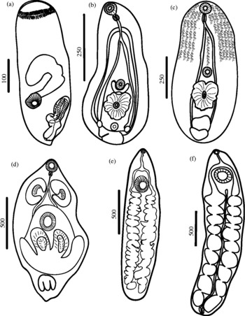
Dollfustrema sp. (fig. 3a)
Description
Based on seven individuals; three from Mulloidichthys vanicolensis, and four (one sent to USNPC, No. 104858) Acanthurus xanthopterus. Body ellipsoidal, 188–450 (288 ± 96) long, 63–130 (106 ± 30) wide. Width as % of body length: 29–45% (38 ± 6%). Tegument with fine spines. Rhynchus a shallow funnel topped with two circlets of short spines 30–57 (48 ± 10) long, 40–95 (70 ± 19) wide. Rhynchus length as % of body length: 12–24 (17 ± 4). Mouth 138–310 (218 ± 67) from anterior end. Pre-mouth distance as % of body length: 67–75% (72 ± 3%). Pharynx 25–42 (31 ± 6) in diameter. Intestine sac-like, 55–155 (101 ± 49), oriented anteriorly. Cirrus sac 128 long (n = 1), 23 width (n = 1). Anterior-most reach of cirrus sac as % of body length: 9%. Excretory vesicle 73–233 (169 ± 62) long.
Excysted metacercariae recovered from lagoon-flat fishes of Palmyra Atoll (all worms in ventral view). (a) Dollfustrema sp.; (b), Mesostephanus sp.; (c) Cyathocotylidae gen. sp.; (d) Cardiocephaloides sp.; (e) Monolicaecum-type; (f) Torticaecum-type. All scale bars are in micrometres.

Hosts
Mullidae: Mulloidichthys vanicolensis (fins); Chaetodontidae: Chaetodon lunula (fins); Pomacentridae: Abudefduf septemfasciatus (eye, fins), Abudefduf sordidus (fins), Pomacentrus adelus (fins); Mugilidae: Crenimugil crenilabris (kidney); Gobiidae: Amblygobius phalaena (liver), Valenciennea sexguttata (muscle); Acanthuridae: Acanthurus triostegus (fins, gills, tissue surrounding eye), Acanthurus xanthopterus (fins); Tetraodontidae: Arothron hispidus (fins).
Comments
The recovered material of Dollfustrema had two circlets of spines around the rhynchus. This characteristic and those of the terminal genitalia correspond well with the generic description provided by Overstreet & Curran (Reference Overstreet and Curran2002). Two species of Dollfustrema Eckmann, 1934 (Dollfustrema bipapillosum Manter et Pritchard, 1961 of Gymnothorax petelli (Bleeker, 1856) and D. strombyrhynchum Manter et Pritchard, 1961 of G. petelli) have been described from Hawaii by Manter & Pritchard (Reference Manter and Pritchard1961). Recently, Nolan & Cribb (Reference Nolan and Cribb2010) described Dollfustrema gibsoni from Gymnothorax woodwardi McCulloch, 1912 from southwest Western Australia. Due to the geographical proximity, any of the species mentioned above could be the adult of the Dollfustrema sp.
Family Cyathocotylidae
Mesostephanus sp. (fig. 3b)
Description
Based on five flattened individuals (one sent to USNPC, No. 104859) from Crenimugil crenilabris. Body pyriform 290–540 (433 ± 127) long and 170–340 (233 ± 65) wide. Tegumental spines not observed. ‘Handle’ of hind-body completely undifferentiated. Oral sucker 35–52 (44 ± 7) long and 40–52 (46 ± 7) wide. Ventral sucker probably non-functional, 27–37 (33 ± 4) long and 14–39 (28 ± 12) wide. Pharynx 17–32 (26 ± 6) long and 17–25 (21 ± 3) wide. Prepharynx absent; oesophagus narrow, 35–70 (47 ± 17) long. Caeca narrow, extends into hind-body. Tribocytic organ fully developed, 70–113 (93 ± 21) long, 67–113 (84 ± 21) wide. Genital rudiment a prominent mass of cells dorso-posterior to tribocytic organ. Excretory system ascending parallel to caeca, forming an extracaecal ring. Excretory pore subterminal.
Hosts
Belonidae: Platybelone argalus (muscle); Mugilidae: Crenimugil crenilabris (brain, heart, muscle), Valamugil engeli, (muscle); Sphyraenidae: Sphyraena barracuda (Edwards, 1771) (gills); Acanthuridae: Acanthurus triostegus (fins).
Comments
These metacercariae were identified as Mesostephanus Lutz, Reference Lutz1935 due to the presence of a spherical oral sucker, a ventral sucker slightly pre-equatorial and smaller than the oral sucker, the voluminous tribocytic organ and the genital primordial excretory vesicle I-shaped. Four species of Mesostephanus have been described for the region: M. haliasturis Tubangui et Masilungan, 1941 for Haliastur indus intermedius Blyth, 1865 from the Philippines (Tubangui & Masilungan, 1941); M. neophocae Dubois et Angel, 1976 for Neophoca cinerea (Péron, 1816) and Mirounga leonina (Linnaeus, 1758) from Australia (Dubois & Angel, Reference Dubois and Angel1976); M. haliasturis and M. scottae Cribb, Barker et Beuret, 1995 for Phalacrocorax sulcirostris (Brandt, 1837) and P. varius (Gmelin, 1789), and M. pelecani Cribb, Barker et Beuret, 1995 for Pelecanus conspicillatus Temminck, 1824 from Queensland, Australia (Cribb et al., Reference Cribb, Barker and Beuret1995). The metacercariae recovered from C. crenilabris could belong to any of these species. Lutz (Reference Lutz1935) reported the presence of Mesostephanus from the booby Sula brasiliensis Spix, 1825 from Brazil. Several species of boobies, such as the brown boobies (Sula leucogaster (Boddaert, 1783)), nest at Palmyra Atoll and could serve as potential definitive hosts.
Cyathocotylidae gen. sp. (fig. 3c)
Description
Based on seven slightly flattened individuals; one (USNPC No. 104860) from Hemiramphus lutkei and six from Hyporhamphus affinis. Body foliaceous to elliptical, slightly folded dorsally at its anterior end; length 680–1080 (877 ± 147), maximum width 320–525 (425 ± 71) at posterior quarter of body. Tegument smooth with granular pigments on anterior part of body, pre-acetabularly. Oral sucker subterminal, long 49–72 (65 ± 8), width 52–72 (62 ± 8). Acetabulum postequatorial, 46–65 long (58 ± 6) and 42–74 width (59 ± 10). Prepharynx absent; pharynx small, elliptical 35–47 long (41 ± 5) and 25–35 (30 ± 4) wide. Oesophagus 50–122 long (76 ± 30). Caeca narrow, close to middle ventral line of body, extending to posterior end. Tribocytic organ fully developed. Developing testes median, in tandem at posterior end, overlapping one to each other, globular. Anterior testis 67–125 long (92 ± 24) and 110–185 (149 ± 28). Posterior testis 52–110 long (84 ± 24) and 112–180 (143 ± 34) wide. Excretory pore subterminal.
Hosts
Hemiramphidae: Hemiramphus lutkei (muscle), Hyporhamphus affinis (eye, fins, muscle); Belonidae: Platybelone argalus (eye, muscle); Mugilidae: Crenimugil crenilabris (muscle), Valamugil engeli (body cavity, heart, kidney, intestine, muscle); Acanthuridae: Acanthurus triostegus (eye, fins, muscle).
Comments
These metacercariae were morphologically different from Mesostephanus due to the foliaceous body shape, granular pigment on anterior part of body, very narrow caeca, and primordia of testes in tandem posterior to tribocytic organ. Based on its general morphology, it might be a cyathocotylid such as Cyathocotyle Mühling, 1896 Holostephanus Szidat, 1936 or Paracoenogonimus Katsurada, 1914. The first intermediate and definitive hosts may be similar to those of Mesostephanus sp.
Family Strigeidae Railliet, 1919
Cardiocephaloides sp. (fig. 3d)
Description
Based on two flattened individuals (one sent to USNPC, No. 104861) from Crenimugil crenilabris. Body oval, 925–1038 long, maximum width at middle 850–1025. Short anterior part of body 140–150 long and 210–240 wide, bearing oral sucker separated by transverse tegumental fold from rest of body. Middle part of body 810–980 long and 1010–1200 wide, with broad longitudinal lateral fields of darker tissue on each side. Posterior part of body 190–270 long and 330–420 wide. Oral sucker subterminal, measuring 110 long and 100–140 wide. Acetabulum situated approximately at middle body, 110–180 long by 160–180 wide. Two well-developed pseudosuckers present about one-quarter into the body, each lateral to gut, centred at level of oesophagus bifurcation. Pharynx 60–80 in diameter. Oesophagus 140–160 long, bifurcating at middle level of pseudosuckers into two caeca ending near caudal end of body. Large, transverse holdfast situated at end of middle part of body, measuring 270–460 long and 510–760 wide. Posterior part of body provided with anlagen of sexual glands and subterminal excretory pore.
Hosts
Mullidae: Mulloidichthys vanicolensis (brain); Kyphosidae: Kyphosus cinerascens (brain); Chaetodontidae: Chaetodon lunula (brain); Pomacentridae: Abudefduf sordidus (brain); Mugilidae: Crenimugil crenilabris (brain, eye, fins, muscle, kidney), Liza vaigiensis (brain); Acanthuridae: Acanthurus triostegus (muscle), Acanthurus xanthopterus (brain); Balistidae: Rhinecanthus aculeatus (brain).
Comments
These metacercariae were originally described in the genus Cardiocephalus Szidat, 1928, and later transferred to the genus Cardiocephaloides Sudarikov, 1959 (Niewiadomska, Reference Niewiadomska2002). These metacercariae were assigned to Cardiocephaloides due to the presence of two pseudosuckers posterolateral to the small pharynx and the large bilobed tribocytic organ posterior to the acetabulum. As metacercariae, these digeneans infect marine fishes from the Sparidae and Scombresocidae families (Niewiadomska, Reference Niewiadomska2002). Several other families of fish at Palmyra Atoll were infected with these metacercariae. The adults of Cardiocephaloides infect marine birds (Lariidae, Spheniscidae and Procellaridae) all over the world (Díaz et al., Reference Diaz, Cremonte and Navone2010), and the cercariae develop in molluscs (Prosobranchia: Nassidae) (Niewiadomska, Reference Niewiadomska2002). Cardiocephaloides musculosus (Johnston, 1904) is the only species that has been recorded as an adult from the crested tern (Thalasseus bergii (Lichtenstein, 1823)) from Australia (Johnston, Reference Johnston1942) and the EIP.
Family Didymozoidae
‘Monolicaecum-type’ (fig. 3e)
Description
Based on one specimen (USNPC 104862) from Amblygobius phalaena. Body unarmed, elongate, 1712 by 440. Parenchyma vesicular. No eye spots or eye-spot pigment. Mouth terminal. Oral sucker 42–62, within body. Acetabulum 182–185, muscular, embedded in body parenchyma, at level of anterior body fourth. Sucker length ratio 1:2.9. Pharynx absent. Oesophagus 155 long, narrow, bifurcation posterior to mid-forebody, 205 from the anterior end. ‘Stomach’ just perceptible. Caeca with narrow and inflated areas, descending in undulating fashion, terminating 77 from posterior extremity. Parenchymal glands fill entire body in zone beneath thin subcuticular longitudinal muscle layer. No reproductive structures. Excretory vesicle postcaecal.
Hosts
Mullidae: Mulloidichthys flavolineatus (fins, muscle); Mugilidae: Liza vaigiensis (intestinal wall); Gobiidae: Amblygobius phalaena (tissue surrounding eye).
Comments
‘Metacercariae’ of Didymozoidae are commonly found in marine fishes as third intermediate or paratenic hosts from the Indo-Pacific region (Køie & Lester, Reference Køie and Lester1985; Cribb et al., Reference Cribb, Pichelina, Dufourb, Bray, Chauvet, Faliexe, Galzinb, Lob, Lo-Yat, Morand, Rigby and Sasal2000). Due to the fact that this metacercaria apparently has a stomach and lacks gland-cells around the oesophagus, it was tentatively recognized as a ‘Monolicaecum-type’ juvenile (Yamaguti, Reference Yamaguti1970; Pozdnyakov & Gibson, Reference Pozdnyakov and Gibson2008). However, due to the lack of development, it was not possible to identify this larva at the genus or species level. In fact, due to the strong morphological changes undergone by didymozoids from juvenile to adult stages, molecular tools or experimental infections are needed for identification. Potential definitive hosts for these metacercariae are marine fishes such Thunnus albacares (Bonnaterre, 1788) and T. obesus (Lowe, 1839) from which Yamaguti (Reference Yamaguti1970) described 14 species of didymozoids from Hawaii. Cribb et al. (Reference Cribb, Pichelina, Dufourb, Bray, Chauvet, Faliexe, Galzinb, Lob, Lo-Yat, Morand, Rigby and Sasal2000) also described adult stages of didymozoids from pelagic fishes of the subfamily Thunninae (Scombridae) for the CIP marine ecoregion. The life cycles of didymozoids in the CIP and EIP are not known. However, Cribb et al. (Reference Cribb, Pichelina, Dufourb, Bray, Chauvet, Faliexe, Galzinb, Lob, Lo-Yat, Morand, Rigby and Sasal2000) presume that most of these life cycles should be pelagic, including molluscs such as heteropods and pteropods, and coral reef fishes such as Lethrinidae and Serranidae as third intermediate hosts.
‘Torticaecum-type’ (fig. 3f)
Description
Based on two broken specimens (one sent to USNPC, No. 104863) from Hemiramphus lutkei). Body unarmed, filiform, 2850–3450 long by 150–180 wide. No eye spots or eye-spot pigment. Mouth terminal. Oral sucker 40–42 by 32–37, within body. Acetabulum 40–76 by 41–67, muscular, embedded in body parenchyma, at level of anterior body fourth. Sucker length ratio 1:1.28–1.81. Pharynx absent. Oesophagus long, narrow, bifurcation posterior to mid-forebody 205 from the anterior end. ‘Stomach’ absent. Caeca with narrow and inflated areas, descending in undulating fashion, terminating 67–132 from posterior extremity. Parenchymal glands fill entire body in zone beneath thin subcuticular longitudinal muscle layer. No reproductive structures. Excretory vesicle postcaecal.
Host
Hemiramphidae: Hemiramphus lutkei (fins); Carangidae: Carangoides orthogrammus (Jordan & Gilbert, 1882) (muscle, water in holding bag); Lutjanidae: Lutjanus monostigma (fins); Mullidae: Upeneus arge Cuvier, 1829 (water in holding bag, scales).
Comments
This ‘metacercaria’ was tentatively recognized as belonging to the ‘Torticaecum-type’ given the absence of stomach, absence of pharynx and lack of gland-cells around oesophagus. See additional comments for ‘Monolicaecum-type’ sp.
Discussion
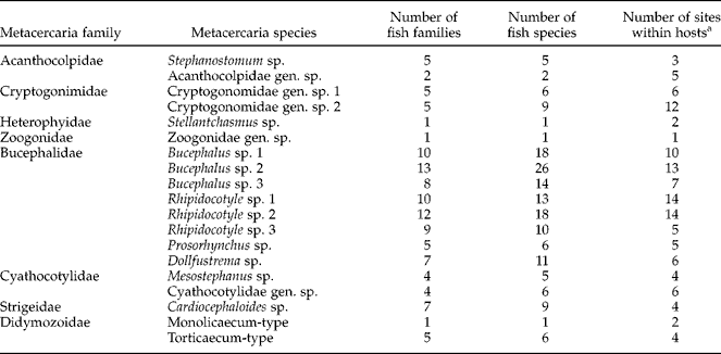
Up to 425 individual host fish were surveyed belonging to 42 species and 33,964 metacercariae of 19 digenean species of 8 families were recovered. Three digenean families were particularly common in the metacercaria assemblage: Bucephalidae, Cryptogonimidae and Cyathocotylidae (table 1). Bucephalid species appear to have the broadest specificity, infecting up to 13 host families, 26 host species, and 14 sites within hosts (tables 1 and 2). Four other digenean families appeared less frequently (Acanthocolpidae, Heterophyidae, Strigeidae and Didymozoidae). All these families are a subset of those previously detected as adults or metacercariae throughout the CIP or EIP marine ecoregions (e.g. Yamaguti, Reference Yamaguti1970; Cribb et al., Reference Cribb, Bray, Barker, Adlard and Anderson1994). Metacercariae in the abundant and less-abundant families originate from first intermediate host infections in bivalves or snails (Yamaguti, Reference Yamaguti1975), both of which are common on Palmyra's lagoon flats (unpublished observations). Trematode species in the common and rarer families use fishes and birds as final hosts (Yamaguti, Reference Yamaguti1975), indicating the role of both fishes and birds as predators on fish at Palmyra.
Metacercaria species generality in terms of number of host families, species and sites within hosts used.
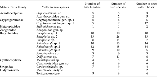
a ‘Sites within hosts’ provides a crude indication of variability in tissue/organ site use, which may vary within or among host species. See metacercaria species descriptions for detailed information.
A few surveys have examined metacercariae infecting marine fishes in the Central and East Indo-Pacific ecoregions (Yamaguti, Reference Yamaguti1970; Køie & Lester, Reference Køie and Lester1985; Cribb, Reference Cribb1998 and references therein). Our study differed from these previous surveys in several respects, and this perspective frames the rest of our discussion.
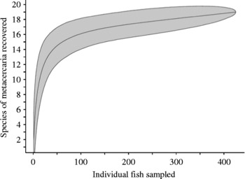
First, the fish assemblage was broadly surveyed, aiming to examine all the potential host species of the lagoon flats. Most other studies have focused on particular host taxa, or have obtained hosts haphazardly (e.g. via sporadic sampling at fish markets). We have thus far sampled 42 of 47 recorded fish species (unpublished data) from Palmyra's lagoon flats. Sampling a major portion of the local fish diversity helps to establish an accurate baseline expectation for the diversity and composition of the parasite community occurring in the entire ecosystem. In fact, a randomized species accumulation curve suggests that we have encountered most of the metacercaria species of the Palmyra Lagoon flats (fig. 4).
A randomized species-accumulation curve (Gotelli & Colwell, Reference Gotelli and Colwell2001) for the metacercaria species detected with varying number of individual hosts processed. The curve represents the average of 10,000 randomizations of the 425 fish in our data set, counting for each randomization the number of encountered metacercaria species for each number of dissected host individuals. The shaded region represents the 95% confidence region, calculated using the standard error and t-distribution. The initial rapid approach to the asymptote indicates that our sampling has captured most of the lagoon-flat metacercaria diversity.

Second, each individual fish was thoroughly and systematically processed, squashing different tissues to count and identify individual metacercariae. This processing facilitated the detection of rare trematode species within individual hosts. This intensive, within-host effort combined with our broad sampling of the fish community to further help to capture adequately the metacercaria diversity of the lagoon flats.
Third, the parasitological survey was conducted on fishes from a largely unexplored habitat: the lagoon flats. Previous efforts in the CIP and EIP marine ecoregions have been restricted to fore-reef habitats. To our knowledge only one of these fore-reef surveys (Lafferty et al., Reference Lafferty, Shaw and Kuris2008) processed individual fish in a manner similar to our own. Lafferty et al. (Reference Lafferty, Shaw and Kuris2008) sampled five fish species from the fore-reef at Palmyra Atoll. From the 110 individuals they processed, they recovered only a single morphospecies of metacercaria. If this represents fore-reef metacercaria diversity, then it is low compared to the 19 species that we documented on the sand flats. Although Lafferty et al. did not broadly sample the fore-reef fish assemblage, data from the present study were re-sampled to make sampling efforts more comparable. Ten thousand random draws (without replacement) of 110 individual fish (matching the number of individuals from Lafferty et al.) from our lagoon-flat data set recovered a minimum of 13 metacercaria species, with a mean ± SD of 16.0 ± 0.9. Additionally, controlling for the number of species examined by randomly sampling 11 individuals (Lafferty et al.'s minimum per species sample size) from 5 of the 14 fish species in our data set with adequate sample sizes, we recovered a minimum of four metacercaria species, with a mean ± SD of 13.0 ± 1.7. Further supporting the hypothesis that fore-reef fish may harbour lower metacercaria diversity, Lafferty et al. (Reference Lafferty, Shaw and Kuris2008) also only encountered a single metacercaria species during their sampling of the fore reef of a nearby Atoll (Christmas Island).
Why might lagoon flats have relatively high diversity and abundance of metacercariae? Compared to the fore reef, the lagoon flats are characterized by shallow, relatively still waters and high densities of probable first intermediate host bivalves and snails (unpublished data). These factors may contribute to greater infectious cercaria densities and higher transmission rates to second intermediate host fishes on the lagoon flats compared to the fore reef.
Another unique attribute of the present study is that it took place in a remote, small, relatively undisturbed atoll. As a trophically intact system, Palmyra Atoll may help establish baseline expectations for undisturbed community structure of both hosts and parasites. This may be particularly useful for efforts to develop parasites as ecological indicators of free-living diversity. For instance, trematode parasites in easily sampled first intermediate hosts can indicate more difficult to sample free-living invertebrates, fishes, birds and mammals (Huspeni et al., Reference Huspeni, Hechinger and Lafferty2005; Lafferty & Dunham, Reference Lafferty and Dunham2005; Hechinger et al., Reference Hechinger, Lafferty, Huspeni, Brooks and Kuris2007). The host specificity documented herein can inform future efforts to use trematode assemblages in first intermediate hosts to indicate surrounding free-living diversity in the CIP and EIP marine ecoregions.
The low variability of the morphological measures used to differentiate metacercariae suggests that each taxon identified is a discrete species. However, cryptic species may, of course, occur within any of our delineated species. This may be most likely for those metacercariae with few developed morphological characters (e.g. the two didymozoids). Further cryptic diversity may be associated with those species characterized by very discrete and disjunct tissue or host use. The descriptions of morphology, and tissue and host use for these metacercariae should help guide future taxonomic research on these trematodes.
In conclusion, metacercariae appear to be abundant and diverse in Palmyra lagoon-flat fishes. The descriptions provided here will facilitate future ecological work by providing names and descriptions. The descriptions and information on host use may also help guide future life cycle and taxonomic research focused on EIP trematodes. Additionally, we hope that the diversity and abundance of metacercariae documented here highlight their importance and the need to include them in future studies of fish–helminth interactions or of the role of parasites in ecosystems.
Acknowledgements
The authors thank The Nature Conservancy staff for their support of research and are particularly indebted to Amanda Meyer (United States Fish and Wildlife Service), Franklin Viola, Brad Kintz, Aaron Kierzek, Jan Eber, Anthony Wilson and Lynette Williams. Gareth Williams and Ingrid Knapp shared their field knowledge. Dr Tomáš Scholz and Dr William Cole made valuable comments on an earlier version of this paper. University of California, Santa Barbara (UCSB), and the US Geological Survey funded travel and accommodation. This study was supported financially by the grant Secretaria de Educación Pública-Programa de Mejoramiento del Profesorado (SEP-PROMEP) ‘Propuesta sobre Calentamiento Global y Cambio Climático de la Red Académica de Instituciones SEP-PROMEP del Sureste: área Sensibilidad Marina’, Fondos Mixtos Conacyt-Gobierno de Yucatán (FOMIX Yucatán) Grant 108929 ‘Sensibilidad y vulnerabilidad de los ecosistemas costeros del sureste de México ante el Cambio Climático Global’ to V.M.V-M. This document is an output from the British Council Mexico Research Links Project funded by the British Council for the benefit of the Mexican Higher Education Sector and the UK Higher Education Sector. The UC-Mexus-CONACyT program provided funds for a sabbatical research fellowship to M.L.A-M. and V.M.V-M. at UCSB during the academic year 2009–10. Any use of trade, product or firm names in this publication is for descriptive purposes only and does not imply endorsement by the US government.