Introduction
Since the birth of the first in vitro produced cattle (Lu et al., Reference Lu, Gordon, Chen, Gallagher and Mcgovern1988), several studies have been carried out to improve the efficiency of in vitro embryo production systems (Brandão et al., Reference Brandão, Maddox-Hyttel, Lovendahl, Rumpf, Stringfellow and Callesen2004; Gonçalves et al., Reference Gonçalves, Barretto, Arruda, Perri and Mingoti2010; La Rosa et al., Reference La Rosa, Camargo, Pereira, Fernandez-Martin, Paz and Salamone2011). In vitro production (IVP) of embryos by parthenogenetic activation (PA), in vitro fertilization (IVF) and somatic cell nuclear transfer (SCNT) are used for different purposes, including basic research in developmental biology, the production of transgenic animals and commercial applications (Gali et al., Reference Gali, Duchi, Crotti, Turini, Ponderato, Colleoni, Lagutina and Lazzari2003; Salamone et al., Reference Salamone, Barañao, Santos, Bussmann, Artuso, Werning, Prync, Carbonetto, Dabsys, Munar, Salaberry, Berra, Berra, Fernández, Papouchado, Foti, Judewicz, Mujica, Muñoz, Alvarez, González, Zimmermann, Criscuolo and Melo2006; Paffoni et al., Reference Paffoni, Brevini, Gandolfi and Ragni2008; Pereira et al., Reference Pereira, Feltrin, Almeida, Carneiro, Avelar, Alcântara Neto, Sousa, Melo, Moura, Teixeira, Freitas, Bertolini and Bertolini2013).
Under the conditions of in vitro culture, pre-fusion gametes and embryos are exposed to different types of stress according to methods for IVP. Oxidative stress and reactive oxygen species damage cell membranes and cause DNA fragmentation and mitochondrial dysfunction (Aitken & Clarkson, Reference Aitken and Clarkson1987; Aitken et al., Reference Aitken, Clarkson and Fishel1989; Takahashi et al., Reference Takahashi, Keicho, Takahasi, Ogawa, Schultz and Okano1999), decreasing the efficiency of IVP systems. Also, epigenetic responses during preimplantation development are affected by in vitro conditions in some species, such as cattle, mouse and man (Barton et al., Reference Barton, Arney, Shi, Niveleau, Fundele, Surani and Haaf2001; Shi & Haaf, Reference Shi and Haaf2002; Santos et al., Reference Santos, Zakhartchenko, Stojkovic, Peters, Jenuwein, Wolf, Reik and Dean2003). In addition, environmental stresses such as ionizing radiation (Ismail & Hendzel, Reference Ismail and Hendzel2008) may also induce DNA damage such as double-stranded breaks (DSBs). In response to these stresses or other types of damage, a group of key proteins cell cycle arrested and allowed DNA repair. The activation of cell cycle checkpoints is crucial to allow appropriate progression through the cell cycle, efficiently maintain the integrity of the genetic material and ensure genomic stability (Fernandes-Capetillo et al., Reference Fernandez-Capetillo, Chen, Celeste, Ward, Romanienko, Morales, Naka, Xia, Camerini-Otero, Motoyama, Carpenter, Bonner, Chen and Nussenzwig2003; Stiff et al., Reference Stiff, O'Driscoll, Rief, Iwabuchi, Lobrich and Jeggo2004; Gawecka et al., Reference Gawecka, Marh, Ortega, Yamauchi, Ward and Ward2013).
The histone H2AX is an H2A family variant with a conserved Ser–Gln–Glu motif at the carboxyl terminal of the protein; the serine of this motif (Ser139) is rapidly phosphorylated by ataxia telangiectasia mutated within minutes after DNA damage (Rogakou et al., Reference Rogakou, Pilch, Orr, Ivanova and Bonner1998; Redon et al., Reference Redon, Pilch, Rogakou, Sedelnikova, Newrock and Bonner2002; Sedelnikova et al., Reference Sedelnikova, Pilch, Redon and Bonner2003). The phosphorylated form of H2AX is referred to as γH2AX, and the γH2AX foci that form at the site of DNA damage serve to recruit repair proteins (Fernandez-Capetillo et al., Reference Fernandez-Capetillo, Lee, Nussenzweig and Nussenzweig2004). In this way, γH2AX plays an important role in DNA repair and chromatin remodelling during the DNA damage response. It has been proposed that γH2AX leads to recombination and conformational changes in chromatin (Fernandez-Capetillo et al., Reference Fernandez-Capetillo, Chen, Celeste, Ward, Romanienko, Morales, Naka, Xia, Camerini-Otero, Motoyama, Carpenter, Bonner, Chen and Nussenzwig2003; Reina-San-Martin et al., Reference Reina-San-Martin, Difilippantonio, Hanitsch, Masilamani, Nussenzweig and Nussenzweig2003), preventing the early separation of broken ends.
Different factors could be involved in the induction of the DNA damage that occurs during the preimplantation period of in vitro manipulated embryos. The DNA damage induced by transgene injection in early embryonic stages is reflected by the formation of γH2AX foci (Bevacqua et al., Reference Bevacqua, Pereyra-Bonnet, Olivera, Hiriart, Sipowicz, Fernandez-Martín, Radrizzani and Salamone2012). Moreover, H2AX phosphorylation can be used as a DNA DSB marker in spermatozoa before intracytoplasmic sperm injection (Li et al., Reference Li, Mizutani, Ono and Wakayama2009). Therefore, the aim of this study was to evaluate the expression pattern of γH2AX, which is an indicator of DNA DSBs, in parthenogenetically activated, in vitro fertilized and cloned bovine embryos at different times of the preimplantation period to quantify the DNA damage and the dynamics of these lesions over time.
Materials and methods
All chemicals and reagents were purchased from Sigma-Aldrich (St. Louis, MO, USA), unless otherwise stated. M199 and the media used for the preparation of donor cells were obtained from Gibco (Carlsbad, CA, USA). Fetal bovine serum (FBS) was purchased from Internegocios (Buenos Aires, Argentina). Bovine oocytes were collected from abattoir-derived ovaries in Buenos Aires Province, Argentina. All trials were conducted in accordance with the guidelines for animal care (ASAB, 2006).
Oocyte preparation
Cumulus–oocyte complexes (COCs) with more than three layers of cumulus cells and with a compact and evenly granulated cytoplasm were cultured in fresh, preincubated bicarbonate-buffered M199 medium that contained 10% FBS, 10 μg/ml follicle-stimulating hormone (NIH-FSH-P1, Follitropin, Bioniche, Caufield Junction Caufield North, Australia), 0.3 mM sodium pyruvate (P2256), 100 μM cysteamine (M9768) and 2% antibiotic-antimycotic (15 240–096; Gibco). The oocytes were incubated under mineral oil (M8410) in 100 μl droplets at 39°C in a humidified atmosphere that contained 6.5% CO2 in air for approximately 21–24 h.
After maturation, the oocytes were isolated for PA and SCNT. Cumulus cells were completely removed by vortexing COCs in 0.1% hyaluronidase (H4272) in Dulbecco's phosphate-buffered saline (DPBS, 14287–072; Gibco) for 2 min and washed three times in HEPES-buffered Tyrode's albumin, lactate and pyruvate (HEPES-TALP) medium. Denuded mature oocytes, which were confirmed by visualization of the first polar body, were immediately used for PA and SCNT, whereas intact COCs were used for in vitro fertilization (IVF).
In vitro embryo production
In vitro matured oocytes were divided in three groups: PA, IVF and cloned embryos derived by SCNT.
PA embryos
In vitro matured oocytes were activated with 5 μM ionomycin (I24222; Gibco) in HEPES-TALP for 4 min at 37°C, followed by washing in HEPES-TALP. The oocytes were subsequently transferred to 1.9 mM 6-dimethylaminopurine (6-DMAP; D2629) in synthetic oviductal fluid (SOF) droplets for 3 h at 39°C. The oocytes were then washed three times in HEPES-TALP to remove the 6-DMAP, and culture was continued as described below.
IVF embryos
In vitro matured oocytes were fertilized in Brackett's fertilization medium, as described by Brackett & Oliphant (Reference Brackett and Oliphant1975). Briefly, bovine semen from two bulls was thawed at 37°C for 30 s. The spermatozoa were washed twice by centrifugation at 490 g for 5 min in Brackett's defined medium. The sperm concentration was adjusted to 15 × 106/ml, and sperm were then co-incubated for 5 h with COCs in 100 μl droplets. The IVF was carried out in an atmosphere of 6.5% CO2 in humidified air at 39°C. After IVF, the presumptive zygotes were washed three times in HEPES-TALP and cultured in vitro as described below.
SCNT embryos
The recipient cytoplast preparation and nuclear transfer procedures were performed as described previously (Oback et al., Reference Oback, Wiersema and Gaynor2003). Briefly, denuded mature oocytes were incubated in a drop of 1.5 mg/ml pronase (P8811) in HEPES-TALP to remove the zona pellucida. Zona-free oocytes were incubated with 1 μg/ml Hoechst 33342 (B2261) and 10 μg/ml cytochalasin B (C6762) for 10 min, after which they were enucleated with a glass micropipette by aspirating the metaphase II chromosomes along with a small volume of the surrounding cytoplasm. Following aspiration, approximately 10 individual fetal fibroblast cells were picked up with a mouth pipette and added to a drop of 10 mg/ml phytohemagglutinin (PHA-P; L8754) in HEPES-TALP that already contained 10 cytoplasts. Fetal fibroblasts were obtained through primary culture from tail biopsies of day 30 fetus. Cytoplasts and donor cells were pushed together with the mouth pipette and incubated for at least 5 min. Then, groups of 10 couplets were transferred into HEPES-TALP without PHA-P. After cell adhesion, the reconstructed embryos were electrofused by two 1.2 kV/cm DC pulses of 30 μs (Electroporator ECM830; BTX Instrument Division; Harvard Apparatus, Holliston, USA). All embryos were activated 2–3 h post-fusion using a combination of ionomycin and 6-DMAP, as described for the PA embryos.
In vitro culture conditions and evaluation of embryo development
Presumptive zygotes were cultured in vitro in 50 μl droplets of SOF supplemented with 2.5% FBS and 0.4% BSA (A6003) under mineral oil (Bevacqua et al., Reference Bevacqua, Pereyra-Bonnet, Olivera, Hiriart, Sipowicz, Fernandez-Martín, Radrizzani and Salamone2012). Cloned embryos were placed individually into microwells using the well-of-the-well (WOW) system based on Vajta et al. (Reference Vajta, Peura, Holm, Paldi, Greve, Trounson and Callesen2000). The embryos were cultured in an incubator with 6.5% CO2 in humidified air at 39°C. The PA and IVF zygotes were transferred to new droplets at days 2 and 5, while for SCNT, 50% of the medium was replaced with fresh medium supplemented with 5% FSB at day 5. For the detection of γH2AX, one-cell (1-cell) embryos were fixed at 16 h after either the beginning of oocyte activation or the spermatozoa-oocyte co-incubation. Two-cell (2-cell) embryos and blastocysts (Bl) were fixed at 48 h and 7 days after the beginning of in vitro culture, respectively. Embryo cleavage was evaluated on days 1–2, and the number of blastocysts was counted on days 7–8 post in vitro culture.
Indirect immunofluorescence analysis
Whole-mount immunofluorescence for quantitative γH2AX detection was performed on the bovine embryos obtained by the three methods (PA, IVF and SCNT) at each of three developmental stages (1-cell, 2-cell and blastocyst). Briefly, embryos were treated to remove the zona pellucida (PA and IVF zygotes), fixed in 4% paraformaldehyde (F1635) in PBS for 30 min and then permeabilized for 30 min at room temperature in PBS that contained 0.1% v/v Triton X-100 (T9284) and 3% FBS. The embryos were then washed three times in 0.1% Tween PBS (PBS-T; Promega, Madison, IL, USA) and blocked and incubated with antiphospho-histone H2AX antibody (1:100 dilution, 05–636, Upstate Biotechnology, Upstate, SC, USA) in 3% FBS 0.1% Tween in PBS for 2 h at 37°C. All embryos were then washed twice in PBS-T, blocked for 30 min and incubated for 45 min at 37°C with a fluorescein isothiocyanate (FITC)-conjugated secondary antibody (F6257, anti-rabbit IgG, 1:200 dilution, Santa Cruz Biotechnology, Santa Cruz, CA, USA) in PBS-T in the dark. After additional washing, the embryos were incubated in PBS that contained propidium iodide (P4170; 1:100 dilution) in the dark for 10 min. Negative controls for γH2AX detection were treated with secondary antibody only. Stained embryos were mounted on slides in 70% glycerol.
Confocal laser scanning microscopy
All embryos were analysed on a Nikon Confocal C.1 scanning laser microscope. An excitation wavelength of 488 nm was selected from an argon-ion laser to excite the secondary antibody, and a wavelength of 544 nm was used to excite the propidium iodide. When necessary, images of serial optical sections were recorded every 1.5–2 μm vertical step along the Z-axis of each embryo. Three-dimensional images were constructed using EZ-C1 2.20 software (Nikon Corporation, Melville, USA).
Image analysis and γH2AX quantification
Images of embryos from each in vitro embryo production method (PA, IVF and SCNT) were obtained for γH2AX quantification. At least eight, 10 and four images derived from independent replicates were collected for 1-cell embryos, 2-cell embryos, and blastocysts, respectively. At the 1-cell stage, only IVF embryos with two pronuclei were used, and the pronuclei were classified as small and large. Both pronuclei were evaluated separately or combined in each structure. For the 2-cell stage, a mean of two nuclei was used. For the blastocyst stage, only 10 independently observed nuclei were analyzed.
The integrated fluorescence intensities of the nuclei were measured using ImageQuantiTL software (GE Healthcare, Amersham Biosciences Corporation, Piscataway New Jersey, USA). After subtracting the background, each nucleus was manually outlined and exposed to quantify the number of γH2AX foci and area of damaged chromatin. The number of γH2AX foci in each image was defined as the total number of spots measured by the software. The area of the nucleus was considered as the area of the spot displaying the number of pixel quantified in the image to produce the measurements. This area in each image was also used to adjust the count with respect to the average nucleus area because nucleus size varied considerably within the cell population.
Data and statistical analysis
All data were expressed as the means of individual values obtained from at least 10 replicates in an in vitro embryo production system. GraphPad software was used for all statistical analyses. The cleavage rate was calculated as the number of cleaved embryos at days 1 to 2 divided by the total number of embryos in culture. Blastocyst rate was calculated as the number of blastocysts at days 7 to 8 divided by the number of cleaved embryos. Cleavage and blastocyst rates were compared among groups by Fisher's exact test.
Differences in the numbers of γH2AX foci and damaged chromatin area per nucleus were analysed using one-way analysis of variance (ANOVA) of log10 transformed data, followed by Tukey's post-hoc test. Differences were considered significant at P < 0.05.
Results
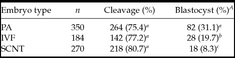
After 10 replications, 804 structures were distributed in different groups for in vitro embryo production: PA (n = 350), in vitro fertilization, IVF (n = 184), and SCNT (n = 270). Cleavage rates were similar for all embryo types (P > 0.05). Totals of 82, 28 and 18 blastocysts were produced in the PA, IVF and SCNT groups, respectively, and the highest proportion of blastocysts was observed in PA embryos (P < 0.05, Table 1).
In vitro preimplantation development in parthenogenetically activated (PA), in vitro fertilized (IVF) and cloned by somatic cell nuclear transfer (SCNT) bovine embryos
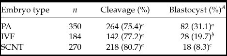
A The blastocyst rate was calculated with respect to the number of cleaved embryos.
a,b,c Within a column, means with a different superscript are significantly different (P < 0.05).
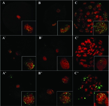
High levels of γH2AX were detected in bovine preimplantation embryos produced by PA (Fig. 1A–C), IVF (Fig. 1A′–C′) and SCNT (Fig. 1A″–C″) at all developmental stages. At the 1-cell stage, IVF embryos exhibited a higher γH2AX focus number (606.1 ± 103.2) and area (12923 ± 3214.1 pixels) than did PA- and SCNT-derived embryos (Table 2). Both pronuclei of IVF embryos exhibited similar γH2AX patterns in terms of γH2AX focus number (377.5 ± 67.7 vs 228.6 ± 42.5; large vs small pronuclei, respectively, P > 0.05) and damaged chromatin area (8698.9 ± 294.8 vs 4224.8 ± 1017.2; large vs small pronuclei, respectively, P > 0.05). In contrast, at the 2-cell stage, both PA and SCNT tended to show greater numbers of γH2AX foci than did IVF embryos (Table 2).
Quantification of γH2AX foci and chromatin area with damage in bovine embryos produced by parthenogenetic activation (PA), in vitro fertilization (IVF) and somatic cell nuclear transfer (SCNT)

a b,cWithin a column, means with a different superscript are significantly different (P < 0.05).
A,B Within a row, means with a different superscript are significantly different (P < 0.05). For all embryos types, 10, eight and four structures were analyzed at the 1-cell, 2-cell and blastocyst stage, respectively, and the numbers of H2AX foci and focus areas were compared. SEM, standard error of the mean.
The phosphorylation of histone H2AX (γH2AX) in bovine preimplantation embryos. Data are presented for parthenogenetically activated (A–C), in vitro fertilized (A′–C′) and cloned by somatic cell nuclear transfer (A″–C″) embryos. The developmental stages analyzed include the 1-cell stage (A–A″), collected at 16 h after initial oocyte activation, spermatozoa–oocyte co-incubation or reconstructed zygote activation; the 2-cell stage (B–B″), recovered after 24 h of in vitro culture; and blastocysts (C–C″), recovered after 7 days of in vitro culture. Embryos were labelled by immunofluorescence using an antibody to γH2AX (green); DNA was counterstained with propidium iodide (red). All images are presented as a juxtaposition of the red and green channels (×400).

Although PA, IVF and SCNT yielded different blastocyst production rates, no differences in the number and area of γH2AX foci were detected among the groups (Table 2). Moreover, when evaluating the dynamics of γH2AX for the different IVP systems, similar numbers of γH2AX foci were observed in PA (1-cell: 163.3 ± 14.2; 2-cell: 367.9 ± 119.7; Bl: 133.8 ± 15.6) and SCNT embryos (1-cell: 200.8 ± 37.2; 2-cell: 682.9 ± 251.0; Bl: 134.5 ± 20.0). An increase in the number of γH2AX foci per nucleus tended to be detected by the 2-cell stage, followed by a reduction at the blastocyst stage. Interestingly, the γH2AX dynamics of IVF embryos were different (1-cell: 606.1 ± 103.2; 2-cell: 260.8 ± 88.0; Bl: 160.0 ± 22.2) from those of the PA and SCNT embryos.
Discussion
Although it has been employed successfully for nearly 3 decades, IVP of embryo by PA, IVF and SCNT still has several limitations that make it less efficient than in vivo production systems. The present study demonstrated that bovine embryos derived by PA, IVF and SCNT exhibited a high number of DNA DSBs by measuring γH2AX accumulation. In addition, the pattern of H2AX phosphorylation exhibited dynamic changes during preimplantation development (1-cell, 2-cell and blastocyst stages) within each treatment, and these dynamics varied among different IVP methods. While IVF embryos exhibited higher numbers of DSBs by the 1-cell stage, PA and SCNT embryos showed a tendency of greater numbers of breaks by day 2. In all embryo types, the numbers of DSBs decreased from the 2-cell stage to the blastocyst stage, indicating that DNA repair mechanisms are functional in blastocysts. In porcine embryos also DSBs occur in a high proportion in structures produced by either IVF or SCNT, proposing that the presence of H2AX foci can be a useful marker of embryo quality of this species (Bohrer et al., Reference Bohrer, Che, Gonçalves, Duggavathi and Bordignon2013).
Initially, it was observed that PA embryos showed greater developmental potential than IVF and SCNT IVP embryos. This effect could be due to the efficiency of this in vitro system, which depends only oocyte competence, while IVF and SCNT embryos are influenced also by spermatozoa quality and donor cell reprogramming, respectively.
In a subsequent step, this work aimed to evaluate the correlation between IVP system efficiency and the induction of DNA damage. Other methodologies may be used to assess DNA damage in cells and embryos (Hosseini et al., Reference Hosseini, Aghaee, Hosseini, Hajian, Forouzanfar, Noorbakhshnia, Gourabi, Shahverdi, Dizaj and Nasr-Esfahani2011; Gawecka et al., Reference Gawecka, Marh, Ortega, Yamauchi, Ward and Ward2013), however the use of γH2AX can explain how embryos respond to such damage. Phosphorylated H2AX was detected in 1-cell embryos, 2-cell embryos and blastocysts, suggesting that the mechanisms involved in the G2/M checkpoint and DNA repair are active in these IVP embryos. It has been demonstrated previously that approximately 2000 histone H2AX molecules near the site of the DSB are phosphorylated at Ser-139 (Pilch et al., Reference Pilch, Sedelnikova, Redon, Celeste, Nussenzweig and Bonner2003), producing one focus that corresponds to a single DSB site in the nucleus (Kaneko et al., Reference Kaneko, Igarashi, Kataoka and Miura2005). Interesting, H2AX was highly phosphorylated throughout development in the absence of any induced DNA damage in mouse embryo produced by in vivo fertilization (Ziegler-Birling et al., Reference Ziegler-Birling, Helmrich, Tora and Torres-Padilla2009), mammalian cell lines and primary cell cultures (McManus & Hendzel, Reference McManus and Hendzel2005). Moreover, H2AX levels ranged significantly during the cell cycle, suggesting that the H2AX phosphorylation is an important event in the fast dividing cells of the early embryo in the absence of any induced DNA damage (Ziegler-Birling et al., Reference Ziegler-Birling, Helmrich, Tora and Torres-Padilla2009), opening the possibility for a role in chromatin assembly and remodelling during these stages.
H2AX is known to be phosphorylated by the ataxia telangiectasia mutated kinase (Rogakou et al., Reference Rogakou, Pilch, Orr, Ivanova and Bonner1998; Kobayashi, Reference Kobayashi2004), and this phosphorylation is one of the first steps in the DNA damage response pathway, as phosphorylated H2AX triggers the assembly of response proteins (Adiga et al., Reference Adiga, Toyoshima, Shimura, Takeda, Uematsu and Niwa2007). γH2AX focus formation is used as a marker of DNA DSBs in irradiated cells, and these foci are even visible at the break and exchange points of metaphase chromosomes (Rogakou et al., Reference Rogakou, Pilch, Orr, Ivanova and Bonner1998). Thus, the γH2AX foci may play an essential role in the efficient recruitment of proteins that are involved in DNA repair.
It was observed that IVF embryos exhibit a higher number of DSBs by the 1-cell stage than PA and SCNT embryos. One interesting observation in this experiment was that this increase was observed not only in the number of γH2AX foci per embryo but also in the number of γH2AX foci in the large IVF pronucleus only. The large IVF pronucleus tended to exhibit a greater number of DSBs than did the IVF female pronucleus (small). The observations from our work are supported by previous studies of male pronucleus formation. Prior to gamete fusion, during spermatogenesis, the postmeiotic spermatid nucleus undergoes a rigorous alteration of its chromatin, with the stepwise removal of most of the nucleosomes from the DNA and their replacement with protamines, which enable the high degree of compaction found in the sperm nucleus (Ward & Coffey, Reference Ward and Coffey1991). After entry into the oocyte, the reversal of this nucleosome-based chromatin state is necessary to enable the formation of a pronucleus that can enter S-phase in conjunction with the maternal pronucleus (Loppin et al., Reference Loppin, Bonnefoy, Anselme, Laurencon, Karr and Couble2005). The spermatid nucleus is devoid of DNA repair machinery upon the onset of protamine-induced compaction at stage 12 of spermatogenesis (Kofman-Alfaro & Chandley, Reference Kofman-Alfaro and Chandley1971; Sega et al., Reference Sega, Sotomayor and Owens1978), 2 weeks before ejaculation. Hence, any DNA lesions present in the paternal nucleus at this time must be repaired after entry into the oocyte, by oocyte-stored factors. Consequently, the large number of γH2AX foci observed in the paternal chromosome (large pronucleus) can be attributed to DNA damage induced during the compaction process, as the activity of phosphorylated H2AX has been demonstrated during chromatin restructuring of the male genetic complement in the zygote (Derijck et al., Reference Derijck, Heijden, Giele, Philippens, Bavel and Boer2006). DNA-damaged spermatozoa can fertilize oocytes and the capacity of early embryos to repair a damaged male genome could be inadequate, suggesting that this damage response may be useful in assessing embryo developmental competence (Grenier et al., Reference Grenier, Robaire and Hales2012).
In addition, during the first cell cycle after fertilization, extensive chromatin remodelling occurs in the zygote, and this appears to be the determining factor for the onset of gene expression (Renard, Reference Renard1998). This remodelling is accompanied by alterations in DNA methylation, histone modification and the recruitment of various chromatin proteins into the pronucleus. The processes of histone modification and methylation affect male and female pronuclei differently (Adenot et al., Reference Adenot, Mercier, Renard and Thompson1997; Mayer et al., Reference Mayer, Niveleau, Walter, Fundele and Haaf2000), even though these structures reside in the same cytoplasm (Liu et al., Reference Liu, Kentaro, Hara and Aoki2005). In mice, it was demonstrated that the male pronucleus has a larger volume, a higher concentration of transcription factors, exhibits a higher level of histone acetylation (Bouniol et al., Reference Bouniol, Nguyen and Debey1995; Adenot et al., Reference Adenot, Mercier, Renard and Thompson1997) and undergoes more extensive DNA demethylation (Mayer et al., Reference Mayer, Niveleau, Walter, Fundele and Haaf2000) than the female pronucleus. The bovine male pronucleus is morphologically distinct from the female version and can be identified based on two distinct features: (1) it is slightly larger than its female counterpart (Beaujean et al., Reference Beaujean, Taylor, McGarry, Gardner, Wilmut, Loi, Ptak, Galli, Lazzari, Bird, Young and Meehan2004) and distal from the polar body; and (2) it is more decondensed than the female pronucleus (Park et al., Reference Park, Jeong, Shin, Lee and Kang2007).
We observed that the number of DSBs tended to increase by day 2 in both PA and SCNT embryos, in contrast with the behaviour observed in IVF embryos. While IVF embryos exhibited a higher γH2AX foci number per nucleus and greater chromatin area with damage at the 1-cell stage, these numbers were reduced at the 2-cell stage, showing that the greatest damage was present in early stages and that repair had occurred by day 2. The epigenetic modification of DNA (methylation of cytosine in the dinucleotide CpG) and/or associated proteins (e.g. histone phosphorylation, acetylation and methylation) regulates gene expression without changing the DNA sequence (Li, Reference Li2002). DNA methylation is recognized as a principal contributor to the stability of gene expression (Bird, Reference Bird2002). The pattern of genomic methylation is stably inherited by differentiated somatic cells; however, genome-wide reprogramming occurs in germ cells and preimplantation embryos (Dean & Walter, Reference Dean and Walter2001). The reprogramming process includes the erasure of existing epigenetic marks and the re-establishment of cell-specific marks to generate cells with nuclear totipotency and broad developmental potential (Dean & Walter, Reference Dean and Walter2001; Meehan, Reference Meehan2003). However, this reprogramming process leads to DNA damage. Thus, we hypothesize that the greater number of γH2AX foci in 2-cell SCNT embryos is observed due to an increased need for nuclear reprogramming.
Finally, blastocysts showed a decreased level of DSBs with respect to 2-cell embryos in all IVP methods. These results indicate that the early embryo stages are more prone to DNA damage and that these lesions are repaired in embryos that reach the blastocyst stage. Poor γH2AX foci formation in early stages is likely to result in defective damage response, leading to improper DNA repair. Thus, poor H2AX phosphorylation in 1-cell and 2-cell stage embryos may serve as a mechanism to eliminate irreparably damaged embryos and prevent the development of defective offspring (Norimura et al., Reference Norimura, Nomoto, Katsuki, Gondo and Kondo1996), as division prior to DSB repair leads to cell death.
In summary, γH2AX is detected at the 1-cell, 2-cell and blastocyst stages in preimplantation bovine embryos produced by PA, in vitro fertilization and SCNT. While IVF embryos exhibited higher numbers of DSBs by the 1-cell stage, no difference was observed between PA and SCNT embryos at any developmental stage. The decrease in the number of DSBs at the blastocyst stage seems to indicate that DNA repair mechanisms are functional in blastocysts.
Acknowledgements
The authors thank the Amancay (Dr Silva) and Nueva Escocia (Dra Saenz) slaughterhouses for providing the biological material and Pablo Do Campo for support with confocal image analysis. V.J.F. Freitas is a CNPq senior investigator (Fortaleza, Brazil), and D.F. Salamone is a CONICET independent investigator (Buenos Aires, Argentina).