
Introduction
The polymerization of silicic acid from aqueous solution has been investigated for more than a century [Reference Graham1], with the majority of studies focused on inorganic systems and the formation of amorphous silica as small particles, sols, and gels. Thorough reviews of these systems are presented in the chemistry, materials, and geological literature [Reference Alexander2, Reference Greenberg and Sinclair3, Reference Bishop and Bear4, Reference Iler5, Reference Markrides, Turner and Slaughter6, Reference Weres, Yee and Tsao7, Reference Tarutani8]. More recently, the geochemical community has recognized the significance of biological silica mineralization in the global biogeochemical cycling of silicon. In modern seas, diatoms, sponges, and radiolaria sequester H4SiO4 and polymerize amorphous silica to produce functional skeletal structures—sometimes within a matter of hours [Reference Volcani, Bendz and Lindqvist9]. Although low Si concentrations prevent a significant amount of silicic acid polymerization in seawater, biosilicification processes by organisms “synthesize” gigatons of amorphous silica each year [Reference Tréguer and De La Rocha10].
Efforts to understand the chemical processes that regulate biosilicification have focused on the composition and structure of macromolecules that are found in association with silicifying organisms. Early investigations sought to probe the nature of biomolecule–silica interactions through bulk analyses of proteinaceous materials in biosilica-associated tissues; studies of this kind suggested that charged amino acids, such as lysine and arginine [Reference Perry and Keeling-Tucker11], and uncharged species (i.e., serine, glycine, and alanine [Reference Hecky, Mopper, Kilham and Degens12, Reference Vrieling, Beelen, van Santen and Gieskes13]) may play critical roles in biosilicification because of their relatively high abundance. More recently, specific silica mineralizing macromolecules have been isolated from both diatoms and sponges. It is now recognized that diatom biosilica is associated with polyamines and/or a class of proteins (silaffins) that contain polyaminated lysine and phosphorylated serine residues. Polyamines have also been implicated in sponge spicule genesis [Reference Matsunaga, Sakai, Jimbo and Kamiya14]; however, the primary workhorse appears to be silicatein, a specialized enzyme that catalyzes the hydrolysis of silicon alkoxides and the condensation of silicic acid [Reference Kröger, Deutzmann and Sumper15, Reference Kröger, Deutzmann, Bergsdorf and Sumper16, Reference Spinthaki, Zerfaß, Paulsen, Hobe and Demadis17, Reference Shimizu, Cha, Stucky and Morse18, Reference Cha, Shimizu, Zhou, Christiansen, Chemelka, Stucky and Morse19]. The catalytic activity of silicatein has been attributed to interactions between the enzyme substrate and histidine and serine residues in its active site.
The prominent involvement of polyamines in diatom and sponge silicification helped to establish the general narrative that amine moieties promote the rate of silicic acid polymerization [Reference Kröger, Deutzmann and Sumper15, Reference Kröger, Deutzmann, Bergsdorf and Sumper16, Reference Spinthaki, Zerfaß, Paulsen, Hobe and Demadis17, Reference Sumper, Lorenz and Brunner20, Reference Patwardhan, Clarson and Perry21, Reference Mizutani, Nagase and Ogoshi22, Reference Mizutani, Nagase, Fujiwara and Ogoshi23, Reference Coradin, Durupthy and Livage24, Reference Pohnert25, Reference Menzel, Horstmann, Behrens, Bärnreuther, Krueger and Jahns26, Reference Patwardhan and Clarson27, Reference Sumper and Kröger28, Reference Knecht and Wright29, Reference Delak and Sahai30, Reference Robinson, Rognlien, Bauer and Simmons31, Reference Belton, Patwardhan, Annenkov, Danilovtseva and Perry32, Reference Kuno, Nonoyama, Hirao and Kato33, Reference Kasotakis and Mitraki34, Reference Mural, Higuchi, Kuno and Kato35, Reference Rimola, Sodupe and Ugliengo36, Reference Lemloh, Altintoprak, Wege, Weiss and Rothenstein37] and inspired a number of efforts to understand the mechanisms by which natural and synthetic polyamines catalyze the condensation of silanol groups (≡Si–OH) [Reference Perry and Keeling-Tucker11, Reference Vrieling, Beelen, van Santen and Gieskes13, Reference Kröger, Deutzmann and Sumper15, Reference Kröger, Deutzmann, Bergsdorf and Sumper16, Reference Sumper, Lorenz and Brunner20, Reference Sumper and Kröger28, Reference Chiovitti, Harper and Willis38] to form siloxane bonds (≡Si–O–Si≡) [Reference Patwardhan, Clarson and Perry21]:
 $$\equiv {\rm{Si}} – {\rm{OH}} + \equiv {\rm{Si}} – {\rm{OH}} = \; \equiv {\rm{Si}} – {\rm{O}} – {\rm{Si}} \equiv + \;{{\rm{H}}_2}{\rm{O}}\quad .$$
However, the mechanisms involved are not well understood [Reference Kröger, Deutzmann, Bergsdorf and Sumper16, Reference Mizutani, Nagase and Ogoshi22, Reference Delak and Sahai30], and factors other than amine availability have been implicated. For instance, Belton et al. [Reference Belton, Paine, Patwardhan and Perry39] investigated several types of polyamines and found that many compounds (both natural and synthetic) had no significant effect on the rate of silicic acid condensation, although certain species were identified that could enhance the rate by as much as 14×. Similarly, Coradin and Livage [Reference Coradin and Livage40] found no measurable differences between silica nucleation rates in solutions with very low concentrations of aspartic acid, serine, lysine, and proline but noted enhanced condensation rates in the presence of small peptides. Liang et al. [Reference Liang, Patwardhan, Danilovtseva, Annenkov and Perry41] found that the ability of polyimidazoles to promote or inhibit silicic acid condensation was dependent on several factors (i.e., polymer chain length, solution pH).
It is evident that factors intrinsic to polyaminated compounds can exert considerable influence on the kinetics of silicate condensation; however, there is further evidence to suggest that synergistic interactions between amine moieties and other functional groups within the organic matrix provide an additional means of control. Belton et al. [Reference Belton, Patwardhan, Annenkov, Danilovtseva and Perry32] found that intrachain amine–amine and amine–methyl interactions affected silicate condensation by regulating the proportion and distribution of protonated amines and the hydrophobicity of the organic matrix. Kröger et al. [Reference Kröger, Lorenz, Brunner and Sumper42] demonstrated that the biological function of silaffins depends on the presence of phosphorylated serine residues alongside polyamine moieties within the peptide backbone. Similarly, Demadis and Neofotistou [Reference Demadis and Neofotistou43] showed the inhibitory effect of cationic polyaminoamide dendrimers on silicate condensation could be reversed by introducing anionic polyelectrolytes to the solution.
Studies such as these provide important insights into the many roles that organic matrices may assume in mediating silica nucleation, polymerization, and aggregation processes in specific systems. However, little is known about the relative import of the kinetic and thermodynamic drivers underlying biosilicification in general. One study designed to quantify these drivers for carboxyl- and amine/carboxyl-terminated surfaces [Reference Wallace, DeYoreo and Dove44] suggests that a nearly 20× increase in the nucleation rate on the introduction of amine moieties is a purely kinetic effect. This result is distinct from similar investigations of calcium carbonate nucleation [Reference Giuffrea, Hamm, Han, De Yoreo and Dove45, Reference Hamm, Giuffre, Han, Tao, Wang, De Yoreo and Dove46], in which the rate is primarily controlled by substrate-specific influences on the interfacial energy, and invites the question of whether nonenzymatic silica templating proteins and macromolecules (e.g., silaffins, polyamines) generally promote silica nucleation by reducing kinetic rather than thermodynamic barriers.
In this experimental study, we explore the idea that small organic acids (amino acids, citrate) regulate the nucleation rate of silica by reducing kinetic barriers. As a test of the experimental method and to separately determine the effect of NaCl on nucleation rate relative to the Control (0.1 M NaCl), we also measure the nucleation rate in 0.7 M NaCl. We also include the tricarboxylic acid, citric acid. Although this small molecule lacks the amine group (NH3) that is implicated as the primary group to promote silica nucleation, the high net negative charge of citric acid is implicated in regulating diatom metabolism and the biomineralization of tests [Reference Coombs and Volcani47]. By fitting a classical nucleation rate model to the rate data, we quantify the thermodynamic and kinetic origins of the faster rates. The findings show NaCl increases rates by lowering the interfacial free energy. In contrast, the amino and citric acids increase rates by lowering the kinetic energy barrier. The molecule-specific effects suggest the potential to tune the kinetics of silicification.
Theoretical model for nucleation rate
Nucleation model
Measurements of induction time combined with parallel chemical characterization provide an opportunity to determine the kinetic and thermodynamic origins of biomolecule and NaCl effects on silica nucleation. A number of studies have considered the relationship between induction time and nucleation kinetics [Reference Toschev, Bardsley, Hurle and Mullin48, Reference Söhnel and Mullin49, Reference He, Oddo and Tomson50, Reference Liu, Wu, Sethuraman and Nancollas51, Reference Teychené and Biscans52]. This study applies the Makrides–Turner–Slaughter (MTS) [Reference Markrides, Turner and Slaughter6] model that was developed to quantify the homogeneous nucleation rate of silica in geothermal brine systems. The MTS approach is based on classical nucleation theory, which states that the steady-state rate (J) of nucleation is given by the following equation:
 $$J = {N_0}{{kT} \over h}\exp \left( { - {{\Delta {G_{\rm{c}}} + {\Delta _{\rm{a}}}{g^{\rm{k}}}} \over {kT}}} \right)\quad ,$$
where N 0 is the number of monomer molecules per unit volume (equal to the total initial concentration of Si, C 0); ΔG c is the free energy change that corresponds to the formation of nuclei of critical size (referred to as the thermodynamic nucleation barrier), Δag k = kinetic barrier to forming a critically sized nucleus, k = Boltzmann constant, T = temperature (K), and h = Planck constant. The thermodynamic terms are further expanded to show [Reference Mann53, Reference de Yoreo, Vekilov, Dove, De Yore and Weiner54]:
 $$\Delta {G_{\rm{c}}} = {4 \over 3}\pi {r_{\rm{c}}}^2\gamma \quad ,$$
where
 $${r_{\rm{c}}} = {{2\gamma \upsilon } \over {kT\sigma }}\quad ,$$
where r c is the critical radius of embryos, γ is interfacial free energy between the solution and critical nucleus, ν is the volume of a molecule of silica in the solid phase (4.5 × 10⁻²³ cm3) [Reference Markrides, Turner and Slaughter6], and σ is the supersaturation of solutions (see Materials and Methods).
Substituting Eqs. (3a) and (3b) into (2) and replacing N 0 with C 0 obtains:
 $$J = {C_0}{{kT} \over h}\exp \left( { - {{{\Delta _{\rm{a}}}{g^{\rm{k}}}} \over {kT}}} \right)\exp \left( { - {{16\pi {\nu ^2}{\gamma ^3}} \over {3{{\left( {kT} \right)}^3}}} \cdot {1 \over {{\sigma ^2}}}} \right) = {C_0}{k^{\rm{o}}}\exp \left( { - {\beta \over {{\sigma ^2}}}} \right)\quad ,$$
where
 $$\beta = {{16\pi {\nu ^2}{\gamma ^3}} \over {3{{\left( {kT} \right)}^3}}}\quad ,$$
and
 $${k^{\rm{o}}} = {{kT} \over h}\exp \left( { - {{{\Delta _{\rm{a}}}{g^{\rm{k}}}} \over {kT}}} \right)\quad ,$$
such that k o (s−1) is the kinetic constant for forming a critical nucleus [Reference Markrides, Turner and Slaughter6, Reference Espenson55]. Equation (4a) gives the explicit dependence of nucleation rate on γ and Δag k through ß and k o, respectively.
To apply Eq. (4a) to the experimental data and obtain estimates of γ and Δag k for the nucleation of silica in the biomolecule and NaCl solutions, we use the MTS model [Reference Markrides, Turner and Slaughter6] for homogeneous nucleation of silicic acid. The model states that if nucleation during the induction time contributes the majority of the nuclei, and nuclei are assumed to be generated continuously throughout the induction time, then:
 $${{{C_0} - {C_\tau }} \over {{C_0} - {C_{\rm{e}}}}} = {{\Delta C} \over {{C_0} - {C_{\rm{e}}}}} = {\pi \over 3}{\left( {{k^{\rm{o}}}\lambda } \right)^3}{\left( {{C_0} - {C_{\rm{e}}}} \right)^2}{\left( {{V^{\rm{s}}}} \right)^2}J{\tau ^4}\quad ,$$
where C τ is the silicic acid concentration at the end of the stable period (τ), ΔC is the detection limit for silicic acid concentration (0.25 mM H4SiO4, 15 ppm SiO2; see Materials and Methods), C e = 1.57 mM H4SiO4, λ is the molecular diameter of silica (3 × 10⁻⁸ cm), V s is the molar volume of the precipitate, where V s = 6.023 × 10⁻²³ ν cm3, and J is the nucleation rate per unit volume. All concentration terms are in units of number of mol/cm3.
Model fit to the data
To evaluate the model, Eq. (4a) is substituted into Eq. (5) to obtain the relationship between τ and σ:
 $${\tau ^4} = \left\{ {\Delta C{{\left( {{\pi \over 3}} \right)}^{ - 1}}{{\left( {{k^{\rm{o}}}} \right)}^{ - 4}}{\lambda ^{ - 3}}{{\left( {{V^{\rm{s}}}} \right)}^{ - 2}}} \right\}{\left( {{C_0}} \right)^{ - 1}}{\left( {{C_0} - {C_{\rm{e}}}} \right)^{ - 3}}{\left\{ {\exp \left( { - {\beta \over {{\sigma ^2}}}} \right)} \right\}^{ - 1}}\quad .$$
This expression can be simplified into the form:
 $$4\ln \tau = {\beta \over {{\sigma ^2}}} - \left( {3\ln \left( {{C_0} - {C_{\rm{e}}}} \right) + \ln {C_0} - K} \right)\quad ,$$
where
 $$K = \ln \left\{ {\Delta C{{\left( {{\pi \over 3}} \right)}^{ - 1}}{{\left( {{k^{\rm{o}}}} \right)}^{ - 4}}{\lambda ^{ - 3}}{{\left( {{V^{\rm{s}}}} \right)}^{ - 2}}} \right\}\quad .$$
Equation (7a) quantifies the relationship between induction time and supersaturation of the initial silicic acid concentration. This expression is evaluated using the second derivative statistical test (JMP, Version 2.0; SAS Institute) to estimate ß and k 0. Substituting these constants into Eqs. (4b) and (4c) obtains the interfacial free energy, γ, and kinetic energy barrier to nucleation, Δag k, respectively, for the Control, NaCl, amino acid, and citric acid solutions.
Results and discussion
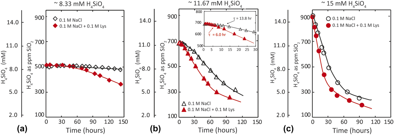
Measurements of nucleation rate show the expected decrease in silica concentration with elapsed time for all solution compositions. Experiments with a lower initial σ exhibit a relatively long latent, or induction, period [Fig. 1(a)] compared with those experiments that begin at a higher initial driving force [compare Figs. 1(b) and 1(c)].
Measurements of silica concentration versus time are used to quantify the inverse dependence of induction time, τ, on supersaturation: (a) 8.33 mM H4SiO4 (510 ppm SiO2), (b) 11.67 mM H4SiO4 (690 ppm SiO2), and (c) 15.23 mM H4SiO4 (914 ppm SiO2). Induction time is defined as the time elapsed between the start of the experiment and a concentration decrease of 0.25 mM H4SiO4 (15 ppm). Inset in (b) illustrates τ = 13.8 and 6.0 h for the 0.1 m NaCl (Control) and 0.1 M NaCl + 0.1 M lysine experiments, respectively.

Dependence of induction time on supersaturation and treatments
Recall the induction time (τ) to the onset of polymerization is defined as the time elapsed from the start of the experiment to a 15 ppm decrease in SiO2 concentration (0.25 mM H4SiO4). For example, Fig. 1(b) (inset) shows that for experiments that began with 690 ppm silica (11.5 mM H4SiO4), the τ = 13.8 and 6.0 h for Control and 0.1 M lysine-bearing solutions, respectively. The accelerating effect of lysine (and other organic acids) on the rate of silica nucleation is particularly evident at the lower silicic acid concentrations and diminishes with increasing silicic acid concentration [compare Figs. 1(a) and 1(c)].
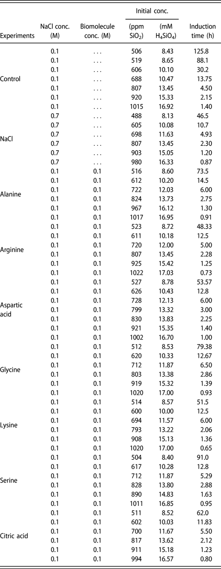
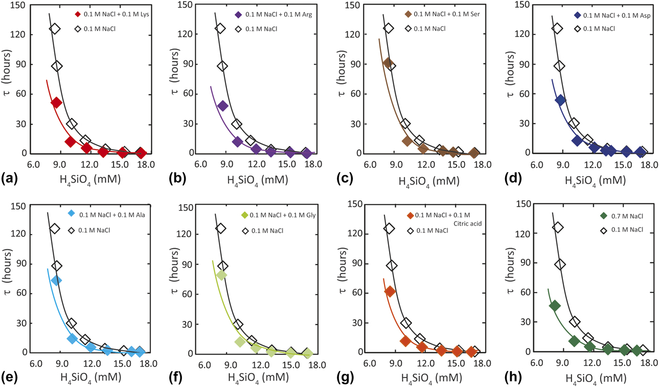
Compared with the Control, all organic acids decrease the induction time to silica nucleation for all supersaturations (Table I). Measurements show that τ decreases as a power function of silicic acid concentration for all Control, amino acid [Figs. 2(a)–2(f)], and citric acid [Fig. 2(g)] solutions. The effects of amino acids and citric acid on τ are biomolecule specific. The τ is also dependent on NaCl concentration as reported previously [Reference Markrides, Turner and Slaughter6]. Experiments conducted in the 0.7 M NaCl solutions also exhibit a shorter induction time compared with the Control.
Summary of experimental conditions and measured induction time, τ.

Experimental measurements for a series of supersaturated solutions illustrate the shorter induction time, τ, determined for the biomolecule and NaCl treatments relative to the Control for (a) lysine, (b) arginine, (c) serine, (d) aspartic acid, (e) alanine, (f) glycine, (g) citric acid, and (h) 0.7 M NaCl.

Theoretical model
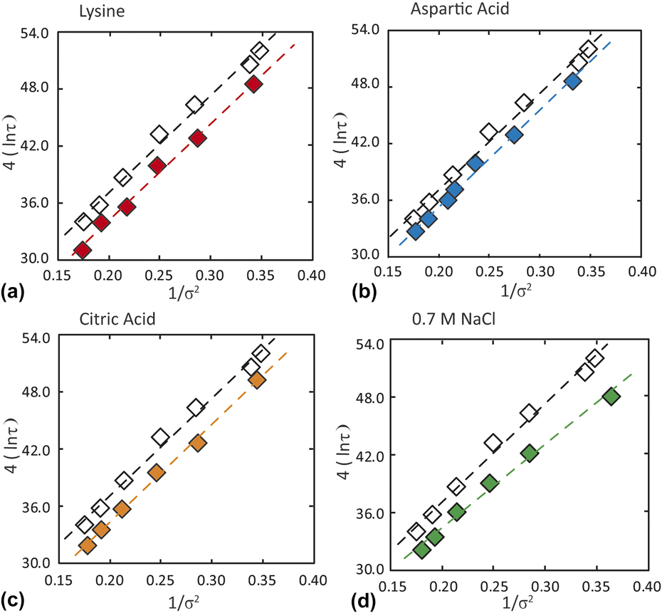
An assumption of the MTS theoretical model is that when most of the nuclei are formed during the transition period, then the relationship between ln(τ) and 1/σ2 should be linear over a limited range of initial concentrations. To test this assumption, we show that measurements from the Control, organic acid, and 0.7 M NaCl-bearing experiments indeed exhibit a linear dependence for all treatments (Fig. 3), thus obeying the MTS continuous nucleation model. By fitting the nucleation rate model to the experimental data, we obtain quantitative insights into the physical basis for faster nucleation rates in solutions with organic acids or a greater concentration of NaCl.
The linear dependence of the logarithm of induction time (τ) on driving force demonstrates agreement with the MTS continuous nucleation model. Organic acids promote faster rates, as measured by shorter induction times, through reductions in the intercept (kinetic barrier) without an effect on slope (interfacial free energy). Typical examples are (a) lysine; (b) aspartic acid, and (c) citric acid. In contrast, greater NaCl concentration (d) enhances rate through reduced slope (thermodynamic barrier). Open symbols = control (0.1 M NaCl) and solid symbols = organic acid or 0.7 M NaCl treatments.

0.1 M NaCl (Control)
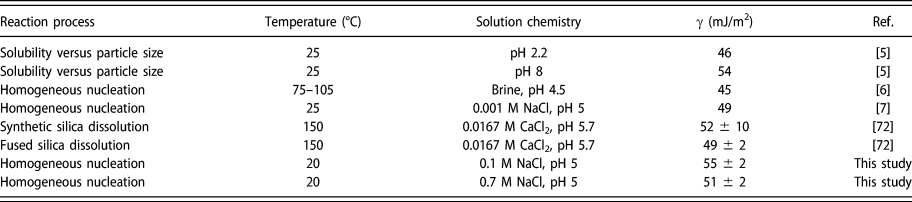
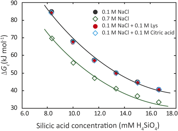
Fitting Eq. (7a) to the Control solution yields an interfacial free energy of γControl = 54.9 ± 1.6 mJ/m2. This value of γ is consistent with previous studies of amorphous and fused silica materials that report 45–54 mJ/m2 (Table II). Substituting γControl into Eq. (3a), Fig. 4 demonstrates the dependence of the free energy barrier to forming of a nucleus of critical size on initial silica concentration.
Summary of reported values of interfacial free energy (γ) for amorphous silica.

Calculated critical nucleation energy barrier for a series of supersaturations shows biomolecules do not affect ΔG c. In contrast, the thermodynamic barrier is reduced when salt concentration is increased from 0.1 to 0.7 M NaCl.

The kinetic energy barrier (Δag k) to silica nucleation for the Control was determined equal to 2.29 × 10⁻¹⁹ J/mol. This value is consistent with the Makrides et al. estimate of ∼1.2 × 10⁻¹⁹ J/mol for silica nucleation in a brine [Reference Markrides, Turner and Slaughter6]. In subsequent discussions, this value is converted to a molar basis and assigned a reference value of 0.0, such that Δag kControl = 0.0 J/mol. Henceforth, estimates of Δag k for all solutions compositions are normalized to this reference (e.g., Table III).
Properties of organic molecules used in this study and estimates of interfacial free energy and kinetic barrier determined in this study.

a α-NH3+ ⇌ α-NH2 + H+ pK values from Ref. Reference Nelson and Cox73.
b Using the formula from Ref. Reference Moore74 and pK values from Ref. Reference Nelson and Cox73.
c Reference Reference Saidel, Goldfarb and Waldman70.
d Reference Reference Van Cappellen and Qiu71.
e pK 1 = 3.13, pK 2 = 4.76, and pK 3 = 6.40.
f Reference Reference Rogacs and Santiago75.
0.7 M NaCl
Analysis of the rate data using Eq. (7a) shows the faster nucleation rates measured in 0.7 M NaCl arise from a reduction in the interfacial free energy, where γ0.7 M NaCl = 51.4 ± 1.7 mJ/m2. This lower value of γ, compared with the Control, is consistent with previous studies of amorphous and fused silica materials (Table II). Equation (4) shows that the reduction in γ has a large effect on rate through the cubic dependence of ß on γ [Eq. (4b)]. Our finding that NaCl modulates nucleation rate primarily through reductions in interfacial free energy is consistent with a study of barium sulfate that used NaCl as a background electrolyte [Reference He, Oddo and Tomson50] and theoretical predictions for the effects of surface adsorption [Reference Poon and Peters56] and ionization on interfacial free energy [Reference Legg, Zhu, Zhang, Waychunas, Gilbert and Banfield57]. Substituting γ0.7 M NaCl into Eq. (3a), Fig. 4 shows the effect of NaCl concentration on ΔG c with free energy values offset to lower values for all silica concentrations. Further analysis of the model shows the kinetic barrier to nucleation is higher compared with the Control, such that Δag k0.7 M NaCl = +140 ± 97 J/mol. Thus, the faster nucleation rates measured in 0.7 M NaCl solutions do not have a kinetic origin.
Also note that interfacial free energy values of 51–55 mJ/m2, calculated from our experiments with solutions that contained 8–16 mM H4SiO4, estimate a critical nucleus radius (r c) of 5.1–7.8 × 10⁻⁸ cm. This size range corresponds to 12–44 Si monomers in a nucleus of critical size (n c). These cluster sizes further support the use of the classical nucleation model [Reference Galkin and Vekilov58].
Our finding that NaCl increases the rate of silica nucleation by lowering the thermodynamic barrier to nucleation leads us to ask the chemical basis for this effect. A plausible explanation is the ability of sodium ions to increase the number of ionized silica groups through electrostatic interactions, thus increasing reactivity. For example, Busey and Mesmer [Reference Busey and Mesmer59] showed that the pK a of silicic acid decreases by ∼0.15 when NaCl concentration increases to 0.7 M. The shift results in a small increase in H3SiO4− concentration (∼1.4 times). Given that the total concentration of H3SiO4− is on the order of 10−7 mol/L for 500–1000 ppm SiO2(aq) solutions for the pH range of these experiments, this highlights the extreme sensitivity of the condensation rate to the presence of H3SiO4−.
The generally accepted reaction for silica nucleation is the condensation of neutral silanol groups as silicic acid or polysilicic acid by the nucleophilic attack of oxygens and the elimination of H2O. When pH < 8, most silanol groups have a neutral charge in simple solutions [Reference Bishop and Bear4] and the rate of nucleation is relatively slow under these conditions. With the addition of 1:1 electrolytes, however, electrostatic interactions between Na+ and silanols promote the formation of ionized groups such that [Reference Iler5]:
 $${\left( {{\rm{HO}}} \right)_3} – {\rm{Si}} – {\rm{OH}} + {\rm{N}}{{\rm{a}}^ + } \leftrightarrow {\left( {{\rm{HO}}} \right)_3} – {\rm{Si}} – {{\rm{O}}^ - }{\rm{N}}{{\rm{a}}^ + } + {{\rm{H}}^ + }\quad ,$$
or
 $${\rm{Poly}} – {\rm{Si}} – {\rm{OH}} + {\rm{N}}{{\rm{a}}^ + } \leftrightarrow {\rm{Poly}} – {\rm{Si}} – {{\rm{O}}^ - }{\rm{N}}{{\rm{a}}^ + } + {{\rm{H}}^ + }\quad .$$
These reactions are increasingly important in the presence of high NaCl concentrations, as shown by a study of pathways to silicic acid polymerization by Shimada and Tarutani [Reference Shimada and Tarutani60]. They demonstrated that the interaction between uncharged and ionized silanol groups is the energetically most favorable. Ionized forms of small oligomers and polysilicic acid are particularly important during the formation of a critical nucleus because monomers and dimers preferentially react with ionized groups, thus increasing molecular size [Reference Iler5]. This enhancement through ionization is consistent with H+ release in Eqs. (8a) and (8b) and the experimental measurements.
Note that ionization equilibria must contribute to the value of interfacial free energy, but the relationship is unclear. If interfacial energy decreases at higher ionic strength, then solubility should also increase by some amount [Reference Söhnel61]. The reduction in the interfacial energy measured in this study is so small that a solubility change would not be detectable within the uncertainties of the molybdate yellow method. However, if solubility increases, the driving force for forming the bulk phase is reduced if all other factors are equal. To some degree, this effect would counterbalance an increased abundance of H3SiO4− in the higher salinity solution.
Amino acids and citric acid
Fitting the rate model to the data, we find that the amino acids and citric acid increase the kinetic rate constant, k o, through biomolecule-specific reductions in the kinetic barrier, Δag k. For the amino acids, the barrier ranges from Δag kalanine = −1009 ± 169 J/mol to Δag klysine = −1685 ± 315 J/mol (compared with the Control). The greatest reduction in the barrier was measured for citric acid with Δag kcitric = −1690 ± 96 J/mol.
None of the biomolecules change the free energy of the silica–water interface. Within the uncertainties of the measurements, γbiomolecules is statistically similar to the Control (Table III), and thus, biomolecules do not promote the rate of nucleation through a thermodynamic effect. Figure 4 shows that ΔG c values from lysine and citric acid experiments are similar to the Control.
To qualitatively illustrate the kinetic effect of biomolecules on the induction time to silica nucleation, Figs. 3(a)–3(c) show the decrease in induction time to nucleation (compared with the Control) through the intercept (k o) and without a change in slope (γ). In contrast, a higher concentration of NaCl (compared with the Control) shows a small reduction in slope, supporting a thermodynamic effect on rate [Fig. 3(d)].
Systematic dependence of kinetic barrier on amino acids
Our measurements showing biomolecule-specific reductions in the kinetic barrier provide an opportunity to probe the nucleation process. We return to the literature to consider three overarching models for how large peptides and polyamines catalyze silica polymerization:
(i) Acid–base catalysis by H+ transfer: Polyamines facilitate silicic acid condensation when protonated (acid) and deprotonated (base) amino groups promote the formation of reactive silanolate groups [Reference Kröger, Deutzmann, Bergsdorf and Sumper16, Reference Mizutani, Nagase, Fujiwara and Ogoshi23, Reference Knecht and Wright29].
(ii) Charge relay facilitates silicic acid condensation: Nucleophilic attraction of an amino group for a silicon center in a silicic acid molecule promotes transfer of a proton from a silicon-bonded hydroxyl to a carboxyl group in the vicinity of the amino group [Reference Kuno, Nonoyama, Hirao and Kato33, Reference Mural, Higuchi, Kuno and Kato35].
(iii) Hydrogen bonding: This process is weaker than electrostatic interactions between charged amino groups and ionized silanol groups [Reference Coradin and Livage40, Reference Gambino, Lombardo, Grassi and Marletta62] but is proposed to promote polymerization [Reference Iler5, Reference Coradin and Livage40, Reference Lomenech, Bery, Costa, Stievano and Lambert63, Reference Gambino, Grassi and Marletta64, Reference Nonella and Seeger65].
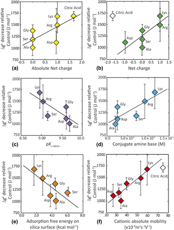
These models share a common theme in the probable relationship between biomolecule charge and functional group properties for promoting silica nucleation. Although the experimental errors associated with estimates of Δag k are large, the general trend indicates that reductions in the kinetic barrier are roughly correlated with a greater absolute charge of the amino acids [Fig. 5(a) and Table III]. This relationship is intriguing and calls for further study, given that previous investigations suggest that positively charged amine groups play a specific role in promoting silica polymerization by bringing silicate oligomers into a configuration that favors nucleation [Reference Coradin, Durupthy and Livage24, Reference Robinson, Rognlien, Bauer and Simmons31, Reference Coradin and Livage40, Reference Yuwono and Hartgerink66]. Indeed, Fig. 5(b) supports this idea by showing that the amino acids promote rate in proportion to their net positive charge. Citric acid, with a net negative charge and without an amine group, does not obey this trend.
The kinetic barrier to nucleation, Δag k, correlates with amino acid properties (Table II): (a) absolute net charge, (b) net charge, (c)  $\left( {{\rm{p}}{K_{\alpha {\rm{ ‐ N}}{{\rm{H}}_3}}}} \right)$, (d) concentration of conjugate base, (e) adsorption free energy of amino acids on hydroxylated silica surface, and (f) absolute ionic mobility, where open symbol for citric acid is shown but not included in the correlation. Error bars about Δag k represent one standard error of the estimate.

To probe the idea that amines promote rates in proportion to the reactivity of the corresponding amine moieties, Fig. 5(c) shows the correlation between Δag k and  $\left( {{K_{\alpha {\rm{ ‐ N}}{{\rm{H}}_3}^ + }}} \right)$ of the corresponding amino acid for the stoichiometry:
 $${\rm{R}} – \alpha – {\rm{N}}{{\rm{H}}_3}^ + = {\rm{R}} – \alpha – {\rm{N}}{{\rm{H}}_2} + {{\rm{H}}^ + }\quad ,$$
where  $\left( {{K_{\alpha {\rm{ ‐ N}}{{\rm{H}}_3}^ + }}} \right)$ is the amine dissociation constant. The relationship suggests that reductions in Δag k are correlated with the presence of amino acids that have larger
 $\left( {{K_{\alpha {\rm{ ‐ N}}{{\rm{H}}_3}^ + }}} \right)$. A similar trend is reported by Delak and Sahai [Reference Delak and Sahai30] in an investigation of tetramethylsiloxane (TMES) hydrolysis and condensation; however, their condensation rate is found to follow sympathetically from the TMES hydrolysis rate and to be catalyzed by acid rather than by amine. Since the tetramethyl orthosilicate (TMOS) silicic acid precursor used in this study was pre-hydrolyzed, this result suggests a specific interaction involving the conjugate base of amino acids that promotes condensation.
Recalling that all experiments began at a steady-state pH = 5, where the conjugate base concentration is larger in the presence of amino acids with a lower  $\left( {{\rm{p}}{K_{\alpha {\rm{ ‐ N}}{{\rm{H}}_3}^ + }}} \right)$, we also consider that nucleation may be driven by an acid–base reaction between the silanols and the conjugate base, R–NH2, of the amine groups. Indeed, reductions in Δag k scale with the concentration of the conjugate base [Fig. 5(d) and Table III]. One possible mechanism that is consistent with this observation involves the abstraction of a proton from silicic acid by the conjugate base, facilitating the concomitant nucleophilic activity of the ionized silanol.
We also ask if the interactions of amino acids with silica surfaces through hydrogen bonding contribute to a faster nucleation rate in proportion to the free energy of adsorption onto a silica surface. A theoretical study reported that nonpolar amino acids, glycine and alanine, and the basic amino acids, lysine and arginine, are the least and the most prone to be adsorbed on the silica surface, respectively [Reference Rimola, Sodupe and Ugliengo67]. They propose that the adsorption mechanism is due to H-bond interactions between the functional groups of the amino acids and terminal silanol groups on the surfaces. By correlating adsorption free energy (ΔG c) of the amino acids [Reference Rimola, Sodupe and Ugliengo67] with the induced reduction in the kinetic barrier (Δag k), determined in this study, Fig. 5(e) shows lysine and arginine indeed increase the rate of silica nucleation more than glycine. The relationship further supports the idea that nucleation rate is influenced by the nature and extent of hydrogen bonding [Reference Iler5, Reference Coradin and Livage40, Reference Lomenech, Bery, Costa, Stievano and Lambert63, Reference Gambino, Grassi and Marletta64, Reference Nonella and Seeger65].
Citric acid provides an independent test
Citric acid, with a net negative charge of −1.65, and without an amine group, causes the greatest reduction in Δag k (Table III) but does not obey the trend in Fig. 5(b). One plausible explanation for the catalytic effect of citric acid may lie in the polyprotic nature of this molecule. The pH conditions of our experiments are intermediate to the three pK a values (3.13, 4.76, 6.39) of citric acid, such that there are both acidic and basic carboxyl groups on the molecule. This is analogous to the amino acids used in the study, whose amine and carboxyl groups also behave as acids (amine) and bases (carboxyl). This leads us to predict that other classes of multiprotic molecules may also have the potential to catalyze nucleation.
Other properties of the simple biomolecules that may affect silica nucleation are steric factors, including conformation, hydration, and mobility. For example, mobility is a bulk property that cannot give mechanistic insights, but the characteristic captures the combined influence of charge, size, orientation, and migration on the properties of biomolecules in an electric field. That is, the kinetic barrier to nucleation may also reflect the rate at which each molecule encounters silicate species in solution (i.e., related to transport properties of the catalyst). By correlating relative Δg k with absolute mobility of the amino acids [Reference Vceláková, Zusková, Kenndler and Gas68], Fig. 5(f) suggests a trend that is roughly inclusive of all organic acids investigated in this study. Citric acid, with the greatest mobility but without an amine functional group, is similar to lysine in its ability to reduce the kinetic barrier to silica nucleation.
These measurements challenge the long-standing dogma that citric acid does not interact with silica. Citrate is widely assumed inert in silicification studies and is frequently used as a pH buffer [Reference Weres, Yee and Tsao7, Reference Patwardhan, Clarson and Perry21]. In hindsight, this assumption appears reasonable in the absence of quantitative measurements. First, amine groups, not carboxyl, are considered to be the primary players in promoting silica reactivity. The second, related to the first, is the assumption that polymerization is promoted by molecules with a greater positive molecular charge. Given the high negative charge of citric acid and its greatest effect on silica nucleation rate, it is clear that net charge is an incomplete indicator.
Conclusions
This quantitative experimental study determined that citric acid and amino acids increase the rate of silica nucleation by lowering the kinetic barrier, Δag k. Citric acid, which lacks an amine group, promotes the greatest rate enhancement. In contrast, the faster rates measured in solutions containing a higher NaCl concentration arise from reductions in the thermodynamic interfacial free energy, γ. While some previous studies have reported faster rates in these types of solutions, to our knowledge this is the first investigation to quantify the kinetic versus thermodynamic origins of the nucleation rate enhancement in biomolecule and NaCl solutions, respectively. The classical nucleation rate model gives good agreement with experimental measurements of silica nucleation rate for all of the systems investigated in this study.
The findings provide a baseline for establishing how functional group chemistry and ionic strength may be tailored to tune the kinetics and thermodynamics of silica nucleation. The relationships also suggest insights for better controls on the synthesis of other covalently bonded structures.
Materials and methods
Preparation of stock solutions
The monomeric/dimeric silicic acid solutions were generated from an organic precursor, TMOS (99.5+%; Sigma-Aldrich, St. Louis, Missouri). To prepare each starting solution with a desired initial silicic acid concentration, 18.6–37.0 μL of TMOS was hydrolyzed in 7.5 mL of 0.03 M HCl for 15 min on an unheated stirring plate at 600 rpm. This procedure produces only monomeric silica species and methanol as the by-product [Reference Wallace, DeYoreo and Dove44]. For an experiment, after combination with the biomolecule stock solution, the final silicic acid concentration was designed to contain 8.33–16.7 mM SiO2 (500–1000 ppm SiO2). Each biomolecule stock solution contained 0.3 M of an organic acid and 0.3 M NaCl as a background electrolyte. The pH of this stock solution was adjusted to ∼4.8 using dilute NaOH or HCl.
Characterization
Prior to the start of the experiment, the total concentration of monomeric and dimeric silicic acid was confirmed using the molybdate yellow colorimetric method, described by Iler [Reference Iler5] (Table I), at an absorption wavelength of 410 nm using UV-VIS Evolution 300 (Thermo Scientific, Madison, Wisconsin). The monomer and dimer forms of silicic acid react with the molybdate reagent in an acidic solution to give a yellow absorbance, whereas larger polysilicic acids do not react. All experimental conditions are summarized in Table I.
The possibility of additive interference with the molybdate yellow method was considered. For each additive, the free silicic acid concentration was measured at t = 0, and all changes in concentration were determined relative to that initial value. Thus, a matrix effect, if there was any, would be constant. Further, published reference data on the influence of NaCl on the molybdate yellow method show only a 5% reduction in absorption at 1 M [Reference Iwaji and Toshikazu69]. For 0.7 M NaCl as the highest concentration in this study, we expect <5% variability. Citric acid and certain amino acids do absorb in the UV, but not in the visible wavelengths. Since our measurements were made at 410 nm, we do not expect any interference of these species with the molybdate yellow method [Reference Saidel, Goldfarb and Waldman70].
Measurements of induction time
Rate experiments were conducted for the conditions summarized in Table I at 20.0 ± 0.1 °C using an incubator (Fisher Scientific, 307C). The experimental control was established by measuring the rate of silicic acid polymerization in 0.1 M NaCl solution. A second type of control (0.7 M NaCl) determined the effect of the background electrolyte on nucleation rate (Table I).
To begin, 7.5 mL of the appropriate silicic acid solution was combined with 5.0 mL of an amino acid stock solution in a 20 mL glass vial while mixing on a stirring plate. These solutions were combined immediately after the TMOS was fully hydrolyzed (see Preparation of Stock Solutions) to minimize any pre-reaction polymerization. The mixed solution was then adjusted to pH 5.0 using dilute NaOH or HCl to obtain a final volume of 15 mL. Thus, the starting solutions contained 0.1 M NaCl, 0.1 M organic acid, and variable silica concentrations (Table I). pH = 5 was used to approximate the physiological pH in the diatom silica deposition vesicle during biosilicification [Reference Vrieling, Beelen, van Santen and Gieskes13]. The solution in the glass vial was gently and continuously mixed on a plate shaker at 125 rpm within the incubator. Aliquots of 10 μL were withdrawn intermittently to analyze for silicic acid concentration. Thus, the concentration change of molybdate-reactive silicic acid indicates the onset of a polymerization process [Reference Iler5]. The silicic acid concentration in the aliquot was measured over the duration of the experiment using the same molybdate yellow colorimetric procedure described previously. pH was constant during all biomolecule experiments, but the pH of 0.7 M NaCl solutions decreases ∼0.1 pH unit.
For this study, the induction time is defined as the time elapsed from the onset of the experiment to a 0.25 mM decrease in H4SiO4 concentration (analytical detection limit [Reference Iler5, Reference Markrides, Turner and Slaughter6]). The duration of the polymerization experiments varied from 4 to 30 days, depending on initial supersaturation. None of the solutions in this study became visibly turbid over the course of the observation period, thus indicating that polymerization of H4SiO4 at pH 5 leads only to small molecular clusters [Reference Mizutani, Nagase and Ogoshi22, Reference Coradin and Livage40].
Supersaturation
Supersaturation, σ, provides the thermodynamic driving force for silica nucleation and is defined as follows:
 $$\sigma = \ln \left( {{{{C_0}} / {{C_{\rm{e}}}}}} \right)\quad ,$$
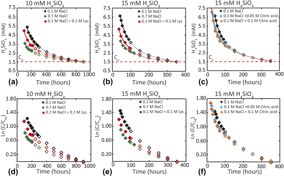
where C 0 = concentration of monomeric and dimeric silicic acid measured in a given solution and C e = 1.57 mM H4SiO4 (94 ppm SiO2) is the equilibrium apparent solubility at 20 °C [Reference Van Cappellen and Qiu71]. Figures 6(a)–6(c) illustrate the approach to equilibrium solubility for the experimental conditions of this study. By determining ln(C/C e) versus time, Figs. 6(d)–6(f) show that all experiments containing NaCl and/or organic acid additives approach 0.0. Thus, the treatments do not affect silica solubility within errors of the analytical measurements.
Typical long-duration experiments show the decrease in silica concentration at two starting H4SiO4 concentrations and NaCl or organic acid solution compositions: (a) 10 mM H4SiO4 with NaCl and 0.1 M lysine, (b) 15 mM H4SiO4 with NaCl and 0.1 M lysine, and (c) 15 mM H4SiO4 with NaCl and two concentrations of citric acid. Approach to equilibrium [ln(C/C e) = 0.0] for treatments that began at (d) 10 mM H4SiO4 and (e, f) 15 mM H4SiO4 for the same treatments as (a, b, c). Solid symbols denote measurements near equilibrium and open symbols indicate calculated fit to the data.

Acknowledgments
This project was funded by the U.S. DOE Office of Basic Energy Sciences Award BES-FG02-00ER15112.