Advanced MRI techniques have enabled the earlier recognition of brain injury and delayed brain development in neonates born with CHD. Reference Licht, Shera and Clancy1,Reference Miller, McQuillen and Hamrick2 White matter injury is the most common brain injury pattern identified in neonates with CHD occurring in nearly 20% of children born with cyanotic cardiac lesions before surgery. Reference Beca, Gunn and Coleman3–Reference Petit, Rome and Wernovsky6 When white matter injury is present after birth in neonates with CHD, it occurs in a distinctive radiographic pattern characterised by discrete, usually punctate, foci of T1 hyperintensity, with or without restriction of water diffusion, with evanescence in the absence of gliosis. Occasionally, the lesions have corresponding T2 hypointensity, though this is an inconsistent feature. Reference Beca, Gunn and Coleman3,Reference Mahle, Tavani and Zimmerman7–Reference Claessens, Khalili and Isgum9
The spatial distribution and burden of white matter injury on MRI correlates with white matter watershed regions where oligodendrocyte precursors predominate and occurs in a background of brain dysmaturity. A similar pattern of white matter injury has also been observed in preterm infants Reference Guo, Chau and Peyvandi8–Reference Tusor, Benders and Counsell10 and has been associated with future adverse neurodevelopmental outcomes, although many of those patients have cystic encephalomalacia and necrosis, typically not seen in the term CHD population. Reference Peyvandi, Chau and Guo11,Reference Lawrence, McGovern and Mejaddam12 Given the correlation between white matter injury and poor cognitive outcomes, there is a need to histologically classify these white matter lesions in order to better understand their aetiology and prognosticative features. There is speculation that in utero conditions of CHD may halt normal oligodendrocyte development and manifest radiographically as these characteristic white matter lesions, Reference Miller, McQuillen and Hamrick2 while others argue that white matter lesions may be the result of embolic strokes or postnatal ischaemic conditions. Reference Beca, Gunn and Coleman3–Reference McQuillen5 Due to the limitations of studying fetuses in utero and scarcity of autopsy data, these white matter lesions in CHD neonates have never before been characterised histologically to our knowledge.
Recently, an artificial womb model has been developed that permits the study of fetuses under normal and pathologic intrauterine conditions. Using this model, we have previously demonstrated that in utero hypoxemia at levels similar to those seen in CHD leads to reduced brain folding, decreased myelination and led to abnormal neuronal migration without necrosis or increased levels of apoptosis. Reference Lawrence, McGovern and Mejaddam13,Reference Partridge, Davey and Hornick14 Utilising this model, we performed an MRI analysis of a fetal sheep brain supported under hypoxemic conditions. Here we report a focus of T1 hyperintensity in the fetal sheep frontal lobe on MRI and the histologic characteristic of this lesion.
Methods
Animal model
Animals were treated according to approved protocols by the institutional animal care and use committee of The Children’s Hospital of Philadelphia Research Institute. All animal experiments were compliant with National Institutes for Health guide for the care and use of Laboratory Animals.
A fetus from a time-dated, mid-gestation ewe was cannulated via its umbilical vessels and attached to a pumpless low resistance oxygenator circuit. They were then transitioned to a sterile fluid environment where they were supported within the extra-uterine transition for neonatal development system. The surgical procedure and maintenance of these fetuses in this system have been described previously in detail. Reference Lawrence, McGovern and Mejaddam13,Reference Partridge, Davey and Hornick14
The fetus was supported under hypoxic (14–16 ml/kg/min) conditions to mimic the levels of hypoxia seen by fetuses with transposition of the great arteries. Reference Sun, Macgowan and Sled15,Reference Lim, Kingdom and Saini16 Fetal oxygen delivery was continuously monitored (LabChart 5, ADInstruments Inc., Colorado Springs, CO, USA) via measurement of weight based umbilical blood flow (HT110 Bypass Meter and HXL Tubing Flowsensor, Transonic Systems Inc., Ithaca, NY, USA), post-membrane saturation (Avoximeter 1000E, Accriva Diagnostics, San Diego, CA, USA), and haematocrit concentration. Oxygen delivery was constantly regulated via blended mixture of nitrogen, medical air, and oxygen whenever oxygen delivery fell outside desire ranges.
The fetus was euthanised (pentobarbitol/phenytoin sodium, 117 mg/kg), and its brain was immediately perfusion fixed with 10% formalin when physiologic umbilical blood flow could no longer be maintained.
MRI acquisition and analysis
After 2 weeks of fixation, the fetal brain underwent MRI using a 3T MAGNETOM trio scanner (Siemens, Erlangen, Germany). Standardised sequences were used, including multiplanar T1 and T2-weighted images, and axial diffusion-weighted imaging. Images were reviewed by aboard certified radiologist and neurologist and evaluated for gross pathologic lesions (AV, DL).
Brain histopathology
Following MRI, the cerebrum and cerebellum were removed from the skull. Coronal slices (10 μm) of the forebrain and cerebellum were cut using a brain matrix (Ted Pella Inc, Redding, CA, USA). The entire brain was examined for areas of structural abnormalities, periventricular leukomalacia, or haemorrhagic/ischaemic lesions. The microtome was used to identify areas of interest identified on MRI.
Paraffin sections (5 μm) were stained for haemotoxylin and eosin. Astroglia were visualised with rabbit glial fibrillary acidic protein antisera (1:400, Z-0334; DAKO, Carpinteria, CA). Microglia were visualised with a rabbit anti-ionised calcium-binding adaptor molecule 1 (Iba-1) antibody (1:1000, 178,846; ABCam, Cambridge, MA, USA). Sections were incubated overnight with anti-rabbit secondary antibodies (1:500, ThermoFisher, Waltham, MA, USA) and developed with horseradish peroxidase (SK-410; Vector Laboratories, Burlingame, CA, USA) for visualisation of primary antibodies. Myelin fibres were stained with Kluver and Barrera Luxol fast blue as detailed in Laboratory Methods in Histotechnology. Reference Prophet17
Results
Extra-uterine transition for neonatal development animal data
A male fetus was cannulated at gestational age 110 days. The fetus was supported in the extra-uterine transition for neonatal development system for 14.5 days. Throughout the duration of support, the animal’s mean oxygen delivery was 16 mL/kg/hr. The mean umbilical blood flow was 131 mL/kg/min, and the mean umbilical artery pressure was 29 mm Hg. In response to the hypoxic environment, the fetus had a mean lactic acid elevation of 5 mmol/L throughout the study.
Qualitative MRI assessment
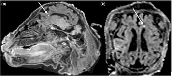
On MRI assessment, the fetus had prominent appearing sulci. Additionally, the fetus had a punctate T1 hyperintense white matter lesion near the anterior superior margin of the frontal horn of the right lateral ventricle (Fig 1). The appearance of this lesion was prototypical of white matter injury seen in neonates with complex CHD.
MRI of periventricular white matter injury in sheep brain. Sagittal ( a ) and axial ( b ) T1-weighted images show a punctate T1 hyperintense lesion near anterior aspect of the lateral ventricle.

Histopathologic assessment
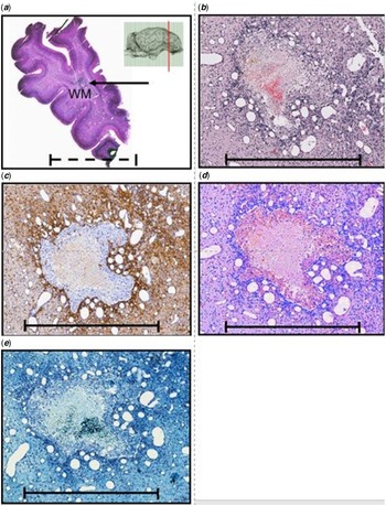
On histologic analysis of the fetal brain, there was a lesion identified in the anterior-superior region of the right frontal horn white matter that correlated to the region of interest on MRI (Fig 2a). On H&E stain, this lesion showed acute on chronic thrombus surrounded by significant micro-vascular proliferation and cystic changes (Fig 2b). In the area immediately adjacent to the lesion, there was significant glial fibrillary acidic protein-labelled astrocyte infiltration (Fig 2c) but no Iba-1-labelled microglial infiltration (Fig 2d). The lesion also lacked myelin fibres (Fig 2e).
( a – e ) The region of T1 hyperintensity identified on MRI in the right frontal lobe. Dotted scale bar = 10 mm; solid scale bar = 1 mm. ( a ) A low powered view of the lesion which is marked with an arrow. The lesion is seen in an area of frontal lobe white matter. ( b ) The same area at 5× magnification in which hypervascularity and an area of thrombosis can be appreciated. ( c ) The same lesion with avid GFAP staining. ( d ) A lack of Iba-1 uptake. ( e ) The absence of myelin staining in the lesion of interest.

Discussion
Utilising an animal model that mimicked the prenatal hypoxemia seen in CHD, we identified a T1 hyperintense lesion in the frontal lobe white matter of a studied fetus. This lesion is similar to those observed on early postnatal MRIs of neonates with CHD and known to be an important marker of future neurodevelopmental outcome. Reference Beca, Gunn and Coleman3,Reference Guo, Chau and Peyvandi8,Reference Claessens, Breur and Groenendaal18 Despites their importance, these MRI lesions have not been previously recapitulated in an animal model of CHD or characterised histologically in neonates with CHD. Other prenatal models of acute, transient ischaemic conditions modelled with acute carotid artery occlusion have generated diffuse areas of T2 hypointensity rather than focal punctate areas of T1 hyperintensity seen in this model of chronic hypoxia where normal cerebral perfusion pressure is maintained without transient perturbations in oxygen delivery throughout the study period. Reference Riddle, Dean and Buser19 Here, for the first time, we were able to reproduce these specific radiographic white matter lesions under prenatal hypoxemic conditions and characterise the lesions histologically by microvascular proliferation and astrocytosis with minimal gliosis. These findings may provide insight into the aetiology of white matter injury in neonates with CHD.
The characterisation of this T1 hyperintense lesion is critical given its association with poor neurodevelopmental outcomes. Here we show that this radiographic lesion, typically recognised postnatally, can occur prenatally. Previous studies in human patients with CHD have only demonstrated these lesions postnatally, but these studies have overwhelmingly been performed with fetal ultrafast T2-weighted imaging. Reference Brossard-Racine, du Plessis and Vezina20,Reference Peyvandi, Lim and Marini21,Reference Claessens, Khalili and Isgum9 When in vivo T1 weighted imaging was performed, its done with low spatial resolution to facilitate some increase in speed that limits the ability to detect small lesions seen in punctate WMI. Since fetal MRIs were performed post-mortem here, T1-imaging could be optimised without time limitations to allow detection of small lesions. Restriction of water diffusion that is a characteristic of acute injury. Identification of this lesion prenatally and its lack of restriction of water diffusion, a characteristic of acute injury, support the notion that many of these lesions may actually be the result of an in utero insult rather than postnatal ischaemia as has been previously speculated. Reference Mahle, Tavani and Zimmerman7 The presence of significant hypervascularity, a chronic compensatory response to thrombus, further suggests these lesions may occur early in gestation after exposure to hypoxemia. White matter hypervascularity has been previously identified in prior experiments under similar prenatal conditions of hypoxia. Reference Lawrence, McGovern and Mejaddam13 Finally, this lesion occurred in an area of relative white matter immaturity, similar to injury patterns observed in preterm infants, supporting the hypothesis that areas of relative brain immaturity may be more vulnerable to ischaemic injury. Reference Licht, Shera and Clancy1,Reference Tusor, Benders and Counsell10,Reference Claessens, Breur and Groenendaal18 Further studies will seek to identify oligodendrocyte precursors to better characterise the background of immaturity. Understanding the aetiology of these lesions will ultimately help better inform the timing and nature of fetal interventions.
In our previously published series on four animals supported under similar conditions of prenatal hypoxia, these lesions were not identified radiographically or histologically in any of the fetuses, including fetuses supported under normoxic conditions and in utero control fetuses. Reference Lawrence, McGovern and Mejaddam13 Interestingly, these lesions are only observed radiographically in 20% of human neonates born with cyanotic CHD as well. Reference Beca, Gunn and Coleman3,Reference Dimitropoulos, McQuillen and Sethi4,Reference Petit, Rome and Wernovsky6 The animal described here had similar umbilical artery blood pressures, fetal substrate delivery, blood gas parameters, and oxygen delivery throughout the study period, supporting that similar cerebral perfusion pressure was maintained throughout the duration of the study period, but notably, umbilical blood flow was lower in the animal described in this report. Unfortunately, though until more studies are completed, the aetiology of this lesion remains unknown and lower umbilical blood flow in the animal of interest is an important confounder in the analysis. Future studies will be aimed at determining if umbilical blood flow, a surrogate of cardiac output, may play a role in the development of these lesions.
The extra-uterine transition for neonatal development system is well suited to study the aetiology of white matter injury in neonates with CHD. This model replicates the chronic hypoxemic conditions seen by fetuses with severe CHD without effecting fetal haemodynamics or substrate delivery. We have previously demonstrated that animals supported in this system under hypoxemic conditions exhibit reduced cortical folding, decreased myelination, and abnormal neuronal migration in patterns similar to those observed in CHD. Reference Lawrence, McGovern and Mejaddam12,Reference Lawrence, McGovern and Mejaddam13 Now we additionally show that this animal model recapitulates another phenotypic marker of CHD, T1 hyperintense lesions on MRI, further solidifying its strength as a model for neurodevelopment in CHD. By recreating the characteristic brain pathology of CHD, this model may be used to test potential fetal interventions in the future.
There are many limitations to this report. Chiefly, since it is focused on one animal, future studies will need to demonstrate reproducibility to better validate the model. Next, since the study period was over two weeks, it is possible that prenatal hypoxemia was not the variable that led to white lesions and the reduced umbilical blood flow in the animal of interest introduces confounding, even if oxygen delivery, umbilical artery pressure, and presumably cerebral perfusion pressure was similar across all animals. Again, future studies will be designed to control for all other fetal variables to better surmise the aetiology of white matter lesions. Next, given a paucity of available brain tissue, we were unable to additionally stain this lesion for calcium or iron to gain additional insight into the aetiology of this lesion and its MRI appearance, including if this lesion may have been the result of a thromboembolic complication. Although this animal was anticoagulated with heparin, and no evidence of thrombus was found on necropsy, this cannot be ruled out without future pathologic analysis. Notably, Usada et al identified an area of white matter necrosis with microglial infiltration in one fetal sheep that experienced a likely thromboembolic event after an episode of cannula occlusion in their fetal sheep model supported for a short period in their artificial placenta model. Reference Usuda, Watanabe and Saito22 This lesion was pathologically distinct from the lesion we identified as it was infiltrated with inflammatory microglia, but it does support that white matter injury may occur during periods of haemodynamic instability in an artificial placenta model. Finally, since the MRI was obtained at necropsy, it is not possible to know when during the study duration the white matter injury occurred. Additional studies will be aimed at obtaining real-time functional brain imaging to better understand the time course of injury.
Despite these limitations, this is the first time that T1 hyperintense white matter lesions have been demonstrated in an animal model of CHD or characterised histologically. Together, these findings offer important insights into the aetiology of white matter injury in CHD that may have important implications for neurodevelopmental outcomes. The recapitulation of this characteristic pattern of white matter injury also further supports that the extra-uterine transition for neonatal development system is a robust animal model that mimics the neurodevelopment of fetuses with CHD, making it an important tool to test future fetal interventions.
Acknowledgements
The authors acknowledge the technical support of Antoneta Radu.
Financial support
This study was made possible by generous support of the Children’s Hospital of Philadelphia.
Conflicts of interest
Alan W. Flake holds multiple patents related to the EXtra-uterine Transition for Neonatal Development technology and is a Clinical Advisor for the Vitara Biomedical Inc. Marcus G Davey holds multiple patents related to the EXtra-uterine Transition for Neonatal Development technology and is the Vice President of preclinical research at Vitara Biomedical Incorporated.
Ethical standards
The authors assert that all procedures contributing to this work comply with the ethical standards of the relevant national guides on the care and use of laboratory animals and has been approved by the EIACUC of the Children’s Hospital of Philadelphia.