Introduction
Members of the genus Oesophagostomum mainly parasitize the large intestine of livestock, wild ruminants and humans (Bremner et al., Reference Bremner, Keith and Winks1976; Lai et al., Reference Lai, Zhou, Huang and Hu2011) with a worldwide distribution (Biffa et al., Reference Biffa, Jobre and Chaka2006; Wang et al., Reference Wang, Qiu, Zhu, Han, Ni, Zhao, Zhou, Zhang and Lun2006; Cenci et al., Reference Cenci, Louvandini, McManus, Dell'Porto, Costa, Araújo, Minho and Abdalla2007; Tariq et al., Reference Tariq, Chishti, Ahmad and Shawl2008; Acevedo-Ramírez et al., Reference Acevedo-Ramírez, Quiroz-Romero, Cruz-Mendoza, Ulloa-Arvizu and Ibarra-Velarde2012). They are called ‘node worms’ due to the yellowish nodules formed on the bowel by the larvae of Oesophagostomum. Oesophagostomum columbianum, O. asperum, O. venulosum and O. kansuensis are considered to be the predominant species in sheep and goats (Roeber et al., Reference Roeber, Jex, Campbell, Campbell, Anderson and Gasser2011; Sweeny et al., Reference Sweeny, Robertson, Ryan, Jacobson and Woodgate2012).
Oesophagostomiasis of small ruminants (sheep and goats) is caused by the ingestion of feed or soil contaminated with the infective larvae of Oesophagostomum species, reducing the productive performance and sometime presenting clinical diseases or even death (Stewart & Gasbarre, Reference Stewart and Gasbarre1989; Sweeny et al., Reference Sweeny, Robertson, Ryan, Jacobson and Woodgate2011). The symptoms caused by infection with O. columbianum or O. asperum are similar. Adult nematodes inhabit the mucosa of the large intestine, leading to anaemia and rapid weight loss. Larval penetration in the mucosa may cause diarrhoea with black-green faeces containing mucus and sometimes blood.
The key to control of Oesophagostomum infection is the accurate identification of causative agents. To date, the traditional morphological approach for the identification and differentiation of Oesophagostomum spp. has limitations in that it is time consuming and requires extensive training (Bott et al., Reference Bott, Campbell, Beveridge, Chilton, Rees, Hunt and Gasser2009), and it can be difficult to identify species with similar morphology, in particular at larval and egg stages (McManus & Bowles, Reference McManus and Bowles1996; Dobson et al., Reference Dobson, Barnes, Birclijin and Gill2002). To overcome these problems, DNA-based technologies utilizing appropriate genetic markers have proved to be useful alternative approaches (Gasser & Newton, Reference Gasser and Newton2000; Blouin, Reference Blouin2002). Previous studies have demonstrated that sequences of internal transcribed spacers (ITS) of nuclear ribosomal DNA (rDNA) provide useful genetic markers for the accurate identification of nematodes, trematodes and cestodes (Orosová et al., Reference Orosová, Ivica, Eva and Marta2010; Atopkin, Reference Atopkin2011; Dai et al., Reference Dai, Liu, Song, Lin, Yuan, Li, Huang, Liu and Zhu2012; Yamada et al., Reference Yamada, Yoshida, Yoshida, Kuraishi, Hattori, Kai, Naqai, Sakoda, Tatara, Abe and Fukumoto2012). Lin et al. (Reference Lin, Zhu, Wei, Deng, Liu, Song, Li and Lun2007, Reference Lin, Ai, Zou, Verweij, Jiang, Li, Song and Zhu2008) demonstrated that ITS rDNA sequences provide reliable genetic markers for the identification and differentiation of Oesophagostomum spp. in pigs.
The objectives of the present study were to characterize the ITS rDNA sequences of O. asperum and O. columbianum from different geographical locations in China, and to develop an effective polymerase chain reaction (PCR) to identify O. columbianum.
Materials and methods
Parasites and isolation of genomic DNA
Adult nematodes of O. asperum and O. columbianum were collected from the large intestine of infected sheep or goats from different geographic locations in China (table 1), washed three or four times with physiological saline, identified according to morphological features described previously (Wang, Reference Wang2002; Bowman, Reference Bowman2009), mainly the anterior end of the parasites (see fig. 1). These samples were fixed in 75% ethanol until genomic DNA (gDNA) could be extracted. Each individual adult was treated with sodium dodecyl sulphate (SDS)/proteinase K at 56°C overnight. The gDNA of each parasite was extracted using the Wizard® SV Genomic DNA Purification System (Promega, Madison, Wisconsin, USA) and eluted in 50 μl elution buffer according to the manufacturer's recommendations.
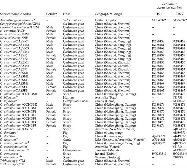
Geographical origins and genders of nematode samples used in the present study, as well as their GenBank™ accession numbers for sequences of the internal transcribed spacer of nuclear ribosomal DNA.

aJefferies et al. (Reference Jefferies, Shaw, Willesen, Viney and Morgan2010); bRomstad et al. (Reference Romstad, Gasser, Monti, Polderman, Nansen, Pit and Chilton1997); cGasser et al. (Reference Gasser, Woods, Huffman, Blotkamp and Polderman1999); dLin et al. (Reference Lin, Zhu, Wei, Deng, Liu, Song, Li and Lun2007); eNewton et al. (Reference Newton, Chilton, Beveridge and Gasser1998); fNewton et al. (Reference Newton, Chilton, Monti, Bjørn, Várady, Christensen and Gasser1997); gSchindler et al. (Reference Schindler, de Gruijter, Polderman and Gasser2005); hChilton et al. (Reference Chilton, Gasser and Beveridge1997).
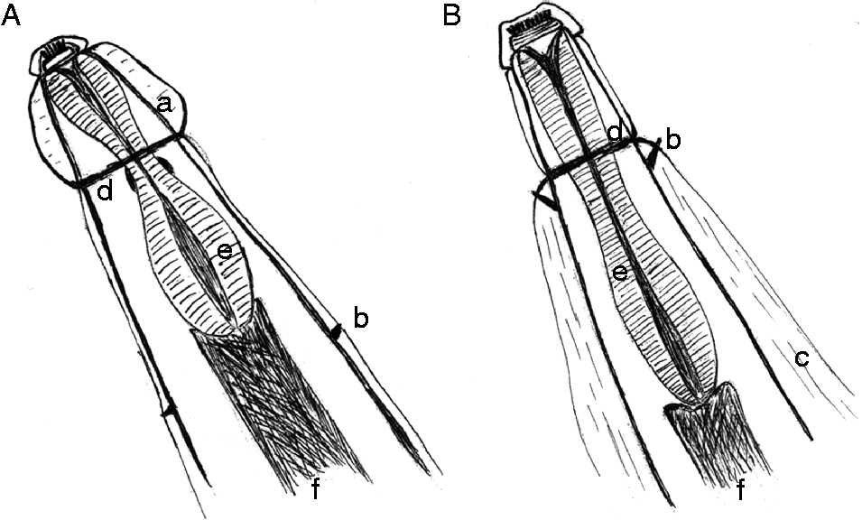
The main morphological differences of the anterior end: (A) O. asperum; (B) O. columbianum. a, Cephalic vesicle; b, cervical papillae; c, lateral alae; d, cervical groove; e, oesophagus; f, intestine.
PCR amplification, DNA sequencing and genetic analysis
The rDNA fragment containing the 3′ end of 18S rDNA, the complete ITS-1, 5.8S, ITS-2 rDNA, and 5′ end of 28S rDNA was amplified from individual nematodes by PCR with the universal primers NC5 (forward: 5′-GTAGGTGAACCTGCGGAAGGATCATT-3′) and NC2 (reverse: 5′-TTAGTTTCTTTTCCTCCGCT-3′) described previously (Zhu et al., Reference Zhu, Gasser, Podolska and Chilton1998). The amount of template used was 1 μl DNA sample in each reaction under the following conditions: denaturation at 94°C for 5 min followed by 35 cycles of 94°C for 2 min, 45°C for 45 s, 72°C for 2 min, and a final extension at 72°C for 10 min. Five microlitres of PCR product was examined by electrophoresis on a 1% (w/v) agarose gel, stained with ethidium bromide and photographed using a gel documentation system (UVItec).
The 5′ and 3′ ends of the ITS-1, 5.8S and ITS-2 rDNA of O. asperum and O. columbianum were determined by comparison with those of O. venulosum (GenBank™ accession no. HQ283349) and O. dentatum (AJ619979). Sequences of these rDNA regions of different isolates were aligned separately using Clustal X 1.83 (Thompson et al., Reference Thompson, Gibson, Plewniak, Jeanmougin and Higgins1997). The intra- and inter-species sequence differences in each of these rDNA regions within the genus Oesophagostomum were determined using Megalign procedure in DNASTAR 5.0 (Burland, Reference Burland2000), and the base composition, transition and transversion were calculated using Mega 4.0 (Tamura et al., Reference Tamura, Dudley, Nei and Kumar2007).
Because O. asperum and O. columbianum had identical 5.8S rDNA sequences (data not shown), the phylogenetic relationships of O. asperum and O. columbianum were reconstructed using ITS-1 and ITS-2 rDNA sequences, respectively, with Angiostrongylus vasorum (GU045372) as the outgroup. Ambiguously aligned regions for each rDNA region were excluded using Gblocks online server (http://molevol.cmima.csic.es/castresana/Gblocks_server.html) with the options for a more stringent selection. The final sequences of the two internal transcribed rDNA regions were used to reconstruct genetic trees among Oesophagostomum species, using maximum parsimony (MP) which was carried out with the PAUP 4.0 Beta 10 program (Swofford, Reference Swofford2002) using the TBR swapping algorithm and ‘branch-and-bound’ search. The consensus tree was obtained after bootstrap analysis, with 1000 replications and values above 50% reported. Phylograms were drawn using the TreeView program version 1.65 (Page, Reference Page1996).
Application of specific PCR for the differentiation of O. asperum and O. columbianum isolates from different geographical regions in China
Based on sequence alignments of ITS rDNA of O. asperum and O. columbianum, a specific PCR assay was established using the primer combination NC5/NC2. The specificity of this PCR assay was determined using some of the common nematode species from goats (table 1), namely Haemonchus contortus, Trichuris spp., Nematodirus sp. and Gongylonema pulchrum, as ‘controls’. The sensitivity of the specific PCR assay was assessed with a DNA sample extracted from a female O. columbianum at concentrations of 53.9, 13.5, 3.4, 0.8, 0.2, 0.05, 0.013, 0.003 and 0.0008 ng/μl, respectively. The detection limit was the minimum DNA amount at which a PCR product was still visible on an agarose gel.
A total of 12 pairs of O. asperum and 12 pairs of O. columbianum adults were obtained from different geographical locations in China (table 1). The gDNA of each isolate was extracted using the method described above, and amplified using this specific PCR. The amplicons (5 μl) were examined by agarose gel (1%) electrophoresis to validate the efficiency of this specific PCR assay.
Results and discussion
All Oesophagostomum isolates were identified to species level (O. asperum or O. columbianum) according to the main morphological differences of the anterior end between O. asperum and O. columbianum (fig. 1): (1) O. asperum has two well-defined cephalic vesicles, but O. columbianum has no cephalic vesicle; (2) O. columbianum has large lateral alae, but O. asperum has no lateral alae; (3) O. columbianum has a pair of strong cervical papillae immediately after the cervical groove, but O. asperum has a pair of tiny cervical papillae after the oesophagus. gDNA was isolated from 37 Oesophagostomum samples, including 21 O. asperum and 16 O. columbianum isolates. The entire ITS fragment, including ITS-1, 5.8S, ITS-2, as well as the primer flanking sequences, was amplified from each individual nematode sample. Each amplicon appeared as a single band of 700–800 bp for both O. asperum and O. columbianum. There was no detectable length difference among different samples of the same species. However, the length of amplicons for O. asperum was longer than those of O. columbianum (fig. 2). These results showed that this PCR assay would provide a useful tool for the rapid identification and differentiation of O. asperum and O. columbianum.
Representative ITS rDNA PCR products of O. asperum and O. columbianum isolates. Lanes: 1–6, O. asperum samples from mainland China (samples OASYM1, OASYF1, OASZM1, OASZF1, OASSM1, OASSF1, respectively); 7–12, O. columbianum samples from mainland China (samples OCHDM1, OCHDF1, OCHDM2, OCHDF2, OCHDM3, OCHDF3, respectively); C, no-DNA control; M, DNA size marker 2000 (ordinate values in bp). Refer to table 1 for sample information.
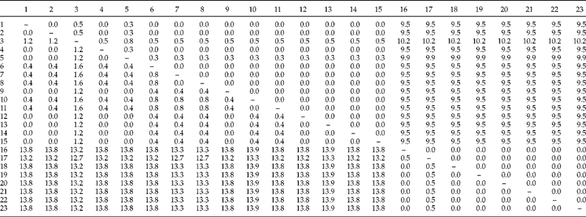
The ITS sequences for the two Oesophagostomum species were obtained by sequencing 23 representative samples (including 15 O. asperum samples and 8 O. columbianum samples). The ITS rDNA sequence of O. asperum was 786 bp in length, consisting of ITS-1 (374 bp), 5.8S rDNA (153 bp) and ITS-2 (259 bp). The length of the ITS rDNA sequence of O. columbianum was 732 bp, containing ITS-1 (361 bp), 5.8S rDNA (153 bp) and ITS-2 (218 bp). The two Oesophagostomum species had identical 5.8S rDNA sequences (data not shown). Sequence variations in the ITS-1 rDNA within O. asperum were 0–0.8%, including three C ↔ T transitions at sequence positions 9, 129 and 219 (data not shown). But no intraspecific variation was detected in the ITS-1 rDNA among O. columbianum isolates. Nucleotide variations in the ITS-2 rDNA among different O. asperum isolates were 0–1.6%, including three transitions (two of A ↔ G and one of C ↔ T), three transversions (A ↔ C, A ↔ T and T ↔ G) and two deletions (A or T) (data not shown). Sequence variations in the ITS-2 rDNA within O. columbianum were 0–0.5%, with only one A ↔ G transition. Sequence differences in the ITS-1 and ITS-2 rDNA between O. asperum and O. columbianum were 9.5–10.2% and 12.7–13.9% (table 2), respectively. Comparison analysis revealed that sequence differences in the ITS-1 and ITS-2 among members of the genus Oesophagostomum were 2.5–11.6% and 6.8–22.3%, respectively (data not shown).
Pairwise comparison of sequence differences (in %) in the first (ITS-1, above the diagonal) and second internal transcribed spacers (ITS-2, below the diagonal) of nuclear ribosomal DNA within and among O. columbianum and O. asperum.
1–23 represent OASZM1, OASZM2, OASZF1, OASYM1, OASYF1, OASYM3, OASYM4, OASYF2, OASYM2, OASSF1, OASSF2, OASSM1, OASSM2, OASSM3, OASSM4, OCHDF1, OCHDF2, OCHDF3, OCHDF4, OCHDF5, OCHDM1, OCHDM2 and OCHDM3 samples, respectively. Refer to table 1 for details of samples.
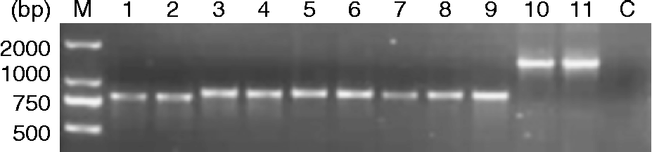
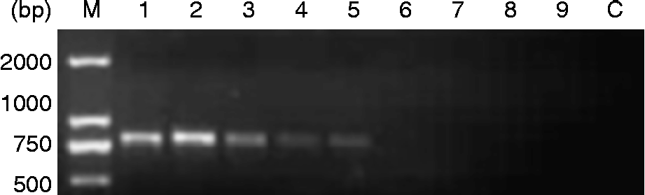
Based on sequence analysis of the ITS rDNA, a specific PCR assay was established to differentiate O. asperum and O. columbianum using primer pair NC5/NC2. The lengths of expected amplicons were 786 bp and 732 bp for O. asperum and O. columbianum, respectively. The specific PCR protocols were optimized by varying the annealing temperatures and titrating the Mg2+concentrations. The optimal annealing temperature was 45°C, and the optimal Mg2+ concentration was 2.5 mm. Under the optimized cycling conditions, the O. columbianum isolates could be easily identified based on the PCR product length (fig. 3). The smallest amount of O. columbianum DNA detectable by the specific PCR assay was 0.2 ng/μl (fig. 4). These findings showed that the PCR assay was specific and sensitive for the detection and delineation of O. columbianum isolates.
Specificity of the specific PCR assay for the detection of O. columbianum. Lanes: 1–2, O. columbianum (samples OCHDM7 and OCHDF9); 3–4, O. asperum (samples OASYM1 and OASZF1); 5–6, Haemonchus contortus (samples HCM and HCF); 7–8, Nematodirus sp. (samples NM and NF); 9, Gongylonema pulchrum (sample GPM); 10–11, Trichuris spp. (samples TM and TF); C, no-DNA control; M, DNA size marker 2000 (ordinate values in bp). Refer to table 1 for sample information.
Sensitivity of the specific PCR for the identification of O. columbianum by agarose gel electrophoresis. Lanes: 1–9, O. columbianum DNA (sample OCHDF9) at concentrations of 53.9, 13.5, 3.4, 0.8, 0.2, 0.05, 0.013, 0.003 and 0.0008 ng/μl, respectively; C, no-DNA control; M, DNA size marker 2000 (ordinate values in bp).
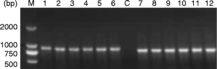
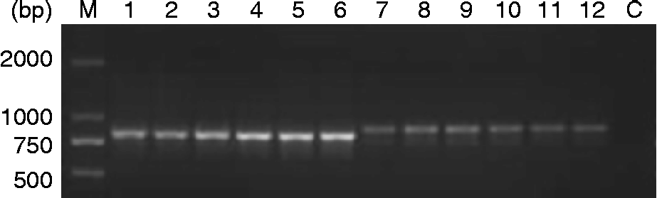
In order to validate the applicability of this specific PCR assay, a total of 48 Oesophagostomum samples from sheep (12 pairs of O. columbianum) and goats (12 pairs of O. asperum) in two endemic provinces (Heilongjiang and Shaanxi provinces) in mainland China (table 1) were amplified by the specific PCR assay. The representative amplification results are shown in fig. 5. There was a visible length difference among amplicons between O. asperum and O. columbianum in that the amplicon length of O. columbianum ITS rDNA was approximately 50 bp shorter than that of O. asperum (fig. 5).
The application of the specific PCR to discriminate O. asperum and O. columbianum isolates from different geographic regions in China. Lanes: 1–6, O. columbianum from mainland China (samples OCHDM4, OCHDF6, OCHDM5, OCHDF7, OCHDM6, OCHDF8, respectively); 7–12, O. asperum from mainland China (samples OASYM5, OASYF3, OASZM3, OASZF2, OASSM5, OASSF3, respectively); C, no-DNA control; M, DNA size marker 2000 (ordinate values in bp). Refer to table 1 for sample information.
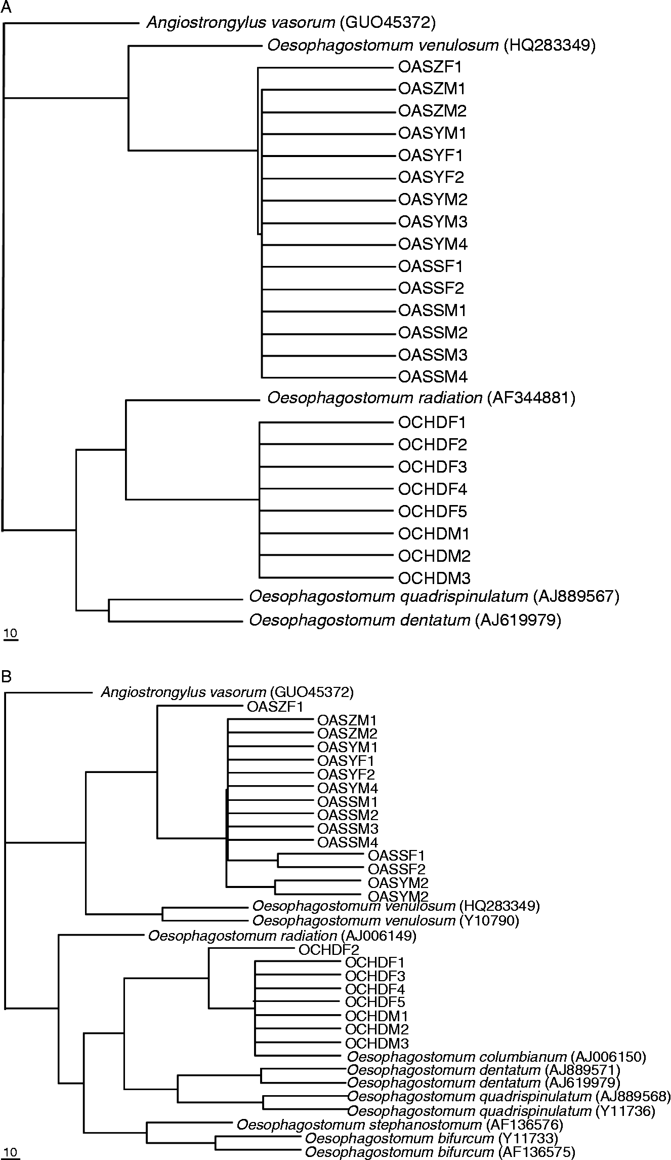
To determine the phylogenetic positions of O. asperum and O. columbianum, phylogenetic relationships among members of the genus Oesophagostomum were reconstructed using maximum parsimony (MP) analysis based on sequences of the ITS-1 (fig. 6A) and ITS-2 rDNA (fig. 6B), respectively, with Angiostrongylus vasorum (GU045372) as the outgroup. On the two phylogenetic trees, all O. asperum isolates clustered together, sistered to O. venulosum of sheep. However, the clustering of O. columbianum isolates was slightly different between the two phylogenetic trees. On the tree based on ITS-1 rDNA sequences, all O. columbianum isolates grouped in one clade sistered to O. dentatum. But on the ITS-2 tree, all O. columbianum isolates grouped with O. columbianum from Australia, located in the sister clade of two pig nodule worms, O. dentatum and O. quadrispinulatum. These results suggested that the ITS rDNA would be suitable for phylogenetic analysis of the genus Oesophagostomum.
Maximum parsimony (MP) trees of Oesophagostomum spp. based on ITS-1 (A) and ITS-2 rDNA (B) using Angiostrongylus vasorum (GU045372) as the outgroup.
Oesophagostomiasis caused by O. columbianum or O. asperum is an important parasitic disease of small ruminants (Eguale et al., Reference Eguale, Chaka and Gizaw2009; Tariq et al., Reference Tariq, Chishti and Ahmad2010). Oesophagostomum columbianum has been identified as the important cause of nodular enteritis, leading to reduced productivity in sheep and goats worldwide, for example in the Kashmir valley of India (Tariq et al., Reference Tariq, Chishti, Ahmad and Shawl2008) and in Ethiopia (Biffa et al., Reference Biffa, Jobre and Chaka2006). Therefore, there is an urgent need to develop effective strategies to control Oesophagostomum infection. Because of the limitations of traditional parasitological methods (Dobson et al., Reference Dobson, Barnes, Birclijin and Gill2002; Bott et al., Reference Bott, Campbell, Beveridge, Chilton, Rees, Hunt and Gasser2009), DNA technology provides an alternative approach. To date, a PCR assay has been developed for the differentiation of O. columbianum and O. venulosum, utilizing genetic markers in ITS-2 rDNA (Bott et al., Reference Bott, Campbell, Beveridge, Chilton, Rees, Hunt and Gasser2009). The present study characterized O. columbianum and O. asperum in China by their ITS rDNA sequences. The lengths of ITS-1 and ITS-2 rDNA of O. asperum were longer than those of O. columbianum. Based on this length difference, an effective PCR approach was developed for the rapid and sensitive differentiation between O. columbianum and O. asperum. Since accurate characterization of parasites at different taxonomic levels is essential for population genetic studies and parasitosis control, the present findings have important implications for studying epidemiology, taxonomy and population biology of Oesophagostomum spp., as well as for controlling oesophagostomiasis.
Acknowledgements
This work was supported in part by grants from the Opening Funds of the State Key Laboratory of Veterinary Etiological Biology, Lanzhou Veterinary Research Institute, Chinese Academy of Agricultural Sciences to G.H.Z. (SKLVEB2011KFKT011) and S.K.Y. (SKLVEB2009KFK003), the Special Fund for Talents in Northwest A&F University (Z109021107 and 2010BSJJ015) and Basic Research Key Program (ZD2012010) to G.H.Z., and Guangdong Entry-Exit Inspection and Quarantine Bureau to Y.D. (2012GDK04). Professor Xing-Quan Zhu at Department of Parasitology, Lanzhou Veterinary Research Institute, Chinese Academy of Agricultural Sciences is thanked for improving the quality of the manuscript.