Introduction
The Papillomaviridae family comprises a heterogeneous group of non-enveloped, double-stranded DNA viruses of 5748–8607 bp in size (Ref. Reference Van Doorslaer1). The analysis of the L1 gene contributed to the classification of Papillomaviruses (PVs) into genera, species, types, subtypes and variants where PV genera in full-length sequence genomes present 23–43% nucleotide sequence similarity, whereas at the L1 sequence level they share less than 60% nucleotide sequence homology (Refs Reference Bernard2–Reference de Villiers4). In addition, species within a genus have 60–70% L1 sequence similarity (Refs Reference Bernard2, Reference Bernard, Calleja-Macias and Dunn3). A novel human papillomavirus (HPV) type has less than 90% similarity to any other HPV genotype (Ref. Reference Bzhalava, Eklund and Dillner5). HPV genomes are further grouped into intratypic variants according to the whole genome sequence, defining variant lineages and sub-lineages by a divergence of 1–10% and 0.5–1%, respectively (Refs Reference Chen6–Reference Mirabello10). HPVs infect the stratified epithelium of skin or mucus membranes and they are classified into five genera (Alphapapillomaviruses, Betapapillomaviruses, Gammapapillomaviruses, Mupapillomaviruses and Nupapillomaviruses) (Refs Reference Bernard2, Reference Bernard, Calleja-Macias and Dunn3, Reference Gheit11–Reference Tsakogiannis13).
The HPVs that infect the mucosal epithelium are classified into the genus Alphapapillomaviruses (alpha-HPVs), and according to their tumourigenic capacity they are subdivided into high-risk (HR-HPV) and low-risk (LR-HPV) genotypes (Refs Reference Bernard2, Reference Bernard, Calleja-Macias and Dunn3, Reference Zur Hausen12). In particular, 15 HPV genotypes have been characterised as high-risk (HPV16, 18, 31, 33, 35, 39, 45, 51, 52, 56, 58, 59, 68, 73 and 82) (Ref. Reference Munoz14). Persistent infection with HR-HPVs over a period of several years, along with associated alterations in the host cell is responsible for cervical cancer development. Cervical cancer is the fourth most common cancer and the fourth leading cause of cancer-related death among women, worldwide (Refs Reference Berman and Schiller15, Reference Ferlay16). With regards to clinical and epidemiological outcomes, HR-HPV16 infection is considered as the most prevalent sexually transmitted viral infection that is observed in more than 50% of cervical cancer incidences, worldwide (Refs Reference Ferlay16–Reference Tsakogiannis19).
The HPV genome is approximately 8 kb in size and is divided into three regions: (i) the long control region (LCR) which contains binding sites of viral regulatory proteins (E1 and E2) and cellular transcription factors, (ii) the early region (E) that encodes for the E1, E2, E4, E5, E6 and E7 genes and (iii) the late region (L) that encodes for the L1 and L2 genes (Refs Reference Tsakogiannis13, Reference Van Doorslaer20). Over the last few decades, the strong tumourigenic capacity of HPV16 DNA triggered the extensive sequence analysis of the viral genome, thus leading to the detection of various nucleotide variations, whereas several of these nucleotide changes have been associated with immune escape, viral persistence and cervical cancer development (Refs Reference Togtema21, Reference Graham and Herrington22). Moreover, specific intratypic nucleotide polymorphisms have been identified and associated with different geographic locations and ethnic groups, while they enabled the clustering of HPV16 DNA into four major phylogenetic branches (Refs Reference Chen23–Reference Ho25). As a result, the HPV16 DNA is grouped into four major variant lineages (A–D) and 16 sub-lineages (A1–4, B1–4, C1–4 and D1–4) the prevalence of which varies geographically. The sub-lineages include, A1–A3 (European variant), A4 (Asian variant), B1–B4 (African type I variant), C1–C4 (African type II variant), D1 (North American variant), D2 (Asian American type I), D3 (Asian American type II) and D4 (Refs Reference Burk, Harari and Chen7, Reference Mirabello9, Reference Schiffman26).
In this review, we discuss in detail the various nucleotide variations that have been detected in HPV16 DNA with a special focus on those that are associated with viral oncogenicity and further shed light on their prevalence among different geographic populations. Finally, we discuss whether the background genomic variability of HPV16 DNA influences viral carcinogenic potential.
E6–E7 genes
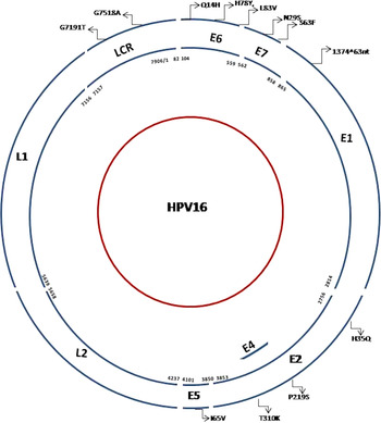
The HPV16 E6 gene is positioned between nucleotides 104 and 559 and it encodes a protein of 158 amino acids that consists of four C-x-x-C motifs which are responsible for the interaction with cellular proteins, transcriptional activation, transformation and immortalisation of infected cells (Fig. 1). In addition, the C-terminal domain of E6 oncoprotein harbours a PDZ-binding motif which is implicated in interactions with proteins containing PDZ domains (Ref. Reference Boulet27). Next to E6 oncogene is placed the E7 gene (nucleotides 562 and 858) that encodes a protein of 98 amino acids. The E7 protein contains three conserved domains, including CR1, CR2 and CR3 (Fig. 1). The CR1 is located in the N-terminal domain of viral protein and it is involved in cellular transformation and pRb degradation (Ref. Reference Boulet27). The CR2 domain includes a pRb-binding sequence LxCxE and a CKII (casein kinase II) phosphorylation site. The CR3 C-terminal acts as a dimerisation domain and it harbours two copies of the C-x-x-C motif which are involved in the interaction of viral oncoprotein with pRB and other host cellular proteins (Refs Reference Boulet27, Reference Jabbar28). The E6 and E7 oncoproteins contribute significantly to the unregulated cell proliferation and cancer development, since they orchestrate the destabilisation of cell cycle control (Refs Reference Martinez-Zapien29–Reference Aarthy31). Briefly, the E6 oncoprotein forms a complex with the E3 ubiquitin ligase E6-associated protein (E6AP) which in turn promotes the recruitment and polyubiquitination of p53 through the ubiquitin-mediated degradation pathway (Ref. Reference Martinez-Zapien29). On the contrary, E7 interacts with the PDZ domain of pRb and it releases the E2F transcription factor from the pRB/E2F suppressor complex. The interference of pRB/E2F suppressor complex promotes the mitotic phase of cell cycle and consequently stimulates an uncontrolled cell proliferation (Refs Reference Aarthy31–Reference Tsakogiannis34).
Organisation of HPV16 genome. The major sequence variations, amino acid substitutions and duplication events are described. The amino acid substitutions E6: Q14H, H78Y, L83V, Ε7: N29S, S63F, E2: H35Q, P219S, T310K and E5: I65V have been involved with increased viral pathogenicity. The E1–1374^63nt duplication has not been associated with cervical cancer development, whereas the G7518A and G7191T in the LCR provide considerable distribution among the different HPV16 isolates in all research studies.

Sequence analysis of the E6 and E7 oncogenes revealed that specific intratypic nucleotide polymorphisms are related to the major HPV16 variant lineages, including, A1 sub-lineage; reference group, A2, (E6: T350G), A4 sub-lineage (E6: T178G, E7: A647G), B lineage (E6: G132C, C143G, G145T, T286A, A289G, C335T, E7: T789C, T795G), C lineage (E6: T109C, G132T, C143G, G145T, T286A, A289G, C335T, G403G, E7: A647G, T789C, T795G) and D lineage (E6: G145T, T286A, A289G, C335T, T350G, A532G, E7: T732C, T789C, T795G) (Refs Reference Wu35–Reference Yamada37).
The most extensively studied nucleotide mutation of HPV16 DNA is the E6 T350G that leads to the amino acid substitution L83V (Refs Reference Togtema21, Reference Zhang38–Reference Zehbe41). Notably, in vitro experiments have revealed that the E6 oncoprotein that harbours the L83V enables the transformation of human keratinocytes more effectively than prototype E6 oncogene and further modifies the expression profile of cervical carcinoma C33A cells (Refs Reference Sichero and Villa42, Reference Zacapala-Gomez43). The L83V is found in the A variant lineage and D variant lineage, whereas several studies have been conducted in order to evaluate its carcinogenic ability (Refs Reference Tsakogiannis39–Reference Zehbe41, Reference Grodzki44, Reference Zhe61, Reference Escobar-Escamilla143, Reference Galati144). In particular, it has been proved that E6 oncoprotein of the D variant lineage transforms keratinocytes more efficiently and exhibits greater migration capacity than that of prototype E6 oncoprotein (Refs Reference Niccoli45, Reference Cuninghame46). The high-tumourigenic capacity of E6 oncoprotein of D lineage is also reflected in clinical studies, as infection with D lineage is strongly related to persistency and higher cancer risk compared with A, B and C lineages (Refs Reference Tsakogiannis39, Reference Moschonas40, Reference Zacapala-Gomez43, Reference Tornesello47). In order to further understand the reasons that the E6 oncoprotein of D clade exhibits higher carcinogenic potential than that of prototype E6 oncoprotein, research interest was focused on the amino acid dissimilarity of the respective proteins. The sequence analysis revealed that the E6 oncoprotein of D variants contains an amino acid substitution pattern Q14H, H78Y and L83V that seems to provide a considerable selective advantage in immortalisation and transformation over the prototype oncoprotein (Refs Reference Niccoli45, Reference Cuninghame46, Reference Vander Heiden, Cantley and Thompson48, Reference Richard49).
The strong oncogenic capacity of E6 oncoprotein of D variant lineage is also involved in host signalling pathways, including the Warburg effect (Refs Reference Vander Heiden, Cantley and Thompson48, Reference Richard49). The Warburg effect is defined as a modified metabolism condition in which cancer cells demonstrate an augmented glucose uptake rate (Refs Reference Cuninghame46, Reference Vander Heiden, Cantley and Thompson48, Reference Liberti and Locasale50). The glucose in turn is preferentially metabolised to lactate through anaerobic respiration which is essential for cell proliferation and tumour development (Refs Reference Vander Heiden, Cantley and Thompson48, Reference Liberti and Locasale50). The E6 oncoprotein of D variant lineage is implicated to the Warburg effect by increasing the levels of the glycolytic enzymes PKM2 and GAPDH, thus offering cancer cells an overall selective advantage to establish an ideal microenvironment necessary for cell proliferation (Refs Reference Cuninghame46, Reference Vander Heiden, Cantley and Thompson48, Reference Richard49). Moreover, it has been observed that under hypoxic conditions the E6 oncoprotein of D clade increases the hypoxia-inducible factor 1-alpha (HIF-1a) concentration and function more efficiently compared with prototype E6 (Ref. Reference Cuninghame46). The activation of HIF-1 factor promotes angiogenesis, erythropoiesis and the regulation of glycolytic enzymes that in turn influence the Warburg effect. The increased expression of HIF-1 by the presence of E6 of D variant lineage is based on the ability of the respective oncoprotein to stimulate the mitogen-activated protein kinase (MAPK)/extracellular related kinase (ERK1/2) signalling pathway (Ref. Reference Cuninghame46). Taking all these aspects into account, it is postulated that the specific pattern of the amino acid substitutions Q14H, H78Y and L83V has negative clinical impact by targeting more efficiently crucial hosts’ pathways involved in cancer development. However, further analyses are required in order to investigate whether the E6 oncoprotein of D clade is involved in other signalling pathways that influence the development of cervical cancer.
Considering the clinical aspects of T350G (L83V), research studies have implied that the corresponding mutation seems to be highly tumourigenic in certain populations, providing conflicting outcomes even between adjacent geographic regions. In particular, a multicentre case–control study showed that the 350T is overrepresented in cervical cancer cases in Central Asia and East Asia, whereas the T350G is prevalent in cervical cancer incidences in Central/South America (Ref. Reference Cornet51).
In European population, the tumourigenicity of T350G shows contradictory outcomes (Table 1). In particular, the nucleotide variation T350G is associated with severe dysplasia in Swedish and French women, whereas no significant correlation between the T350G variation and the development of advanced cervical dysplasia was found in women from Greece, Italy and the Czech Republic (Refs Reference Tsakogiannis39–Reference Zehbe41, Reference Grodzki44, Reference Tsakogiannis52). In contrast, infection with the E6 350T variant is more likely to be persistent compared with the T350G variant in Danish patients. In the respective population group, the T350G has not been associated with advanced cervical disease (Ref. Reference Gheit53) (Table 1).
Association of the sequence mutation T350G (L83V) in the HPV16 E6 gene with the development of cervical dysplasia among the different investigated populations

The statistical analysis that was performed in each study as well as the cell functions that were influenced by the mutation T350G are described.
One possible explanation of the observed contradictory outcomes in the studies assessing the consequence of T350G might be the different epistatic interaction of the T/G alleles of E6 350G position with the genetic background of the circulating HPV16 variant lineages in a given population. In particular, the position 350 has been found to be polymorphic T/G in HPV16 A lineage, monomorphic T in HPV16 B and C lineages and monomorphic G in HPV16 D lineage (Ref. Reference Nicolas-Parraga54). The epistatic interaction of T/G alleles with the genetic background of HPV16 isolates may also explain the higher tumourigenicity of D lineage when compared with that of A sub-lineages.
The prevalence of T350G in different populations influences the association of the respective mutation with cervical cancer risk (Refs Reference Wu35–Reference Yamada37, Reference Yamada55). A recent study that analysed more than 7000 genomes of the different sub-lineages globally revealed that A1 (reference group) is the most widespread with more than 70% of all isolates in Europe, South/Central America, North America, South Asia and Oceania, whereas A2 sub-lineage is detected in 10–20% of isolates from Europe, North America and Oceania (Ref. Reference Clifford56). Moreover, A3 and A4 sub-lineages are the most commonly detected HPV16 isolates in East Asia (50 and 20%, respectively), and B and C lineages are present almost exclusively in Africa (Ref. Reference Clifford56). Considering the American population, the D variant lineage (monomorphic G for 350 position) is more common in South/Central and North America and is associated with an increased cervical cancer risk (Refs Reference Burk, Harari and Chen7, Reference Mirabello9, Reference Mirabello10, Reference Clifford56). In North America, the D variant lineage and A4 sub-lineage (monomorphic T for 350 position) are more frequent and are also associated with an increased risk for cervical cancer development (Refs Reference Burk, Harari and Chen7, Reference Mirabello9, Reference Mirabello10, Reference Clifford56). In Europe, the most prevalent sub-lineage in cervical cancer incidences was the A1 followed by A2. In East Asia, the sub-lineages A3 and A4 were associated with an elevated cancer risk (Ref. Reference Clifford56). As a result, the higher prevalence of T350G in specific geographic areas may explain the reasons that T350G is associated with an increased cancer risk in South/Central America and in various European countries, whereas no association has been found in East Asia where A3 and A4 sub-lineages are dominant.
The genetic heterogeneity of host genes plays a pivotal role in patients’ vulnerability towards the development of cervical cancer, because specific HLA haplotypes and p53 polymorphisms have been associated with specific E6 variants and subsequently with a cervical cancer risk (Refs Reference Moschonas40, Reference Burroni57–Reference Bao59). Accordingly, the carcinogenic ability of T350G should be considered along with the genetic background of circulating HPV16 variant lineages as well as with the genetic background of the investigated ethnic cohorts.
Apart from the T350G mutation, additional nucleotide variations have been found to be prevalent in E6 and E7 genes among different geographic populations. In particular, a previous study in Greek population revealed 14 novel non-synonymous nucleotide changes [E6: A184G (I27M), G201A (C33Y), G219A (R39Q), T302A (L67I), A336G (H78R), G514A (M137I), C539T (R146C); E7: C565T (H2Y), T596A (M12K), G685A (A42T), G813A (M84I), G814A (G85S), G815A (G85D), C818T (T86I)] and two synonymous mutations [E6: A280T (P59P), C523T (C140C)] in A lineage (Ref. Reference Tsakogiannis39). However, these changes were not associated with dysplasia and cancer of the cervix. In addition, a previous analysis in patients from northeast China who were diagnosed with invasive cervical carcinoma revealed one novel nucleotide variation in the E6 gene [E6: T341G (C80G)] (Ref. Reference Shang60). In a more recent analysis that was conducted in patients from southern and northern China, 11 new non-synonymous mutations were found [E6: C168G (T22S), G176A (D25N), T178G (D25E), T183G (I27R), A275G (N58D), A276G (N58S), T295G (D64E), C360A (T86K), A442C (E113D); E7: A645C (L28F), A646C (N29H)] (Ref. Reference Zhe61). These changes were not associated with cancer. The analysis of polymorphic variability of the E6 and E7 genes in the Indian population showed a new nucleotide mutation in the E6 gene [E6: T527A (S142T)] that is identified along with E6 T350G nucleotide mutation (Ref. Reference Pande62), whereas two new non-synonymous mutations [E6: C279G (Q66P), A305C (K75R)] were found in Romanian women with or without T350G (Ref. Reference Plesa63). However, these changes were not associated with cervical cancer development. A recent study that was conducted in Poland in cervical cancer cases revealed three non-synonymous mutations within the E6 gene [E6: G122A (E7K), C153T (T17I), G188A (E29K) that were strongly associated with the European T350G variant lineage in low-grade squamous intraepithelial neoplasia (LSIL) (C153T, G122A) and cancer (G188A) cohort (Ref. Reference Szostek64).
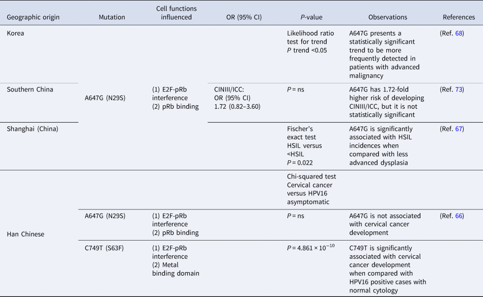
The E7 gene has been found to be more conserved compared with the E6 gene (Refs Reference Mirabello10, Reference Wu35, Reference Shang60–Reference DeFilippis, Ayala and Villarreal72, Reference Zhe61). Notably, three HPV16 E7 nucleotide variations have been extensively investigated in the Asian population including, A647G (N29S), C749T (S63F) and T846C (S95S) (Table 2). The prevalence of these changes in cervical cancer cases varies among the different studied populations (Refs Reference Wu35, Reference de Boer65–Reference Zhao67). In particular, the N29S was found to be more prevalent in cervical cancer incidences compared with HPV16 positive asymptomatic individuals (control group) in Korean patients (Ref. Reference Song68). No considerable difference between cancer cases and the control cohort was observed when the respective mutation was studied in patients derived from southern China and Japan (Refs Reference Fujinaga69, Reference Chan73). Interestingly, another study that was conducted in women from Shanghai suggested that N29S increases the development of high-grade squamous intraepithelial lesion (HSIL) (Ref. Reference Zhao67). Moreover, an analysis that was performed in the Han Chinese population suggested that N29S, S63F and S95S are found in cervical cancer cases with the S63F to be considerably associated with the development of cervical cancer (Ref. Reference Zhou66) (Table 2). This is the first study proposing that S63F is linked to cervical cancer development. The prevalence of this mutation varies among different populations including 14.3 % for Sichuan in central China (Ref. Reference Stephen70) 59.5 % for Korea (Ref. Reference Song68), 0.9 % for Germany (Ref. Reference Nindl74) and 36.4 % for Tanzania (Ref. Reference Eschle75).
Association of A647G (N29S) and C749T (S37F) in the HPV16 E7 gene with the development of cervical dysplasia among the different studied Asian populations

The statistical analysis that was conducted in each study as well as the cell functions that were influenced by the corresponding mutations are summarised.
The fact that the E7 gene is more conserved compared with E6 was also reflected in molecular evolutionary studies. In particular, it has been suggested that the E6 oncogene is evolving under positive Darwinian selection, whereas there is no evidence of positive selection for the E7 oncogene (Refs Reference Tsakogiannis39, Reference Carvajal-Rodriguez71, Reference DeFilippis, Ayala and Villarreal72). Nevertheless, sequence and evolutionary analysis of HPV16 isolates from Greek patients revealed that the novel amino acid substitution at codon 85 (G85S/D) of the E7 oncoprotein is under positive selective pressure (Ref. Reference Tsakogiannis39). The corresponding mutation was preferentially identified in HSIL cases and it is located within the C-terminal CR3 domain of E7 oncoprotein. Interestingly, a previous study that introduced in vitro mutations into the CR3 of HPV16 E7 oncoprotein revealed that the E7 mutant that contains the amino acid change G85A interacts with pRb as effectively as the wild-type E7, although it demonstrates reduced ability to promote the mitotic phase of cell cycle (Ref. Reference Todorovic76). Moreover, it was observed that the G85A E7 mutant interacts efficiently with p21Cip1/WAF1, whereas it exhibits a reduced ability to interact with other cellular targets which are essential for cell cycle promotion. These outcomes suggested that E7 mutants significantly eliminate the oncogenic capacity of viral oncoprotein (Ref. Reference Todorovic76). As a result, the high conservation of the E7 protein might provide the virus an overall selective advantage to establish a persistent infection, possibly because of its crucial function to interact with fundamental cellular factors responsible for cell cycle control.
E1 gene
The HPV16 E1 gene is the largest viral open reading frame (ORF) and it is located between nucleotides 865 and 2814 (Fig. 1). The PV E1 gene is highly conserved among the different HPV genotypes and encodes a protein 649 amino acids long which is the only viral protein with enzymatic function (ATPase and 3′–5′ helicase activities) (Ref. Reference Bergvall, Melendy and Archambault77). The viral protein is expressed during the early stage of virus life cycle and consists of three functional domains comprising (i) the N-terminal regulatory domain that harbours a nuclear localisation signal and a nuclear export signal, (ii) the central DNA-binding domain (DBD) that interacts with the viral DNA replication origin into the LCR, (iii) the C-terminal ATPase/helicase domain that also contains sequences implicated in E1 oligomerisation (Refs Reference Bergvall, Melendy and Archambault77, Reference Cote-Martin78). As it was described above, the viral E2 protein recruits E1 molecules in the viral DNA replication origin, thus forming a functional double E1 hexameric helicase which in turn triggers DNA replication (Refs Reference Bergvall, Melendy and Archambault77, Reference Garcia-Vallve, Alonso and Bravo79–Reference Egawa81). Moreover, the E1 protein recruits various cellular replication factors to the viral DNA replication origin in order to establish an operating replication machinery including, α-DNA polymerase, replication A proteins (RPA), topoisomerases I and II, nuclear proliferation cell antigen (PCNA) and the replication factor C (RFC) (Refs Reference Bergvall, Melendy and Archambault77, Reference Castro-Munoz82–Reference Parker85). Regardless of viral replication, E1 protein also interacts with cellular host factors that are associated with the regulation of epigenetic procedures, such as histone H1 and Ini1/hSNF5 (Refs Reference Swindle and Engler86, Reference Lee87).
Recently, it has been reported that specific variations into the E1 gene are related to the different HPV16 intratypic variant lineages including the B lineage (C1096G, T1366A, C1377T, T1486C, C1624T, C1744T, A1842G, C2041T, G2220C, T2586C and T2631A), C lineage (T921C, C1096G, G1163A, T1200A, T1366A, C1377T, C1416T, C1426G, T1486C, C1624T, C1744T, A1842G, C2041T, G2220C, C2237G, G2249A, C2262T, C2287T, C2344T, T2355C, T2586C, A2608C and T2631A) and D lineage (T921C, A1041G, C1096G, G1163A, T1200A, T1366A, C1377T, C1426G, T1486C, C1624T, A1668G, C1744T, A1842G, C2041T, G2220C, C2237G, G2249A, C2262T, C2287T, C2344T, T2586C, A2608C, T2631A and G2650A) (Ref. Reference Tsakogiannis88). Moreover, it was suggested that these sequence variations enable the classification of viral DNA into four major phylogenetic branches (Ref. Reference Tsakogiannis88). A study that was conducted in Han Chinese patients confirmed the presence of the above polymorphisms in the non-European variant lineages, whereas additional changes were described in European (A1–A3 sub-lineages) and Asian (A4 sub-lineage) variants that appear in the Chinese population. In particular, the nucleotide variations T933A and T1014G were observed only in the A4 sub-lineage, the nucleotide variation A2547G was detected only in the A1–A3 sub-lineages, whereas the sequence changes G2160A and T2232C were detected in all members of the A lineage (Ref. Reference Yao89). In contrast, these changes were not noticed in Greek patients.
The extensive sequence analysis of the HPV16 E1 gene revealed that the nucleotide variations that were detected in the N-terminal domain [T921C (T19T), T933A (A23A), T1014G (D50E), A1041G (L59L), C1096G (Q78E), G1163A (G100E) and T1200C (A112A)], the DNA- binding domain [T1366A (C168S), T1407G (S181R), C1426G (Q188E), T1486C (L208L), T1522A (S220T), C1624T (L154L) and C1744A (L294M)] and the C-terminal domain C2041T [(L393L), G2220C (E452D), T2232C (F456F), C2237G (T458S), G2249A (R462K), C2262T (G466G), C2287T (L475L), T2567G (I568S), T2586C (S574S), A2608C (R582R) and T2631A (P589P)] were only identified in cervical cancer cases in the Chinese population, whereas in Greek patients these changes were preferentially identified in HSIL incidences. The nucleotide variations in the E1 C-terminal domain G2073A (K403K), T2169C (D435D), T2189C (I442T), A2453T (N530I), C2454T (N530N) and A2587T (R575W) that were detected in European variants of Greek population were observed exclusively in HSIL cases (Refs Reference Tsakogiannis88, Reference Yao89). As a result, the differences that are noted between Chinese and Greek populations are required to be further investigated in order to explain whether this can be because of genetic background, epistasis or even because of genetic drift. Then we may elucidate whether these sequence alterations may be used as valuable biomarkers that could predict the development of cervical disease.
Interestingly, in the HPV16 E1 gene the region between nucleotides 1311–1374 (E1-1374^63nt duplication) is found to be duplicated in tandem, leading to a duplication of the region between amino acids 150 and 170 (21 amino acids) into the N-terminal domain of E1 protein. To date, this duplication has been observed in patients derived from China, Croatia, Slovenia and Greece and was strongly associated with the E6 T350G nucleotide variation (Refs Reference Dong, Zhou and Pfister90–Reference Tsakogiannis93). This duplication is significantly associated with the E1 sequence variation A1053C (E63D), as well (Ref. Reference Tsakogiannis93). The A1053C is located upstream of the E1-1374^63nt duplication resulting in the amino acid substitution E63D (Ref. Reference Tsakogiannis93). It is important to highlight that the E1-1374^63nt duplication was preferentially identified in LSIL cases thus related to a reduced viral tumourigenicity (Refs Reference Dong, Zhou and Pfister90–Reference Tsakogiannis93). To date, the impact of E1-1374^63nt and A1053C (E63D) in the function and structure of E1 protein is unknown. Notably, a previous study simulated the three-dimensional structure of the E1 protein with the duplication of 21 amino acids (Ref. Reference Sabol91). The structure prediction models suggested that this duplication partially overlaps the probably phosphorylated threonines at positions 153 and 155, thus modifying phosphorylation regulation of the E1 activity (Ref. Reference Sabol91). Nevertheless, no further studies including in vitro experiments have been conducted in order to reinforce these findings and subsequently to clarify the role of this sequence alteration in E1 protein function.
E2 gene
The PV E2 protein is a virus-encoded transcriptional and replication factor that plays a pivotal role in PV life cycle. The HPV16 E2 ORF is situated between nucleotides 2756 and 3853 and encodes a protein with 366 amino acids (Fig. 1) (Refs Reference McPhillips, Ozato and McBride94, Reference McBride95). The viral protein comprises an N-terminal transactivation domain (TAD), a hinge region and a C-terminal DBD. The E2 as a homodimeric protein binds to a sequence motif (ACCGN4CGGT) in the LCR of viral DNA and it serves as a transcription activator or suppressor (Refs Reference McBride95, Reference D'Abramo and Archambault96). The function of E2 protein as transcription activator or suppressor lies mainly on E2 protein levels as well as on the binding of E2 protein to specific LCR sites (Refs Reference McBride95, Reference D'Abramo and Archambault96). Furthermore, the E2 protein has a fundamental impact on viral DNA replication, as well. In particular, the TAD of the E2 protein interacts with the helicase domain of the viral E1 protein, whereas the E2 protein in turn assembles more E1 molecules in the viral DNA replication origin, hence generating a functional double E1 hexameric helicase that coordinates the replication of viral episomal DNA (Refs Reference Swan36, Reference D'Abramo and Archambault96, Reference Gauson97).
The sequence analysis of the HPV16 E2 gene revealed specific intratypic nucleotide polymorphisms that have been associated with the major HPV16 variant lineages including, A1–A3 sub-lineages (A2938G, T3384C and C3684A), A4 sub-lineage (G2828A, C3159A, T3224A, T3524C, G3249A, T3384C, G3449A, T3524C, C3684A and C3787A), B lineage (C2860A, C3159A, C3161T, A3181C, G3182A, G3249A, A3362G, C3377G, A3425C, G3449A, C3516A, T3566G, T3664C, C3684A, T3694A, G3778T, T3784G, C3787A and T3858C), C lineage (C2860A, C3043T, C3159A, C3161T, G3182A, G3249A, A3362G, C3377G, G3441A, G3449A, C3516A, T3517C, A3538C, T3566G, C3684A, T3694A, T3706C, G3778T, C3787A, T3805G and T3858C) and D lineage (C2860A, C3159A, A3181C, G3182A, T3224A, G3249A, A3362G, C3377G, T3387C, G3449A, C3516A, T3517C, A3538C, T3566G, T3664C, C3684A, T3694A, T3706C, G3778T, C3787A, T3805G and T3858C). Notably, these nucleotide variations enabled the classification of HPV16 genome into the four major phylogenetic branches (Refs Reference Tsakogiannis98, Reference Kahla99).
Considering the distribution of nucleotide variations throughout HPV16 E2 gene, it has been observed that the region that encodes for the N-terminal TAD of the E2 protein seems to be conserved among the different intratypic variant lineages (Refs Reference Graham and Herrington22, Reference Tsakogiannis98, Reference Kahla99). This phenomenon might provide E2 protein a selective advantage that retains the efficient binding potential of E2 protein to E1 and consequently preserves the replication competence of E2 protein (Ref. Reference Graham and Herrington22). Notably, a specific nucleotide change at position 3410 (C3410T) was found to induce the amino acid substitution P219S in the hinge region of E2 protein. This amino acid change was significantly associated with severe cervical dysplasia and cervical cancer development (Refs Reference Graham and Herrington22, Reference Tsakogiannis98–Reference Eriksson100). Moreover, it is important to highlight that the P219S is detected among all HPV16 variant lineages (Refs Reference Graham and Herrington22, Reference Tsakogiannis98–Reference Eriksson100). The strong association of P219S with advanced cervical disease might be attributed to structural changes that the respective amino acid substitution induces in the hinge region of E2 protein. In particular, the P219S may modify the three-dimensional structure of E2 protein, hence influencing the association between N-terminal transactivation and DNA binding domains (Refs Reference Graham and Herrington22, Reference Tsakogiannis98–Reference Eriksson100).
Additional nucleotide variations within the HPV16 E2 have been implicated in viral pathogenicity in specific geographic populations. In particular, the nucleotide mutation C3684A (T310K) that is located in the DNA binding domain appears to eliminate the function of E2 protein as transcription suppressor, thus modifying the expression of E6 and E7 oncogenes in Tunisian patients (Ref. Reference Kahla99). In addition, the nucleotide variation C2860A (H35Q – TAD) was strongly correlated with cervical cancer development in specimens obtained from the Han Chinese population (Ref. Reference Dai101). Although the corresponding mutations have been described by previous analyses, these are the first studies that associated C3684A and C2860A with the development of severe dysplasia (Refs Reference Tsakogiannis98–Reference Eriksson100).
Although numerous nucleotide variations in the HPV16 E2 gene have been described and associated with cervical lesions, only a limited number of novel sequence variations has been recorded. Women from Texas participated in a study that revealed only one new silent nucleotide variation at position 3205 (T3205A: Gly silent – TAD) that was not further related to viral oncogenicity (Ref. Reference Swan36). A similar study in Greece revealed two new changes within the part of the E2 sequence that encodes for the TAD (G2791A: I15L, G2823T: S23) and one novel silent nucleotide mutation that was detected into the part that encodes for the hinge region of E2 protein at position 3361 (C3361T: S202 silent) (Ref. Reference Tsakogiannis98). These changes were not associated with viral aggressiveness, as well. Finally, in cervical cancer incidences from Tunisian women one novel sequence variation was revealed at position 3790 (A3790T) that leads to the amino acid substitution I347F in the DNA binding domain (Ref. Reference Kahla99). This mutation was detected in four out of 18 cervical cancer cases (Ref. Reference Kahla99).
E4 gene
The E4 gene is completely located within the E2 ORF and differs significantly in size among the various HPV genotypes (Ref. Reference Doorbar102). The HPV16 E4 gene lies between nucleotides 3338–3571 (249 bp), covering the E2 sequence that encodes for the hinge region of the E2 protein (Fig. 1). The E1^E4 protein is generated by a spliced mRNA with the first five amino acids to be encoded by the E1 gene (Refs Reference Doorbar102–Reference Shiraz104). The viral protein is accumulated in the upper epithelium layers during the late phase of viral life cycle and it interacts with the cytokeratin filament network in the host cell. This interaction results in the collapse of the epithelial cell intermediate filament network, thus facilitating the release of the newly formed viruses (Refs Reference Doorbar102–Reference Wang105). Moreover, the overexpression of the E1^E4 protein induces G2/M cell cycle arrest (Refs Reference Davy106–Reference Davy109). Finally, the E1^E4 influences the function of the E2 protein by stabilising its structure, whereas it can modify the replication competence of the E1 protein (Refs Reference Egawa81, Reference Griffin110, Reference Davy111).
Although the E4 gene is a small region of the viral DNA, little is known concerning its sequence variability. To date, the sequence analysis of the HPV16 E4 gene revealed numerous nucleotide variations, several of which have been related to the following non-European HPV16 variant lineages, including the B lineage (A3362G, C3377G, G3449A, C3516A, C3556T and T3566G), C lineage (C3361T, A3362G, C3377G, G3449A, C3516A, C3517T, A3538C and T3566G) and D lineage (A3362G, C3377G, T3387C, G3413A, G3449A, C3516A, C3517T, A3538C and T3566G) (Refs Reference Eriksson100, Reference Tsakogiannis112). No specific nucleotide variations have been associated with the A variant lineage. It has been suggested that the corresponding nucleotide variations facilitate the clustering of HPV16 DNA into the four major phylogenetic branches (Ref. Reference Tsakogiannis112).
Considering the sequence analysis of E4 ORF, six silent and nine missense nucleotide variations have been described (Refs Reference Eriksson100, Reference Tsakogiannis112). In particular, a study in Greek women revealed three silent (A3362G: A7A, C3377G: L12L, T3387C: L16L) and two missense nucleotide variations (G3360C: A7P, C3361T: A7V) in the region that encodes the N-terminal domain of the HPV16 E1^E4 protein (Ref. Reference Tsakogiannis112). The missense sequence variation G3360C (A7P) appeared in cases characterised as D and A variant lineages, whereas the C3361T (A7V) was only detected in samples that were grouped into the C variant lineage. There is no evidence to support the association of these sequence changes with the severity of cervical malignancy (Ref. Reference Tsakogiannis112). It is important to highlight that the N-terminal domain of the E1^E4 protein contains a ‘leucine-cluster’ domain (LLXLL) that is essential for the interaction of the E1^E4 with the keratin network. It has been proved that the integrity of the first 16 amino acids of N-terminal domain is crucial for an effective LLXLL-keratin association (Refs Reference Doorbar102, Reference Wang105, Reference Roberts113, Reference McIntosh114). As a consequence, the accumulation of amino acid substitutions at position 7 of E1^E4 protein should be further investigated in order to determine whether this variability is detected in other geographic populations and to elucidate the impact of this position on viral carcinogenic potential.
The protein region between amino acids 17 and 45 contains the ‘arrest domain’ of the E1^E4 protein that is involved in G2/M cell cycle arrest (Refs Reference Davy106, Reference Davy109, Reference Davy111). In this specific region three silent (C3410T: T23T, G3413A: P24P, G3449A: P33P) nucleotide variations have been reported (Ref. Reference Tsakogiannis112). The nucleotide mutation C3410T induces the amino acid substitution P219S in the hinge region of the E2 protein, whereas it is considered as a silent mutation according to the E4 ORF (Refs Reference Graham and Herrington22, Reference Tsakogiannis98, Reference Kahla99, Reference Tsakogiannis112). The amino acids at positions 22 and 23 of E1^E4 protein harbour a single cyclin-dependent kinase phosphorylation site that contributes to G2/M cell cycle arrest, whereas point mutation at position 23 has been proved to influence the ability of viral protein to interfere G2 arrest (Refs Reference Davy106, Reference Davy109, Reference Davy111). As a consequence, the position 3410 of HPV16 DNA seems to be a spot of viral DNA which confers dual impact on HPV16 carcinogenic ability. In particular, it modifies the three-dimensional structure of E2 protein, whereas it concurrently sustains the integrity of E1^E4 ‘arrest domain’ which is essential for the G2 cell arrest. Therefore, the corresponding viral position offers to the viral genome an overall selective advantage to promote the E6/E7-mediated viral DNA amplification.
The region of E4 gene that encodes the C-terminal domain of viral protein seems to be polymorphic, because seven missense nucleotide variations (C3516A: L59I, C3517: L59T, A3538C: Q66P, C3556T: S72F, T3566G: H75Q) have been identified (Refs Reference Eriksson100, Reference Tsakogiannis112). As it was described above, these changes have been associated with non-European variant lineages. The C-terminal domain of viral protein contributes to the disruption of keratin filament and the release of the newly formed viruses (Ref. Reference Roberts113). Notably, Tsakogiannis et al. (Ref. Reference Tsakogiannis112) revealed that these changes are preferentially identified in HSIL patients. Nevertheless, there are no available data concerning the association of these sequence changes with the severity of cervical dysplasia, thus far. However, considering the strong impact of non-European variants on viral carcinogenicity as well as the role of C-terminal domain in the function of E1^E4 protein, we postulate that these changes might be further examined in order to answer whether they can be used as prognostic tools.
E5 gene
The HPV16 E5 gene is regarded as an oncogene even though it has not been extensively investigated. E5 is located between nucleotides 3850 and 4101, encoding a small hydrophobic protein of 78 amino acids (Fig. 1) (Refs Reference Gutierrez-Xicotencatl115–Reference Wetherill117). The viral oncoprotein forms a hexamer that acts as ion channel pores located in the endoplasmic reticulum and Golgi apparatus membranes (Ref. Reference Oetke118). The E5 oncoprotein coordinates cancer development by modifying several processes including, immune evasion, inhibition of apoptosis and uncontrolled cell proliferation (Ref. Reference DiMaio and Petti119). The E5 protein influences the immune response against viral infection and reduces the expression of surface HLA I, by retaining the histocompatibility complex in the Golgi apparatus (Refs Reference Ashrafi120, Reference Campo121). The E5 protein can also impair the extrinsic apoptosis pathways mediated by tumour necrosis factor-related apoptosis-inducing ligand and FasL (Fas ligand) in human keratinocytes (Ref. Reference Kabsch and Alonso122). Moreover, the E5 protein triggers cell proliferation either by blocking p21 or by stimulating the EGF-induced ERK/MAPK pathway (Ref. Reference Pim, Collins and Banks123). The function of the viral oncoprotein is mainly defined in the early stages of viral infection, whereas its impact on the progression of cervical malignancy seems to be restricted, as viral DNA is usually found to be integrated into the host chromosome through disruption of E1, E2, L1 or L2 viral genes (Refs Reference Tsakogiannis13, Reference Tsakogiannis124, Reference Tsakogiannis125). Consequently, low E5 transcript levels or even Ε5 transcription arrest is generally observed in advanced disease (Ref. Reference Hafner126).
Even though E5 oncoprotein targets different host molecular pathways, the sequence variability of E5 ORF as well as the prevalence of particular sequence variations in specific populations has not been broadly studied. Specific nucleotide variations are associated with the different variant lineages, including A1–A3 sub-lineages (A4042G), A4 sub-lineage (A4042G and A4077T), B lineage (T3850C, T3867A, G3868A, C3991T, A4042G/T and T4089C), C lineage (T3850C, C3991T, A4042G/C and T4089C) and D lineage (T3850C, A3967G, C3991G, G4017A, A4042G and T4089C) (Refs Reference Plesa63, Reference Eriksson100). A previous analysis that was carried out in Romanian patients showed eight novel missense mutations T3904A (F19I), T3910C (V21A), G3919C (C24S), T3928C (L27P), A3978C (I44L), T3988C (L47S), T3991G (L48A) and T4033C (V62A), and one silent mutation A4001T that were found only in A variant lineage (A1–A3 sub-lineages) (Ref. Reference Plesa63).
Previous analyses showed that the position 4042 of E5 gene was highly polymorphic in all variant lineages. In particular, the corresponding position in A and D variant lineages is detected as A4042G (I65V), in C variant lineage as A4042G/C (I65V/L), whereas in B variant lineage as A4042G/T (I65V) (Refs Reference Plesa63, Reference Eriksson100). This sequence mutation has been mostly identified in severe dysplasia and cervical cancer cases (Refs Reference Plesa63, Reference Eriksson100). The wide distribution of the respective sequence alteration among patients led to the assumption that position 65 of E5 oncoprotein seems to be associated with viral persistence and increased viral aggressiveness (Ref. Reference Plesa63). The effect of the respective change in the function and structure of viral oncoprotein and its prevalence in different geographic populations remains unclear.
Long control region
The HPV16 LCR of approximately 850 bp in size is located between the L1 and E6 genes (nucleotide positions: 7156–7906, 1–82) (Fig. 1). The LCR harbours the early p97 promoter and numerous regulatory binding motifs that are recognised by host and viral factors which are essential for the regulation of E6 and E7 transcription and for the effective replication of viral DNA (Refs Reference Stunkel and Bernard127, Reference Kammer128). Several host transcription factors have been recognised to bind to specific LCR positions, including AP1, NF1, TEF1, OCT1, YY1, BRN-3a, NF-IL6, KRF-1, NF-κB, FOXA1 and GATA3. Their function either triggers or suppresses p97 promoter activity (Refs Reference Dong129–Reference Cripe132). With regards to viral factors, the E2 protein interacts as a dimmer with particular sequence motifs and serves as transcriptional suppressor or activator, whereas four E2 binding sites have been recorded and enabled the classification of LCR into three functional segments, containing the 5′ segment, the central segment and the 3′ segment (Refs Reference O'Connor133, Reference Ribeiro, Caodaglio and Sichero134). The 5′ segment harbours the termination signal of transcription (pA), polyadenylation sites for late transcripts and an E2 binding site (Ref. Reference Yamada37). The central segment is flanked by two E2 binding sites and it serves as epithelial cell-specific enhancer. The central segment also harbours the vast majority of host transcription factors binding sites (Refs Reference Dong129–Reference Cripe132). Finally, the 3′ segment includes two E2 binding sites, the E1 binding site and the p97 early promoter (Refs Reference O'Connor133, Reference Ribeiro, Caodaglio and Sichero134).
Nowadays, the sequence analysis of LCR is widely used for the classification of HPV16 DNA into the four major phylogenetic branches (Refs Reference Fang135–Reference Ramas140). Specific intratypic nucleotide polymorphisms have been associated with A4 sub-lineage (A7729C and G7841A), B lineage (G7488A, C7688A, C7763T, C7785T and G7833T), C lineage (A7484C, C7488A, C7668T, C7688A, C7763T, C7785T, G7825A, G7833T, A7836C and G7838G) and D lineage (A7484C, G7488A, C7668T, C7688A, A7728T, T7742G, C7763T and C7785T) (Ref. Reference Yamada37). Considering the impact of LCR on viral replication and transcription regulation, research interest has focused on the sequence and transcriptional analysis of HPV16 LCR in order to elucidate whether the prevalence of specific sequence changes in the LCR influence virulence (Refs Reference Cornet136, Reference Moussavou141).
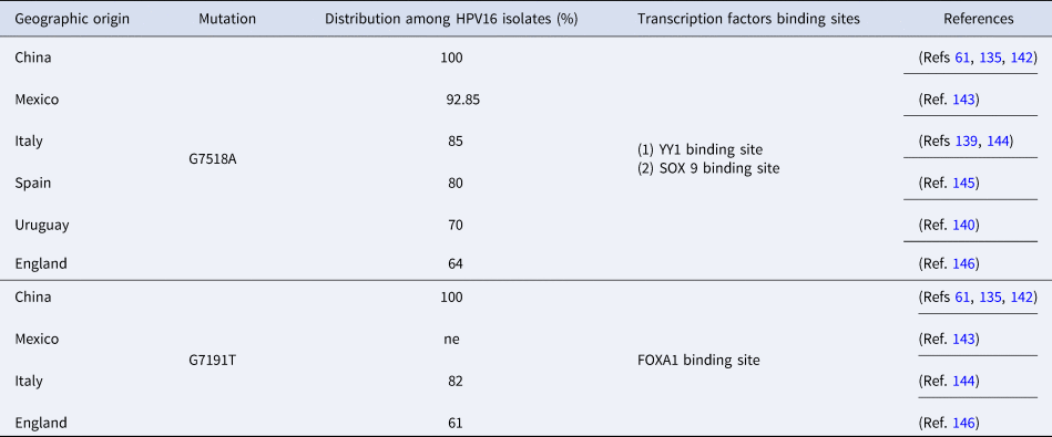
Outcomes derived from sequence analysis of LCR revealed that the nucleotide variation G7518A is widely distributed among the different HPV16 variant lineages. The G7518A was found to be the most prevalent nucleotide polymorphism among the examined populations, including 100% for China (Refs Reference Zhe61, Reference Fang135, Reference Xi142), 92.85% for Mexico (Ref. Reference Escobar-Escamilla143), 85% for Italy (Refs Reference Martinelli139, Reference Galati144), 80% for Spain (Ref. Reference Mosmann145), 70% for Uruguay (Ref. Reference Ramas140) and 64% for England (Ref. Reference Marongiu146) (Table 3). The G7518A is located in the binding site of transcriptional factor YY1 (ying yang), which is a negative regulatory protein that acts as an epithelial-specific transcription enhancer (Refs Reference Pande62, Reference Dong129). Sequence changes in YY1 binding sites provide to the viral genome increased carcinogenic capacity (Refs Reference Pande62, Reference Dong129). It has been predicted that the G7518A is also placed in the binding site of the transcriptional repressor SOX 9 (sex-determining region Y-box 9), which is implicated in suppression of cervical cancer through the activation of p21WAF1/CIP1 (Refs Reference Xi142, Reference Wang, Lian and Zheng147).
Distribution of the sequence variations G7518A and G7191T of the ΗPV16 LCR among the different examined populations

The G7518A is located in the binding sites of transcription factors YY1 and SOX 9, as well. The G7191T is placed in the binding site of FOXA1 transcription factor.
The HPV16 LCR nucleotide variation G7191T was also frequently detected among the different studied populations, including 100% for China (Refs Reference Zhe61, Reference Fang135, Reference Xi142), 82% for Italy (Ref. Reference Galati144), 61% for England (Ref. Reference Marongiu146) and Mexico (Ref. Reference Escobar-Escamilla143) (Table 3). The G7191T was predicted to be placed at the binding site of FOXA1 transcription factor (Fork head box protein A1) (Ref. Reference Xi142). The FOXA1 is a crucial transcription factor that is considerably involved in the regulation of various types of cancer, including breast, liver, prostate, lung and endometrium cancer (Refs Reference Xi142, Reference Wang, Lian and Zheng147–Reference Jin151). Although the HPV16 LCR sequence alterations G7518A and G7191T influence the binding site of fundamental host transcriptional factors, no association of the respective mutations with the development of cervical lesions has been found (Ref. Reference Escobar-Escamilla143).
Research findings derived from Asia also revealed that different nucleotide variations in the LCR are prevalent in diverse studied population groups. A study conducted in women from Shanghai showed that T6329G and A6657G are the most frequent sequence changes among patients with the position 6329 to be a potential binding site for TATA-binding protein (Ref. Reference Zhao152). In addition, two new nucleotide variations in the LCR (G7429A: 41.9% and G7874C: 39.5%) were found to be the most prevalent in patients derived from Thailand (Ref. Reference Pientong153). Moreover, it was estimated that the G7429A is located in the fourth binding site of E2 protein (residues 7453–7464), whereas the G7874C is located in the third binding site of E2 protein (residues 7860–7871). Despite the fact that HPV16 LCR is regarded as the most polymorphic region of viral DNA, no strong association between specific genome alterations and the development of severe cervical dysplasia has been reported. It is possible that the nucleotide mutations that are observed in LCR might be neutral. On the contrary, random nucleotide changes that are accumulated in each patient might influence differently the transcriptional capacity of viral genome and consequently may affect the progression of cervical malignancy. However, the personalised mapping of LCR nucleotide changes in each individual patient could be crucial regarding patients’ susceptibility towards cervical cancer development.
Discussion
HPV16 is a strong carcinogenic factor, because it is detected in more than 50% of cervical cancer cases. It has been estimated that half of women with at least 2 years of persistent HPV16 infection will develop precancerous lesions within the next 5 years (Refs Reference Tsakogiannis13, Reference Ferlay16, Reference Kjaer154). The tumourigenic hallmark of HPV16 DNA lies mainly on the function of E6 and E7 oncoproteins and numerous analyses have proved that mutations particularly within E6 increase the oncogenicity of viral DNA (Refs Reference Cuninghame46, Reference Tsakogiannis52, Reference Zhe61, Reference Szostek64, Reference Zhou66). In the present review, we describe a remarkable variability in the HPV16 early and LCR regions, considering research outcomes derived from different investigated populations. However, it was summarised that specific nucleotide variations and amino acid substitutions are implicated in viral aggressiveness, including the early genes E6: Q14H, H78Y, L83V, Ε7: N29S, S63F, E2: H35Q, P219S, T310K and E5: I65V (Fig. 1).
Nowadays, the most broadly investigated nucleotide variation of HPV16 DNA is the T350G (L83V) on the E6 gene, implicated in the development of severe dysplasia in various geographic populations. In vitro experiments demonstrated that the coexistence of nucleotide changes G145T (Q14H), C335T (H78Y) along with T350G (L83V) considerably augment the tumourigenic capacity of the viral oncoprotein (Refs Reference Niccoli45, Reference Cuninghame46). In contrast, a high level conservation has been described in the E7 gene, underlying its fundamental impact on cell cycle control destabilisation. Nevertheless, two nucleotide variations [A647G (N29S), C749T (S63F)] have been related to an increased cervical cancer risk, in Asian populations (Refs Reference Song68, Reference Fujinaga69). Similarly, the E1 gene is considered a highly conserved region of HPV16 genome because of its essential function in viral replication. In the E1 ORF numerous nucleotide variations and duplication events have accumulated in clinical samples, thus shifting the initial perception about the E1 gene (Refs Reference Bergvall, Melendy and Archambault77, Reference Tsakogiannis88, Reference Yao89) (Fig. 1). The impact of E1 genetic variations on viral tumourigenic capacity is yet to be investigated.
The C3410T (P219S) mutation in the E2 gene has been associated with a higher risk of cervical cancer development, whereas other studies in Asian populations involved the nucleotide changes C3684A (T310K) and C2860A (H35Q) in the development of severe cervical dysplasia (Refs Reference Kahla99, Reference Dai101) (Fig. 1). Furthermore, the polymorphic variability of E4 and E5 genes in different geographic populations remains rather unclear, because only a limited number of studies have focused on the extensive sequence analysis of the respective viral regions. To date, no nucleotide variations have been involved in the development of cervical disease concerning E4 gene. Conversely, the nucleotide change A4042G/T (I65V) in the E5 oncogene seems to be mainly identified in specimens diagnosed with advanced cervical dysplasia (Refs Reference Plesa63, Reference Eriksson100, Reference Tsakogiannis112). Finally, the LCR is regarded as the most polymorphic region of viral DNA, with numerous novel nucleotide variations to be continually reported in diverse research studies. Notably, the G7518A and G7191T have been found to be the most frequently detected nucleotide variations among HPV16 isolates, although their carcinogenic potential needs to be further clarified (Refs Reference Fang135, Reference Martinelli139, Reference Xi142). Recently, a bioinformatics tool named HPV16-Genotyper has been developed to detect the cancer-related variations of viral DNA and also automates the entire computational process of HPV16 lineage genotyping and the detection of potential recombination events (Ref. Reference Nikolaidis155).
Despite the prevalence of particular cancer-associated mutations, the background genomic variability of HPV16 DNA appears to play a decisive role in viral carcinogenic potential, hence supporting the theory that each HPV16 isolate may act as a single virus with different tumourigenic capacity (Ref. Reference Mirabello9). The genetic signature of each HPV16 isolate is crucial for the progression of cervical dysplasia and cervical cancer development. This phenomenon explains the conflicting outcomes derived from different studies concerning the implication of specific mutations in the development of cervical disease. The genetic signature of HPV16 strains might be related to the quasi-species theory, although it is a distinguishing feature of RNA viruses (Refs Reference Kukimoto156, Reference Domingo, Sheldon and Perales157). However, we cannot exclude the possibility that this theory might be applicable for HPV16 in the future. Additional factors should be taken into consideration in order to lead to more accurate conclusions about cervical cancer risk, such as multiple HR-HPV infections, viral DNA integration, the methylation status of HPV DNA and mutations into the host target proteins (Refs Reference Tsakogiannis13, Reference Tsakogiannis18, Reference Tsakogiannis34, Reference Kottaridi158–Reference Tsakogiannis160).
Finally, highly conserved regions of the viral DNA have been extensively characterised, including the complete E7 gene and the regions that encode the N-terminal domain of E1^E4 protein and the N-terminal domain of E2 protein. The high level of conservation of these positions seems to provide the virus with an overall selective advantage to establish a persistent infection, possibly because of their crucial role in viral life cycle. As a consequence, it is suggested that this high conservation may constitute these proteins suitable targets for future research and therapeutic approaches.
To the best of our knowledge, this is the first extensive literature review on a large number of nucleotide variations and amino acid substitutions of the early genes and LCR that have been reported for HPV16, thus far. In the present review, we provide important insights regarding the variability of HPV16 DNA, postulating that the genetic signature of HPV16 isolates can determine cervical cancer risk. As a result, the personalised mapping of HPV16 sequence variations in each individual patient can enhance the tumourigenic capacity assessment of the viral DNA and define the clinical characteristics of HPV16 infection that is viral persistence, recurrence or cessation.
Acknowledgements
This study was supported by the Hellenic Anticancer Institute, a private non-profit organisation. The funding source had no involvement in study design, conduct of research or manuscript publication. The authors thank the president of the Hellenic Anticancer Institute Mr. M. Vrontakis and the members of the Board for their continuous support.
Conflict of interest
All authors declare that they have not conflicting or dual interests.