Introduction
Ebstein’s disease, also known as Ebstein anomaly, primarily manifests as a congenital morphological abnormality affecting the valves and ventricles of the right heart. Reference Dearani and Danielson1 Clinical presentation varies based on the degree of tricuspid valve dysplasia, pulmonary valve hypoplasia, and the size and function of the right ventricle, ranging from cases requiring no therapeutic intervention throughout life to cases of fetal death. Reference Herrera-Bello, Ávila-Vanzzini, Fritche-Salazar, Kuri-Alfaro, Gaxiola-Macias and Cossio-Aranda2 Several severity indices have been reported, with the right atrium index, also referred to as the Celermajer index, being among the most widely used. Reference Celermajer, Cullen, Sullivan, Spiegelhalter, Wyse and Deanfield3 This index calculates the ratio of the combined area of the right atrium and atrialized right ventricle to that of the functional right ventricle and the left heart in a four-chamber view at the end of the diastole. An right atrium index >1.5 indicates the most severe form of Ebstein’s disease and, without neonatal intervention, significantly increases mortality risk. Reference Celermajer, Cullen, Sullivan, Spiegelhalter, Wyse and Deanfield3 Survival rates have significantly improved with improved surgical and perinatal management; however, one-third of patients diagnosed with Ebstein’s disease prenatally die before hospital discharge. Reference Freud, McElhinney and Kalish4 Severe cases often necessitate surgical intervention in the neonatal period due to circulatory instability immediately after birth. Reference Reemtsen, Fagan, Wells and Starnes5,Reference Elzein, Subramanian and Ilbawi6 However, some patients with seemingly severe Ebstein’s disease can survive the neonatal period with medical management alone, and the initial surgery may be postponed until infancy or later stages of development. Neonatal cardiac surgery entails a higher frequency of complications than surgeries performed later in life. Whenever feasible, delaying the initial surgery until the patient reaches an older age is recommended. Reference Elassal, Al-Radi and Debis7 However, to the best of our knowledge, no study has yet examined the indicators that predict the necessity of neonatal surgery in patients with severe Ebstein’s disease. This retrospective study aimed to identify a predictive index for neonatal surgery in patients with severe Ebstein’s disease.
Materials and methods
Study population
This retrospective study included patients who had severe Ebstein’s disease with an right atrium index >1.5 diagnosed prenatally and were born and treated after gestational management at our hospital from 2018 to 2023. Patients whose right atrium index was <1.5 or those who had less than severe tricuspid valve regurgitation shown by prenatal and postnatal echocardiography were excluded. The severity of tricuspid valve regurgitation was evaluated based on the recommendation in the report by the American Society of Echocardiography. Reference Zoghbi, Enriquez-Sarano and Foster8 The study retrospectively examined the laboratory indices and clinical course of two groups of patients: those who underwent surgery in the neonatal period and those who did not. The study protocol conformed to the ethical guidelines of the 1975 Declaration of Helsinki. The Ethical Committee of Kanagawa Children’s Medical Center approved the protocol, and all patients or their parents provided informed consent.
Measurement of the systolic time-to-diastolic time ratio by echocardiography
According to previous studies, the systolic time-to-diastolic time ratio was measured by extracting the Doppler waveform of tricuspid valve regurgitation and measuring the systolic time, which is the duration from the rise to the convergence of the tricuspid valve regurgitation waveform, and the diastolic time, which is the duration from the convergence of the tricuspid valve regurgitation waveform to the rise of the subsequent tricuspid valve regurgitation waveform in each cardiac cycle. Reference Friedberg and Silverman9 The average values of three consecutive heartbeats were obtained, compared, and verified. We measured the systolic time-to-diastolic time ratio at several time points, but we mainly compared the systolic time-to-diastolic time ratio at birth between the groups. The fetal systolic time-to-diastolic time ratio that we used was not the umbilical artery systolic time-to-diastolic time ratio that is typically reported in fetal ultrasound reports, but instead, we measured and analysed the prenatal tricuspid systolic time-to-diastolic time ratio the same as during the postnatal period.
Statistical analysis
Statistical analyses were performed using R Studio (version 2023.12.0 + 369). Welch’s t-test was used for continuous variables that were normally distributed based on the Shapiro–Wilk test. For other continuous variables that were not normally distributed, Wilcoxon’s rank sum test was performed. Fisher’s exact probability test was employed for categorical data, whereas Spearman’s product-rate correlation test was applied for binary correlations. The Youden method was used to determine threshold values using receiver operating characteristic curves.
Results
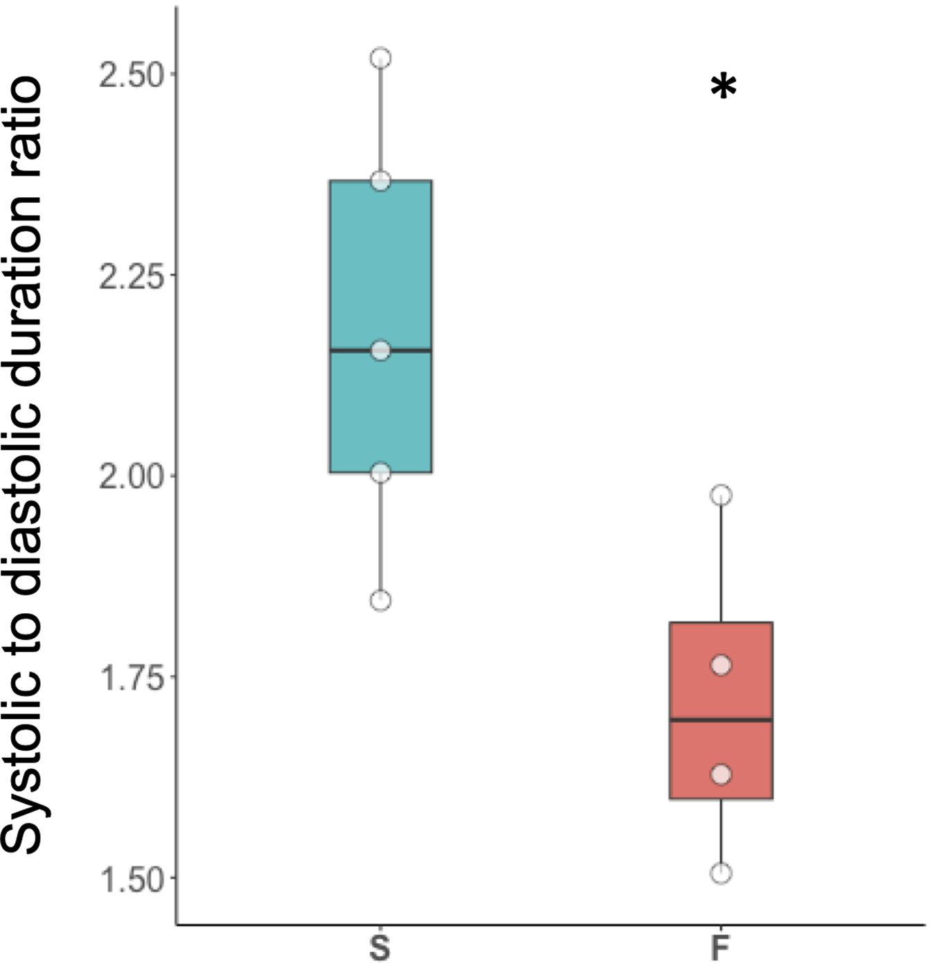
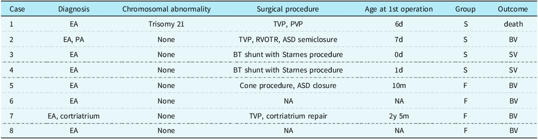
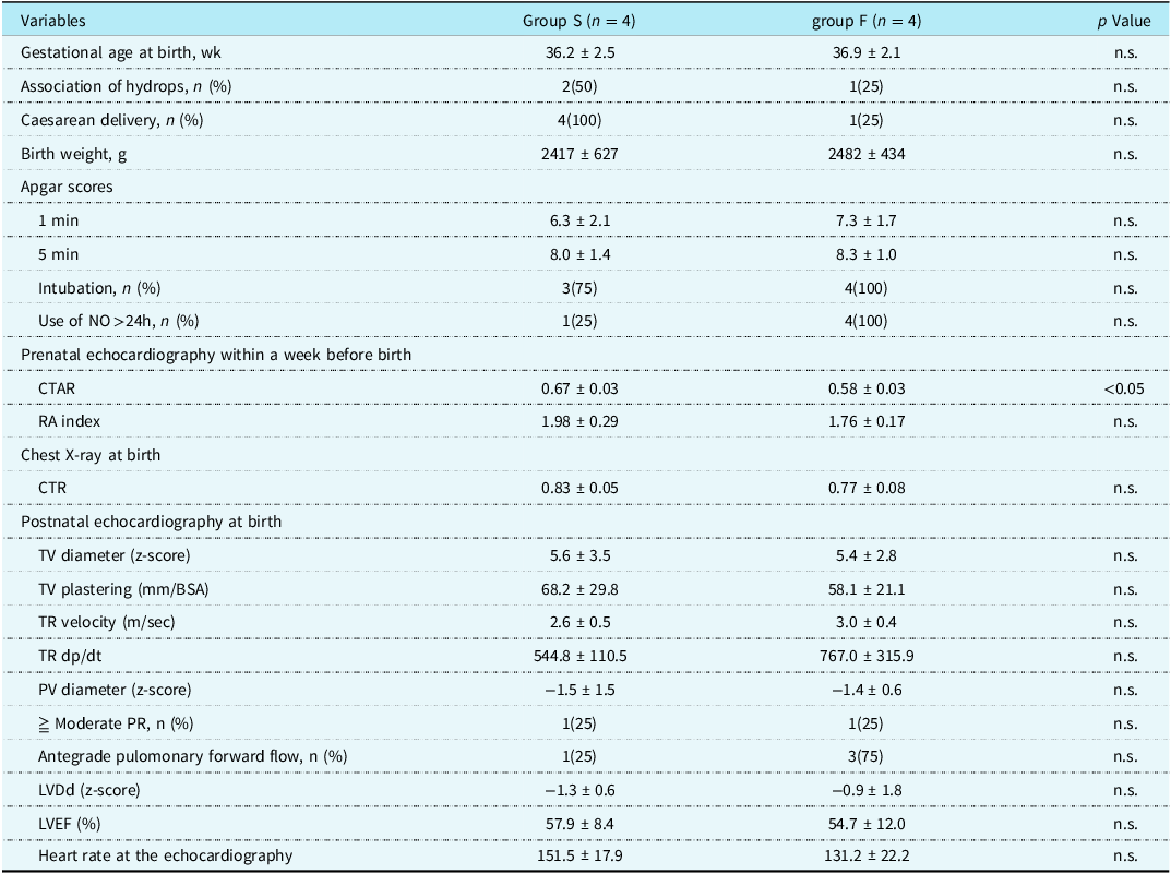
Four patients underwent surgery (Group S) during the neonatal period, whereas four did not (Group F). Table 1 presents the surgical procedures and timing for each group. Among the cases in Group S, two out of four cases were intended for biventricular repair, with one resulting in early postoperative mortality associated with trisomy 21. In Group F, two patients underwent tricuspid valve repair in late infancy or at a later stage, whereas the remaining two, who were not subjected to surgery, were placed under observation. In Group S, surgical intervention was prompted by circulatory collapse attributed to severe right heart failure, including functional and anatomical pulmonary valve atresia. Demographic data for each group are outlined in Table 2, revealing a higher incidence of caesarean births in Group S, whereas the use of nitric oxide for >24 h after birth was more common in Group F. The systolic time-to-diastolic time ratio, an index of ventricular function calculated from the tricuspid valve regurgitation waveform of echocardiography at birth, was significantly prolonged in Group S [2.18 ± 0.31 vs. 1.72 ± 0.20 in Group F (p = 0.043)] (Figure 1). Representative tricuspid valve regurgitation waveforms for each group are provided in Figure 2.
S/D ratios in groups S and F. Distribution of neonatal S/D ratios at birth. The solid line represents the median, the box represents the interquartile range, and the whiskers represent the entire range. *P = 0.043.

Representative TR waveforms for each group obtained from the apical 4 chamber view. The duration of systole was measured from the duration of the TR (pink bars). The interval between the end of TR and the onset of regurgitation demarcates the diastole duration (green bar). S/D ratio = systolic/diastolic time ratio; TR = tricuspid regurgitation.

Clinical characteristics and surgical procedures of patients with severe Ebstein’s anomaly (EA)

ASD = artrial septal defect; BT = Blalock–Taussig; BV = biventricular circulation; NA = not applicable; PA = pulmonary atresia; PDA = patent ductus arteriosus; PVP = pulmonary valvuloplasty; RVOTR = right ventricle outflow tract repair; SV = single ventricular circulation; TVP = tricuspid valvuloplasty.
Demographic data for patients who underwent surgery during the neonatal period (Group S) and patients who did not (Group F)

Data expressed as mean ± SD, unless otherwise specified.
CTAR = cardiothoracic area ratio; CTR = cardiothoracic ratio; LVDd = left ventricular end-diastolic diameter; LVEF = left ventricular ejection fraction; NO = nitric oxide; PR = pulmonary valve regurgitation; PV = pulmonary valve; RA = right atrium; TR = tricuspid valve regurgitation; TV = tricuspid valve.
Although the heart rate at the time of the tricuspid valve regurgitation measurement tended to be higher in Group S, the difference was not statistically significant. The investigation of the correlation between the heart rate and systolic time-to-diastolic time ratio in each group revealed a relatively strong correlation in Group F but not in Group S, although the difference was not statistically significant.
The receiver operating characteristic curve showed an area under the curve of 0.94, with the threshold value of the systolic time-to-diastolic time ratio determined as 1.99 using Youden’s method. The predictive model demonstrated a sensitivity of 100% and specificity of 75% in anticipating the necessity for neonatal surgery. To observe the change in the systolic time-to-diastolic time ratio over time, we plotted its values before birth, at birth, up to the initial surgery for Group S, and up to 1 month after birth for Group F (Figure 3). Notably, no discernible difference was observed between the groups before birth. However, postnatally, the systolic time-to-diastolic time ratio exhibited an increasing trend in Group S and a decrease in Group F.
Perinatal changes in the S/D ratio. The S/D ratio before birth, at birth, up to the first surgery for group S, and up to 1 month after birth for group. No obvious difference was observed between the groups before birth; however, after birth, the S/D ratio tended to increase in group S, and decrease in group F. Data of group S after birth is partially truncated due to the surgery. S/D ratio = systolic/diastolic time ratio.

Discussion
Various indices have been proposed for prognosticating Ebstein’s disease, primarily focusing on prognosis or determining single or biventricular circulation. Reference Andrews, Tibby, Sharland and Simpson10–Reference Ikegawa, Kim and Kawataki12 In this study, we aimed to identify a predictive index specifically targeting the necessity for neonatal surgery, and we found the systolic time-to-diastolic time ratio at birth to be a promising candidate. The systolic time-to-diastolic time ratio <2 helps to identify patients with severe Ebstein’s disease based on a high right atrium index, but who would not have clinical decompensation and might not need neonatal intervention. This novel finding holds clinical significance, allowing us to anticipate cases where neonatal surgery, often associated with high perioperative complications, might be avoidable.
Previous studies have reported that indices, such as tricuspid valve regurgitation velocity and dp/dt, recognised as useful prognostic indicators of life outcomes, are less discriminative than the systolic time-to-diastolic time ratio, as evidenced by the lack of significant difference between Groups S and F in our study. The systolic time-to-diastolic time ratio has been reported to correlate with contractility and the diastolic index in various conditions, such as dilated cardiomyopathy, pulmonary hypertension, hypoplastic left heart syndrome, and Fontan surgery, making it a valuable prognostic indicator. Reference Friedberg and Silverman9,Reference Friedberg and Silverman13–Reference Meinel, Koestenberger, Sallmon, Hansmann and Pieles16 The threshold of the ratio differs, ranging from 1.0 to 1.6, depending on the disease and population in question.
We delve into how the systolic time-to-diastolic time ratio becomes prolonged in severe Ebstein’s disease. Prolonged S, shortened D, and increased heart rate are believed to be the three contributing factors to the elevated systolic time-to-diastolic time ratio. First, the prolonged ejection duration, stemming from reduced contractility and impaired pulmonary artery closure both functionally and anatomically, leading to S prolongation. Furthermore, the compromised diastolic capacity of the right ventricle, along with the compression on the left ventricle by the right ventricle, contributed to impaired diastolic function, which may manifest as a shortening of D. Additionally, the heart rate naturally increases as a compensatory mechanism for low cardiac output. Since some reports suggest that heart rate affects systolic time-to-diastolic time ratio, increased heart rate in these patients could elevate systolic time-to-diastolic time ratio. Reference Spitaels, Arbogast, Fouron and Davignon17,Reference Boudoulas, Geleris, Lewis and Rittgers18 Consequently, the systolic time-to-diastolic time ratio is believed to be prolonged in severe Ebstein’s disease due to a combination of these three factors, reflecting the collapse of cardiac function. Supporting this, when examining changes in the systolic time-to-diastolic time ratio before and after birth, as well as before birth with placental and circulatory assistance, the difference in the systolic time-to-diastolic time ratio is not significant between Groups S and F. Following birth, the systolic time-to-diastolic time ratio in the surgical group was prolonged, reflecting maladaptation to postnatal circulation, whereas in the non-surgical group, it was shortened, reflecting adaptation.
The systolic time-to-diastolic time ratio has some extra advantages over other indexes. One is that it is simple to measure during echocardiography. The patients with severe Ebstein’s anomaly have evident tricuspid valve regurgitation, and it was easy to measure by Doppler echocardiography. Another advantage is that the systolic time-to-diastolic time ratio may also be useful after birth. As we demonstrated, this index exhibited an increasing trend in Group S and decreases in Group F. Therefore, the systolic time-to-diastolic time ratio is increasingly predictive when tracked over time.
Limitations
Our study was conducted with a small sample size; with a larger sample size, significant differences between the two groups in other indices might emerge. However, the systolic time-to-diastolic time ratio showed a significant difference even with a small sample size, which indicates its potential advantage as a discriminative function over other indices. According to previous studies, heart rate may influence the systolic time-to-diastolic time ratio to some extent, but we could not find a significant difference in heart rate between the groups or a significant correlation between heart rate and the systolic time-to-diastolic time ratio in each group. Reference Spitaels, Arbogast, Fouron and Davignon17,Reference Boudoulas, Geleris, Lewis and Rittgers18 We performed a multiple regression analysis in which we set the systolic time-to-diastolic time ratio and heart rate as explanatory variables, but the results were not statistically significant, probably due to the small sample size. However, a future study with a larger sample size might reveal the statistical significance and may enable us to correct the systolic time-to-diastolic time ratio for heart rate as a confounder and check whether the statistical significance would remain unchanged. Relatedly, since heart rate and some variables that we tested might be dependent on the systolic time-to-diastolic time ratio, we should employ the Bonferroni correction to adjust for multiplicity. However, the size of the study sample was not sufficient to maintain statistical power through the correction. Therefore, a future study with a larger sample size is needed. Furthermore, this study was retrospective, and certain well-known cardiac function indices, such as the Tei index, were not evaluated. However, a previous study showed that the systolic time-to-diastolic time ratio serves as a superior prognostic indicator to the Tei index. Reference Rösner, McElhinney, Diab, Friedberg and Lui15 Although our study provides valuable insights into predictive indices for neonatal surgical necessity in severe Ebstein’s disease, future research with larger sample sizes is warranted to further explore potential differences between groups in additional cardiac function indices. Additionally, prospective studies could investigate the comparative effectiveness of the systolic time-to-diastolic time ratio against established indices like the Tei index, shedding further light on its prognostic utility in this patient population.
In conclusion, for patients with severe Ebstein’s disease, an systolic time-to-diastolic time ratio <2, assessed from the tricuspid valve regurgitation waveform on echocardiography at birth, supports monitoring without surgical intervention through the newborn period and beyond.
Acknowledgements
We would like to thank Editage (www.editage.jp) for English language editing.
Author contribution
Conceptualisation: S.S., S.O.; methodology: S.S., S.O., N.S.; data acquisition: S.S., F.I., T.T., D.H., T.I., T.W., writing— original draft: S.S.; writing—review & editing: S.S., S.O.; supervision: S.Y., H.U.
Financial support
This research received no specific grant from any funding agency, commercial, or not-for-profit sectors.
Competing interests
None.
Ethical standard
All procedures performed to treat our patients were in accordance with the ethical standards of the institutional and/or national research committee and the 1964 Helsinki Declaration and its later amendments or comparable ethical standards.
Informed consent
Informed consent was obtained from our patients or their parents.
Data availability statement
The data that support the findings of this study are not openly available due to reasons of sensitivity and are available from the corresponding author upon reasonable request.