Right ventricular outflow tract stenting is a gradually evolving alternative to surgically placed systemic to pulmonary artery shunt or ductal stenting.Reference Dryzek, Moszura and Goreczny1,Reference Sunhee, Hong and Jeong2 This is classically described in ventriculoseptal defect with pulmonary stenosis physiology with compromised flow to the pulmonary vascular bed. It usually involves accessing the outflow tract antegradely from inferior caval vein across the tricuspid valve. We describe right ventricular outflow tract stenting in tricuspid atresia with critical pulmonary stenosis and discuss the encountered problems and optimised solutions.
Case report
A 2-day-old male neonate was referred to our heart centre for further evaluation and management of a complex CHD. He was born via vaginal delivery of non-consanguineous parents at term, with a body weight of 3.4 kg. The 5-minute APGAR score of 8 was normal. However, after 8 hours of birth, respiratory distress became obvious. Management started with oxygen application by nasal prongs and antibiotic treatment due to positive sepsis screening. Echocardiography revealed a tricuspid atresia. At admission at our heart centre, physical examination showed a central cyanosis with an oxygen saturation (SaO2) of 45%, heart rate 132 beats/minute, blood pressure 74/37 mmHg, and respiratory rate 46 breaths/minute.
Transthoracic echocardiography showed situs solitus, bilateral caval veins (superior caval vein) with drainage of the left superior caval vein to the coronary sinus, multi-fenestrated, borderline restrictive atrial communication with interatrial septum bowing to the left side, and normal pulmonary venous drainage. The tricuspid atresia was associated with atrioventricular and ventriculoarterial concordance and infundibular and valvular pulmonary valve obstruction (pulmonary stenosis). The foramen bulbo-ventriculare was unobstructed corresponding with a morphology of TA-IB. The pressure gradient across the right ventricular outflow tract was 45 mmHg. Mitral valve and left ventricular function were normal. There was left aortic arch, and no flow was detected across the arterial duct. Nevertheless, prostaglandin E1 was continuously infused in a dosage of 0.05 µg/kg/minute. Clinical stabilisation was initially tried by beta-blocker treatment, starting with oral propanolol, later on by a continuous infusion of metoprolol. In view of hypoxaemia (SaO2 between 28 and 40%) and still septic signs, written informed consent was obtained from the parents for catheter-based treatment. Surgical aorto-pulmonary shunt was considered having higher morbidity in view of newborn with sepsis.
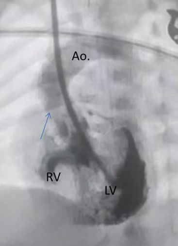
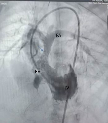
At the 5th day of life, percutaneous catheterisation was performed for right ventricular outflow tract stenting under general anaesthesia. Right femoral artery access and right jugular vein access were chosen, for placement of a 4Fr and 5Fr Terumo® sheaths, respectively, under ultrasound guidance. Femoral vein access could not be established. After application of heparin (100 U/kg), left ventricular angiography was performed in left anterior oblique projection of 30° via a 5Fr Cordis Right Judkins right catheter advanced from right internal jugular vein crossing the atrial septum and finally placed within the left ventricle. Left ventricular angiogram demonstrated morphologically normally developed left ventricle with a good ventricular function filling aorta, non-restrictive foramen bulbo-ventriculare, hypoplastic right ventricle, obstructed right ventricular outflow tract, and pulmonary stenosis with a tiny flow to the pulmonary branches (Fig 1). The right ventricular outflow tract was measured with a length of 16 mm and a diameter of 3.5 mm. Attempts to cross the right ventricular outflow tract from jugular access remained unsuccessful; therefore, retrograde catheterisation was performed from the femoral artery, through the aortic valve by utilising a 4Fr Cordis Judkins right catheter and a Terumo® 0.035” curved glide wire. After stabilising the wire in the left pulmonary artery, Judkins right was exchanged with a 4Fr Terumo Glide catheter followed by placing a 0.014” Hi torque All-star guidewire (Abott) and 0.014” Hi torque Whisper Extra support guide wire (Abott) as Buddy wire. Abott’s Xience expedition stent (4 × 18 mm) was positioned within the right ventricular outflow tract (Fig 2) under fluoroscopic and echocardiography guidance. While crossing the right ventricular outflow tract, a transient bradycardia with a 1:1 atrioventricular conduction was observed. Final left ventricular angiography showed an improved flow across the stented right ventricular outflow tract and pulmonary artery branches (Fig 2). Echocardiography and SpO2 of 85% (room air) indicated a good interventional result. The patient was extubated the following day and was closely monitored until full oral feeding was established. Pre-discharge echocardiography on the 4th day after the procedure showed a well-positioned right ventricular outflow tract stent with a flow velocity-related gradient of 35 mmHg, associated with an adequate cardiac function and stable clinical condition.
LV angiogram done via hand injection in left anterior oblique view. Catheter course being superior caval vein to right atrium to left atrium to left ventricle. Showing tiny antegrade flow (marked by arrow). Note the presence of moderate ventricular septal defect filling into the right ventricle. LV, left ventricle; RV, right ventricle; Ao, aorta.

LV angiogram done via hand injection in left anterior oblique view. Catheter course being superior caval vein to right atrium to left atrium to left ventricle. Showing stent in situ across the right ventricular outflow tract with good antegrade flow (marked by arrow). LV, left ventricle; RV, right ventricle; Ao, aorta; PA, pulmonary artery.

Discussion
To our knowledge, we describe here the first newborn with TA-IB, in whom a right ventricular outflow tract stent could successfully be placed, as a first-stage palliation. Surgical aorto-pulmonary shunt procedures remain the treatment of choice palliating neonates with cyanotic congenital cardiac malformations,Reference Sunhee, Hong and Jeong2,Reference Potapov, Alexi-Meskishvili, Dahnert, Ivanitskaia, Lange and Hetzer3 but our reported experience expands the alternatives of catheter-based interventions to TA-IB. Bentham et al in their study showed that ductal stenting is emerging as a preferred alternative to a surgical shunt for neonatal palliation with an evidence for greater post-procedural stability and improved patient survival to destination surgical treatment.Reference Bentham, Zava and Harrison4 Recent studies are being done for the use of right ventricular outflow tract stenting as an alternative to aorto-pulmonary shunts.Reference Dryzek, Moszura and Goreczny1,Reference Alkhulaifi, Lacour-Gayet and Serraf5 Right ventricular outflow tract stenting alleviates both sub-valvar and valvar pulmonic stenosis with good results in the high-risk infants in patients with tetralogy of Fallot.Reference Castleberry, Gudausky and Berger6 Quandt et al in their study comparing right ventricular outflow tract stenting with modified Blalock–Taussig shunt concluded that right ventricular outflow tract stenting promotes better pulmonary arterial growth and oxygen saturations compared with modified Blalock–Taussig shunt in the initial palliation of Fallot-type lesions.Reference Quandt, Ramchandani and Stickley7 Our anecdotal report augments further the indication for right ventricular outflow tract stenting in a complex cardiac morphology with a neonatal single ventricle pathophysiology (TA-IB), but should not underscore the risk of such a challenging procedure. Since the ductus was closed in our case, the options were to consider either surgery or right ventricular outflow tract stenting. Right ventricular outflow tract stent had an advantage of an improved pulmonary blood flow without distorting the pulmonary arteries which is particularly important for newborns with a single ventricle physiology. Another risk in right ventricular outflow tract stenting in tricuspid atresia is the risk of an atrioventricular block. We observed only a transient bradycardia with preserved 1-to-1 conduction, but it is not predictable and one needs to be cautious. Apparently, a good subset of patient with wide foramen opening enabled us to park a stent distal to the ventriculoseptal defect.
In summary, our successful retrograde approach of right ventricular outflow tract stenting in a newborn with a complex TA-IB morphology with an unrestrictive interventricular communication can be considered as an alternative to surgical shunts. The approach was technically challenging, but the outcome was exceptionally satisfactory with rise in oxygen saturation and clinical improvement. The safety of the procedure however needs to be confirmed in multiple case studies.
Acknowledgements
None.
Financial Support
This research received no specific grant from any funding agency, commercial, or not-for-profit sectors.
Conflicts of Interest
None.
Ethical Standards
The authors assert that all procedures contributing to this work comply with the ethical standards of the relevant national guidelines on human experimentation and Helsinki Declaration of 1975, as revised in 2008.