Introduction
Since the first successful report of the production of somatic cell-cloned sheep (Wilmut et al., Reference Wilmut, Schnieke, McWhir, Kind and Campbell1997), a number of clones has been produced after nuclear transfer (NT) of somatic cells from various mammalian species (Campbell et al., Reference Campbell, Fisher, Chen, Choi, Kelly, Lee and Xhu2007). Although Zhou et al. (Reference Zhou, Renard, Le Friec, Brochard, Beaujean, Cherifi, Fraichard and Cozzi2003) reported the successful production of clones by NT of fetal fibroblast cells in rats, there has been no report of successful rat somatic cell nuclear transfer (SCNT) (Hayes et al., Reference Hayes, Galea, Verkuylen, Pera, Morrison, Lacham-Kaplan and Trounson2001; Iannaccone et al., Reference Iannaccone, Taborn and Garton2001; Kato et al., Reference Kato, Hirabayashi, Aoto, Ito, Ueda and Hochi2001; Hirabayashi et al., Reference Hirabayashi, Kato, Ishikawa and Hochi2003; Tomioka et al., Reference Tomioka, Mizutani, Yoshida, Sugawara, Inai, Sasada and Sato2007; Popova et al., Reference Popova, Bader and Krivokharchenko2009). For successful reprogramming, the importance of premature chromosome condensation of somatic cells in recipient oocyte cytoplasm remains unclear (Popova et al., Reference Popova, Bader and Krivokharchenko2009), but it is now established that somatic cell nuclei must be introduced into oocytes at the second metaphase (MII) stage, in which there is a high level of maturation promoting factor (Du et al., Reference Du, Sung, Tian and Yang2002; Tani et al., Reference Tani, Kato and Tsunoda2003).
The potential of enucleated MII-stage oocytes receiving somatic cells to develop in vitro and in vivo is enhanced if SCNT mouse oocytes are cultured for several hours before parthenogenetic activation (Wakayama et al., Reference Wakayama, Perry, Zuccotti, Johnson and Yanagimachi1998). In contrast, however, the exposure of somatic cells to MII oocyte cytoplasm decreases the developmental potential of bovine SCNT oocytes (Sung et al., Reference Sung, Shen, Jeong, Xu, Chang, Cheng, Wu, Lee, Broek, Faber, Tian, Yang and Du2007).
We previously demonstrated that rat oocytes preserved in MG132-supplemented medium for 1 to 4 h have a high potential to develop into blastocysts (62–77%) if the oocytes are first treated with 10 mM strontium in KSOM with 1.71 mM calcium for 6 h (Mizumoto et al., Reference Mizumoto, Kato and Tsunoda2008); a finding that differs from other reports (Jiang et al., Reference Jiang, Mizuno, Mizutani, Sasada and Sato2002; Mizutani et al., Reference Mizutani, Jiang, Mizuno, Tomioka, Shinozawa, Kobayashi, Sasada and Sato2004). Therefore, we examined the developmental potential of SCNT rat oocytes produced in MG132-supplemented media during oocytes recovery, enucleation, introduction of somatic cells and preincubation before parthenogenetic activation. Although more than 83% of SCNT oocytes cleaved, the potential to develop into blastocysts was low (2%). Abnormal spindle formation was observed in SCNT oocytes preincubated for 2 h in MG132 and may be one reason for the poor developmental potential of rat SCNT oocytes (Mizumoto et al., Reference Mizumoto, Kato and Tsunoda2008).
In the present study, we examined the effect of preincubation time before activation on SCNT rat oocytes that had been produced in the presence of MG132 with regard to spindle formation and the potential to develop into blastocysts. Treatment of MII oocytes with nocodazole and demecolcine increases maturation promoting factor activity (Kubiak et al., Reference Kubiak, Weber, Pennart, Winston and Maro1993; Tani et al., Reference Tani, Shimada, Kato and Tsunoda2006) and induces spindle depolymerization (Kato & Tsunoda, Reference Kato and Tsunoda1992; Yin et al., Reference Yin, Tani, Yonemura, Kawakami, Miyamoto, Hasegawa, Kato and Tsunoda2002; Kawakami et al., Reference Kawakami, Tani, Yabuuchi, Kobayashi, Murakami, Fujimura, Kato and Tsunoda2003); therefore, we examined the developmental potential of rat SCNT oocytes preincubated with nocodazole and demecolcine before activation.
Materials and Methods
All experiments and protocols were performed in strict accordance with the Guiding Principles for the Care and Use of Research Animals adopted by the Kinki University Committee on Animal Research and Bioethics. All chemicals were purchased from Sigma-Aldrich Chemical Co. unless otherwise stated.
Oocyte preparation
Cumulus–oocyte complexes were recovered 15 h after injection of human chorionic gonadotropin from the oviducts of 3- to 5-week-old Sprague-Dawley female rats (Clea Japan, Inc.) superovulated by injection of 20 IU pregnant mare serum gonadotropin and human chorionic gonadotropin 48 h apart. Cumulus–oocyte complexes were denuded in M2 medium (Fulton & Whittingham, Reference Futon and Whittingham1978) supplemented with 300 IU/ml hyaluronidase and washed several times in M2 medium. The denuded oocytes were maintained in KSOM (Erbach et al., Reference Erbach, Lawitts, Papaioannou and Biggers1994) until manipulation (37°C, 5% CO2 in air). All media used for oocyte preparation (M2, M2 with hyaluronidase, KSOM) contained 5 μM MG132 (stock: dissolved in DMSO at 10 mM) to inhibit natural activation.
Production of NT oocytes
Nuclear transfer was performed as described previously (Mizumoto et al., Reference Mizumoto, Kato and Tsunoda2008). The methods were performed essentially according to the Honolulu technique (Wakayama et al., Reference Wakayama, Perry, Zuccotti, Johnson and Yanagimachi1998). Oocytes were enucleated in M2 medium supplemented with 1 μg/ml cytochalasin B (CB) (stock: dissolved in DMSO at 1 mg/ml). Enucleated oocytes were maintained in KSOM until NT at 37°C, 5% CO2 in air. For donor cells, cumulus cells removed during the oocyte preparation step and maintained in M2 medium were centrifuged and, once the outer layer was removed, mixed with clinical grade polyvinylpyrrolidone just before NT (Rybouchkin et al., Reference Rybouchkin, Kato and Tsunoda2006). Donor cells were injected into recipient oocytes in M2 medium. Except when indicated, all the media used for NT (M2, M2 with cytochalasin B and KSOM) contained 5 μM MG132.
Reconstructed oocytes were transferred into KSOM supplemented with 5 μM MG132 and 0.05 μg/ml demecolcine (stock: dissolved in DMSO at 10 μg/ml) or 0.025 μg/ml nocodazole (stock: dissolved in DMSO at 3 mg/ml) and maintained at 37°C, 5% CO2 in air until parthenogenetic activation.
Oocyte activation and culture
Reconstructed oocytes were washed four times with M2 medium and incubated for 10 min in KSOM to remove the MG132, demecolcine or nocodazole. The oocytes were then activated with 10 mM SrCl2 and 5 μg/ml cytochalasin B-supplemented KSOM at 37°C, 5% CO2 in air (Mizumoto et al., Reference Mizumoto, Kato and Tsunoda2008). Activated oocytes were washed with M2 medium and transferred into a 50 μl droplet of culture medium for 1-cell rat embryos (mR1ECM) (Miyoshi et al., 1994) for culture (10 embryos per droplet). The embryos were cultured for 5 days with mR1ECM and for an additional day with mR1ECM supplemented with 5% fetal bovine serum at 37°C, 5% CO2 in air.
Spindle and chromosome staining
Oocytes were fixed for 30 min with 3.7% paraformaldehyde in phosphate-buffered saline (PBS) and then washed with PBS supplemented with 1% bovine serum albumin (BSA; PBS + BSA) for 30 min. The oocytes were permeabilized for 30 min with PBS + BSA supplemented with 0.5% Triton X-100. The oocytes were washed again and blocked for 30 min with PBS + BSA. These treatments were performed at room temperature. Oocytes were incubated with 1% fluorescein isothiocyanate-conjugated α-tubulin antibody in PBS + BSA overnight at 4°C and washed for 30 min with PBS + BSA. The oocytes were then stained with 10 μg/ml propidium iodide-supplemented PBS + BSA for 1 min to detect the chromosomes. Samples were mounted with glycerol on glass slides and observed under a fluorescence microscope (Nikon UV eclipse E800m microscope) using an F-R filter.
Experimental design
Experiment 1
In this experiment, we examined the effects of duration of SCNT oocyte preincubation in MG132, demecolcine and nocodazole prior to activation on the spindle morphology and chromosome segregation status. After NT in the presence of MG132, SCNT oocytes were cultured in KSOM supplemented with MG132, demecolcine, or nocodazole for 0, 30, 60 and 120 min. Spindle morphology was then categorized as: type I, no spindle formation; type II, spindles shorter than MII oocyte spindles; type III, spindles similar to MII oocyte spindles; type IV, spindles elongated. Because NT was performed within 15 min in the presence of MG132, all SCNT oocytes were exposed to MG132 for less than 15 min before culture. After parthenogenetic activation, NT oocytes with a pseudopronucleus (or pseudopronuclei) were cultured to examine their developmental potential.
Experiment 2
In Experiment 1, there was a high rate of cleavage of oocytes activated immediately after NT. Because the oocytes in this group were exposed to MG132 for less than 15 min before activation, the duration after NT in Experiment 2 was set up much shorter. Oocytes were recovered and enucleated in the presence of MG132, but cumulus cells were injected into oocytes in MG132-free M2 medium. Reconstructed oocytes were then washed with M2 medium and activated within 5 min after starting the injection. Therefore, the exposure time of SCNT oocytes to MG132 was zero. The oocytes were cultured with or without 5 nM trichostatin A (TSA) during activation for 6 h. After activation, oocytes were divided into two groups according to the number of pronuclei: oocytes with one pronucleus (PN) and oocytes with more than two pronuclei. The developmental potential of NT oocytes was compared with that of oocytes exposed to MG132 for less than 15 min.
Statistical analysis
The developmental data were analysed by chi-squared analysis. A p-value of less than 0.05 was considered to be statistically significant.
Results
Effects of duration of SCNT oocyte preincubation on spindle morphology and the chromosome segregation status
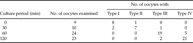
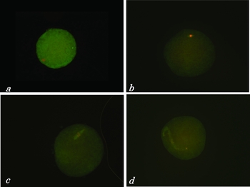
The spindles of NT oocytes cultured in KSOM supplemented with MG132 elongated gradually with an increase in the duration of culture in MG132 (Table 1). Immediately after NT, spindle formation was not observed in most oocytes (8/9) whereas condensed chromosomes were observed in all oocytes (Fig. 1a). Highly condensed chromosomes were observed and although spindles bagan to polymerize after 30 min incubation in MG132, the spindles were shorter than those of MII oocytes (Fig. 1b). After 60 min incubation, the spindle length in 79% of the oocytes (19/24) was the same as that in MII oocytes, but 21% of the oocytes (5/24) had elongated spindles and chromosomes had dispersed into the cytoplasm (Fig. 1c). After 120 min incubation, the spindles in most of the oocytes had elongated further and the chromosomes were dispersed along the spindles (Fig. 1d). In all oocytes cultured with demecolcine and nocodazole for 120 min, the chromosomes were condensed and spindles did not form.
Effect of duration of culture in MG132-supplemented medium on spindle morphology in SCNT rat oocytes.
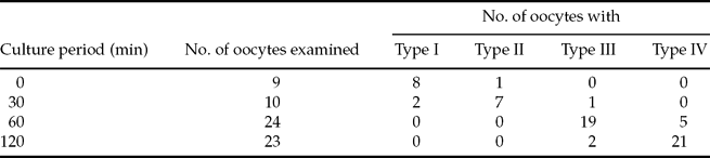
Type I, no formation; Type II, length shorter than MII oocyte spindle; Type III, length similar to MII oocyte spindle; Type IV, elongated.
Spindle morphology (green) and chromosome segregation status (red) in SCNT rat oocytes cultured in MG132-supplemented medium for 0 min (a), 30 min (b), 60 min (c) and 120 min (d).

Effects of duration of SCNT oocyte preincubation on developmental potential
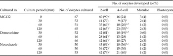
The proportion of SCNT cleaved oocytes that were activated soon after NT in the presence of MG132 was slightly or significantly higher compared with other groups (90% vs 65–79%; Table 2). The cleavage rates of oocytes cultured with MG132 for 60 min, in which spindles started to elongate and cultured for 120 min, in which spindles were elongated, were slightly lower compared with those of oocytes cultured for 30 min (69% and 65% vs 79%). The cleavage rates of oocytes cultured with demecolcine or nocodazole, in which the chromosomes were highly condensed and the spindles disappeared, were not affected by the culture duration (61–81% for demecolcine; 72–86% for nocodazole). The potential of SCNT oocytes to develop to the 4- to 8-cell stage was not different among groups (17–40%). The potential to develop into compacted morulae was low (0–4%), however, and blastocyst formation was not observed in any of the groups.
Effects of duration of culture in MG132, demecolcine and nocodazole on in vitro developmental potential of SCNT rat oocytes.

a–cValues with different superscripts in the same column differ significantly (p < 0.05).
Effects of MG132 and TSA treatment on the developmental potential of SCNT oocytes
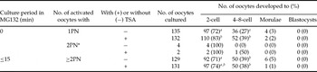
The number of pronuclei formed 6 h after parthenogenetic activation in SCNT oocytes depended on the MG132 preincubation period. When oocytes receiving cumulus cells in the absence of MG132 were activated within 5 min after injection, 98% (267/273) of oocytes formed a single pronucleus. When SCNT oocytes that were exposed to MG132 for less than 15 min were activated, however all of them formed more than two pronuclei (260/260). The proportion of oocytes with one pronucleus cleaved was not different from that of oocytes with more than two pronuclei (72% vs 71%). The developmental potential of oocytes with more than two pronuclei to develop to the 4- to 8-cell stage, however, was significantly higher than that of oocytes with one pronucleus (39% vs 27%). A few SCNT oocytes developed into morulae irrespective of the number of pronuclei that formed (0–5%), but none of them developed into blastocysts.
Treatment of SCNT oocytes with TSA enhanced the potential of oocytes with one pronucleus (83% vs 72%), but not the oocytes with more than two pronuclei (74% vs 71%), to develop to the 2-cell and 4- to 8-cell stages, but it did not enhance the development of oocytes to the morula and blastocyst stages.
Discussion
Unlike embryonic nuclei, somatic cell nuclei are only reprogrammed in oocytes that contain high maturation promoter factor activity (Tani et al., Reference Tani, Kato and Tsunoda2001; Lee & Campbell, Reference Lee and Campbell2006). Cloned animals have been produced after NT of somatic cells from various mammalian species to enucleated MII oocytes (Campbell et al., Reference Campbell, Fisher, Chen, Choi, Kelly, Lee and Xhu2007). Rat MII oocytes are naturally activated in vitro (Keefer & Schuetz, Reference Keefer and Schuetz1982) due to a decrease in cdc2 kinase activity (Ito et al., Reference Ito, Hirabayashi, Kato, Takeuchi, Ito, Shimada and Hochi2005). This characteristic of rat oocytes may be one of the reasons why it is difficult to produce somatic cell cloned rats. In only one report, Zhou et al. (Reference Zhou, Renard, Le Friec, Brochard, Beaujean, Cherifi, Fraichard and Cozzi2003) succeeded in producing live cloned rats by using oocytes treated with MG132, a proteasome inhibitor, to inhibit natural activation. There are no other reports, however, of successful rat SCNT even with MG132-treated oocytes (Hayes et al., Reference Hayes, Galea, Verkuylen, Pera, Morrison, Lacham-Kaplan and Trounson2001; Iannaccone et al., Reference Iannaccone, Taborn and Garton2001; Kato et al., Reference Kato, Hirabayashi, Aoto, Ito, Ueda and Hochi2001; Hirabayashi et al., Reference Hirabayashi, Kato, Ishikawa and Hochi2003; Tomioka et al., Reference Tomioka, Mizutani, Yoshida, Sugawara, Inai, Sasada and Sato2007; Popova et al., Reference Popova, Bader and Krivokharchenko2009). Our previous report (Mizumoto et al., Reference Mizumoto, Kato and Tsunoda2008) demonstrated that the potential of MII oocytes treated with MG132 to develop into blastocysts was increased by parthenogenetic activation (62% to 77%) but was low after NT of cumulus cells (2%).
When enucleated oocytes receiving cumulus cells in the presence of MG132 were incubated in MG132-supplemented medium for 30 min, the nuclear membrane was broken down, chromosomes were condensed and spindles began to form around the chromosomes (Fig. 1b). The spindles continued to elongate with an increase in the culture duration and, in approximately half of oocytes, the chromosomes were distributed along the spindles at 120 min after incubation (Fig. 1d). Because elongation of the somatic spindles was not observed when SCNT oocytes were preincubated in medium without MG132 (unpublished observation), the elongation was considered to be due to the MG132. The mechanisms underlying the elongation are not clear, but might be due to the formation of additional asters that cause the assembly of multipolar spindles in mitotic cells (Ehrhardt & Sluder, Reference Ehrhardt and Sluder2005). As the formation of spindles is regulated by the polymerization and degradation of various proteins, treatment of SCNT oocytes with MG132 might disrupt normal spindle formation.
Abnormal spindle formation in SCNT oocytes is a possible reason for the low developmental potential of SCNT rat oocytes. The cleavage rates of SCNT oocytes preincubated with MG132 for 60 min and 120 min (in which 21% and 91% of SCNT oocytes, respectively, showed elongated spindles) were significantly lower than that of oocytes activated immediately after NT (Table 2). The proportion of SCNT oocytes that developed to the 4- to 8-cell stage after activation without exposure to MG132, however, was significantly lower than that of oocytes exposed to MG132 for less than15 min (27% vs 39%, Table 3). Because most of the oocytes that were not treated with MG132 formed one pronucleus 6 h after activation, it is possible that the nuclear membrane of the SCNT oocytes in this group was not broken down. In this case, donor cell chromosomes would not have been directly exposed to the reprogramming factor(s) such as phosphorylated TCTP (Tani et al., Reference Tani, Shimada, Kato and Tsunoda2007) in the oocyte cytoplasm, which could lead to the low developmental potential. On the other hand, the higher developmental potential of SCNT oocytes activated after less than 15 min of exposure to MG132, after which spindles were not formed and premature chromosome condensation occurred, was expected, but none of these oocytes developed into blastocysts. For successful mouse cloning, SCNT oocytes are routinely incubated enucleated MII oocytes for at least 40 min (Kishigami et al., Reference Kishigami, Wakayama, Thuan, Ohta, Mizutani, Hikichi, Bui, Balbach, Ogura, Boiani and Wakayama2006a) and up to 2 h (Wakayama et al., Reference Wakayama, Perry, Zuccotti, Johnson and Yanagimachi1998; Rybouchkin et al., Reference Rybouchkin, Kato and Tsunoda2006) to stimulate reprogramming of donor nuclei before parthenogenetic activation. Fifteen minutes of incubation before activation might not be sufficient for reprogramming of rat somatic cell nuclei.
Effects of treatment of rat SCNT oocytes with MG132 and TSA on the oocyte developmental potential.

a ,b Values with different superscripts in the same column differ significantly (p < 0.05).
*Excluded from statistical analysis. PN, pronucleus.
One idea to promote reprogramming of donor nuclei is to culture SCNT oocytes whose spindle formation was inhibited for several hours before parthenogenetic activation. Because the potential of rat MII oocytes to develop into blastocysts was high when they were activated after four hours of treatment with demecolcine, during which the chromosomes were condensed without spindle formation (Mizumoto et al., Reference Mizumoto, Kato and Tsunoda2008), we examined the developmental potential of SCNT rat oocytes preincubated with demecolcine and nocodazole for 30 min to 120 min. The rates of cleavage and development to the 4- to 8-cell stage in SCNT oocytes treated with demecolcine and nocodazole, however, were decreased. The reprogramming factor(s) might not facilitate reprogramming of condensed chromosomes.
Histone acetylation levels in gametes are low, but increase after fertilization. When somatic cells with high histone acetylation levels are injected into enucleated oocytes, rapid deacetylation occurs soon after NT (Rybouchkin et al., Reference Rybouchkin, Kato and Tsunoda2006). To prevent histone deacetylation of somatic cell nuclei, SCNT mouse oocytes were treated with TSA, a histone deacetylation inhibitor. Treatment of SCNT oocytes with TSA increases the developmental potential of SCNT oocytes in the mouse (Kishigami et al., Reference Kishigami, Mizutani, Ohta, Hikichi, Thuan, Wakayama, Bui and Wakayama2006b; Rybouchkin et al., Reference Rybouchkin, Kato and Tsunoda2006), bovine (Ding et al., Reference Ding, Wang, Zhang, Wang, Guo and Zhang2008) and porcine (Zhang et al., Reference Zhang, Li, Villemoes, Pedersen, Purup and Vajta2007). The present study confirmed, however, our findings described in a previous report (Mizumoto et al., Reference Mizumoto, Kato and Tsunoda2008) that the developmental potential of rat SCNT oocytes was not improved after TSA treatment. The reason for this discrepancy is not clear, but in the present study the reprogramming of somatic cell nuclei was insufficient due to the short exposure period in MII oocytes.
For successful rat somatic cell cloning, therefore, two steps might be required: (1) to culture the somatic cell nuclei for long enough time in MII oocyte cytoplasm to enhance nuclear reprogramming; and (2) to induce normal spindle formation with normal chromosomal construction.
Acknowledgements
This work was supported by grants from the Ministry of Education, Science and Culture (20650063, 21028022) and from the Mitsubishi Foundation.