




No CrossRef data available.
Article contents
The ratio of flow in the superior and inferior caval veins after construction of a bidirectional cavopulmonary anastomosis in children
Published online by Cambridge University Press: 18 April 2005
Abstract
In patients who have undergone a superior cavopulmonary anastomosis, the superior caval venous flow provides the only, or the most important, pulmonary blood supply, while the inferior caval venous blood is not oxygenated, being mixed with the pulmonary venous blood before entering the systemic circulation. In healthy children, the contribution of superior caval venous flow to total cardiac output has been shown to decrease during growth. Patients who have undergone a superior cavopulmonary anastomosis, however, often have a higher oxygen saturation than predicted by the age-matched ratio of superior to inferior caval venous flows. This study was designed, therefore, to assess the ratio of flows in the superior and inferior caval veins subsequent to a superior cavopulmonary anastomosis. We carried out 18 magnetic resonance imaging studies with velocity-mapping and heart catheterisations so as to assess the contribution of superior caval venous flow to total cardiac output. Patients were divided into 3 groups according to their age. There were five aged from 8 to 24 months, eight aged from 24 to 48 months, and five older than 48 months. No significant difference could be found in the ratios of superior-to-inferior caval venous flow, nor of superior caval venous-to-systemic flow, between the 3 groups. The ratio of venous flows was 0.89 ± 0.34 in those aged from 8 to 24 months, 1.09 ± 0.42 in those from 24 to 48 months, and 1.25 ± 0.27 in the older patients (F analysis of variance 1.06, p 0.37). The ratio of superior caval venous-to-systemic flow was 0.46 ± 0.08 in the youngest patients, 0.50 ± 0.09 in those aged from 24 to 48 months, and 0.55 ± 0.05 in the older patients (F analysis of variance 0.76, p 0.49). These findings suggest that the hemodynamics of a cavopulmonary anastomosis may affect the normal decrease of superior caval venous flow with age. This could be related to a redistribution of flow, with a proportionally higher flow to the head and upper body after construction of a superior cavopulmonary anastomosis. Since increasing cyanosis and progressive exercise intolerance are the main indications for creation of a total cavopulmonary connection, these findings should be taken into account when determining the timing for completion of the Fontan circulation.
Information
- Type
- Original Article
- Information
- Copyright
- © 2003 Cambridge University Press
A bidirectional cavopulmonary anastomosis is frequently performed as part of a staged approach towards an ultimate Fontan circulation in patients with a functionally single ventricle. This procedure provides satisfactory arterial saturations of oxygen, and minimizes the volume load on the functionally single ventricle.1–3 In patients with a superior cavopulmonary anastomosis, the superior caval venous flow then provides the only, or certainly the most important, supply of blood to the lungs. The ratio of the superior to inferior caval venous flow is then a major determinant of the systemic arterial saturation of oxygen.4, 5 The contribution of the superior caval venous flow to total cardiac output in normal children has been shown to decrease during growth, from almost half in the newborn period to the reported adult value of about one-third at the age of 6 years.6, 7 If children who underwent a cavopulmonary anastomosis would have the same decrease in superior caval venous flow during growth, they should develop increasing cyanosis with age.8 Salim et al.,4 using the Fick principle, calculated the ratios of pulmonary to systemic flows in 29 children who had undergone a cavopulmonary anastomosis. At a mean age of 2.95 ± 1.65 years, those patients without systemic-to-pulmonary collateral vessels had a ratio of flows of 0.53, which was similar to the previously reported echocardiographically derived ratio of superior caval venous flow to systemic flow in normal children of the same age.4, 7 To our knowledge, the ratio of superior to inferior caval venous flow has not been studied during growth, and during the follow-up, of patients with a cavopulmonary anastomosis. In our clinical experience, some patients show a higher arterial oxygen saturation than might be expected on the basis of the predicted ratios of superior to inferior caval venous flows for their age, which cannot be explained by the presence of systemic-to-pulmonary collateral vessels increasing the pulmonary flow. We hypothesized that there might be a redistribution of the systemic venous flows after construction of a cavopulmonary anastomosis, resulting in a higher ratio of pulmonary to systemic flows, and a better arterial saturation of oxygen. The aim of our study, therefore, was to measure the ratios of superior to inferior caval venous flows in patients of different age who had previously undergone a bidirectional cavopulmonary anastomosis, using data derived from cardiac catheterisation and magnetic resonance imaging velocity mapping. The latter technique has previously been shown to be an accurate non-invasive method for measurement of volumetric venous and arterial flows.6, 9–11
Materials and methods
Patients
We enrolled 22 patients who had undergone a bidirectional cavopulmonary anastomosis. Because of clinical indications, these patients had undergone a magnetic resonance imaging scan, and catheterization of the right and left heart. Indications were evaluation prior to completion of the Fontan circulation for increasing cyanosis or exercise intolerance, or an interventional catheter procedure. Of the 22 patients, three, two with only a cavopulmonary anastomosis and one with accessory pulmonary flow through a Blalock-Taussig shunt, were studied twice with magnetic resonance imaging at different time intervals after construction of the cavopulmonary anastomosis. In total, therefore, data from 25 examinations were available for analysis.
All the operations had been performed by one surgeon. In 8 patients, a bidirectional Glenn anastomosis had been constructed, consisting of an end-to-side anastomosis of the superior caval vein on the right pulmonary artery. In the other 14 patients, a hemi-Fontan procedure had been performed, including patch closure of the distal end of the superior caval vein and the right atrium. During surgery, the azygos vein had been ligated in all patients. There was no accessory source of flow to the lungs in 9 patients, 3 patients had a cavopulmonary anastomosis and accessory pulmonary flow through a modified Blalock-Taussig shunt, and 10 patients had a cavopulmo-nary anastomosis and residual forward flow from the ventricle through a banded or stenotic pulmonary trunk.12–14 The additional contribution to pulmonary flow, however, was always small, as there was no significant difference in arterial saturations of oxygen between the patients with and without an accessory source of pulmonary blood. The anatomical diagnoses and surgical procedures are summarized in Table 1. In the sixth and seventh patients, the previously constructed Blalock-Taussig shunts were transsected surgically at the time of the cavopulmonary anastomosis. All patients were in sinus rhythm. The patients were divided into three groups according to their age at the time of the study. Thus, five studies came from patients who had been aged from 8 to 24 months, 3 with a cavopulmonary anastomosis only, and 2 with accessory flow through a Blalock-Taussig shunt. Their mean age at the time of the study was 16.6 ± 6.9 months, with a mean interval of 7.8 ± 5.8 months after the cavopulmonary anastomosis. The second group of 10 studies came from patients aged from 4 to 48 months, 4 studies coming from those with only a cavopulmonary anastomosis, 2 with accessory flow through a Blalock-Taussig shunt, and 4 with accessory forward flow from the ventricle. In these patients, the studies had been performed at a mean age of 31.9 ± 6.5 months, with a mean interval of 17.1 ±12.4 months after the cavopulmonary anastomosis. The final series of 10 studies came from patients older than 48 months, 4 from those with a cavopulmonary anastomosis only, and 6 from those with accessory forward flow from the ventricle. These patients had been examined at a mean age of 56.6 ± 7.7 months, with a mean interval of 28.3 ± 9.5 months from the cavopulmonary anastomosis. Informed consent was obtained from the parents in all patients. The protocol for investigation was approved by the local ethical committee.
Table 1. Summary of anatomical diagnoses and surgical procedures.

Methods
Magnetic resonance imaging
Magnetic resonance imaging was performed on the day prior to cardiac catheterization, using sedation with propofol infusion, the patients breathing spontaneously through a laryngeal mask using 21% oxygen. The heart rate and saturations of oxygen were continuously monitored. Scanning was performed on a 1.5 Tesla system (Magnetom Vision; Siemens AG, Erlangen, Germany) with a phased-array body coil using prospective electrocardiographic triggering. As previous studies have demonstrated that total caval venous flow does not vary throughout the respiratory cycle, respiratory gating was omitted.6 To reduce the influence of respiration, data were recorded during 224 consecutive heart cycles, and subsequently averaged to obtain flowcurves over one R-R interval. The imaging time for each flow study was from 1.5 to 3 min. For the anatomic evaluation and localization, HASTE (half Fourier single shot turbo spin echo) and turbo spin-echo images were obtained in the transverse and coronal planes. Quantitative measurements were obtained with a flow-sensitive cine gradient echo sequence using phase-contrast velocity mapping with a velocity encoded magnetic gradient in the direction of flow.11 The sequence parameters consisted of a repetition time of 21 msec, echo time 3.8 msec, 192 × 256 matrix, f lip angle of 25°, slice thickness of 6–8 mm in the 8–24 months group and of 10 mm in the older children, and velocity encoding of 200–250 cm/sec for measurements of aortic flow and of 75 cm/sec for those of venous flow. The maximum number of temporal phases obtained depended on the heart rate. This sequence yielded both phase and magnitude images. Through-plane flow was measured on images oriented perpendicular to the direction of flow. Measurements of inferior caval venous flow were taken at the level between the confluence of the hepatic veins and the junction with the right atrium. For superior caval venous flow, measurements were obtained 1 vessel diameter above the cavopulmonary anastomosis. Descending aortic flow was measured at the level of the diaphragm, and flow across the ascending aortic between the sinutubular junction and the brachiocephalic trunk. The borders of the vessel were manually outlined on the magnitude images. The software supplied with the magnetic resonance scanner was used to determine the quantitative flow as the product of the cross-sectional area of the vessel and the mean rate of flow. The total examination time was approximately 40 min.
We made several basic assumptions when calculating the flows. Thus, we considered total systemic flow to represent superior and inferior caval venous along with flow from the coronary sinus. Ascending aortic flow was considered equal to superior and inferior caval venous flows together with flow from the coronary sinus and aortopulmonary collaterals, or flow through a Blalock-Taussig shunt. The measurement of flow from the coronary sinus, however, was omitted, as it cannot be measured accurately and only accounts for 2% of total systemic output. Systemic flow was calculated as superior plus inferior caval venous flows. The ratio of pulmonary to systemic flows after the cavopulmonary anastomosis was considered equal to the ratio of superior to inferior caval venous flow.
Cardiac catheterisation
Catheterisation of the right and left heart was performed via femoral and jugular approaches. Standard measurements of pressure and saturation were taken in the superior and inferior caval veins, the pulmonary arteries and veins, the left atrium, left ventricle and aorta. In 9 out of the 11 patients with a cavopulmonary anastomosis without accessory pulmonary flow, the ratio of pulmonary to systemic flows was calculated according to the Fick principle.4 In the 2 remaining patients, the sampling was unreliable, precluding calculation of the ratio using the Fick principle. Data calculated from flows were compared with the measurements of flow obtained using magnetic resonance imaging. Biplane angiograms were obtained in the superior caval vein to assess the cavopulmonary anastomosis, the integrity of the pulmonary vascular bed, and the presence of veno-venous collateral vessels.15 Aortography was performed to show the presence of systemic-to-pulmonary collateral vessels.
Statistics
Differences between the mean values were calculated by univariate analysis of variance. To compare the data from catheterisation and magnetic resonance imaging, we used the method of Bland and Altman.16 The SAS computer software (SAS institute, Inc. Cary, North Carolina) was used for statistical analysis. The significance level was set at p < 0.05, and data are presented as mean values with the standard deviation.
Results
Magnetic resonance imaging
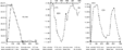
We obtained reliable data suitable for further analysis in 18 out of the 25 studies (Table 2), 11 coming from patients having a cavopulmonary anastomosis without accessory pulmonary flow, three from those with an accessory Blalock-Taussig shunt, and the others from those having accessory flow from the ventricle. We deemed the other seven studies, all from patients with accessory pulmonary flow, inadequate because of artifacts, 6 of these coming from patients with residual forward flow from the ventricle and the other from a patient with a Blalock-Taussig shunt. In 3 patients, flow curves were inaccurate due to operation clips. In 4 patients, the artifacts were ascribed to residual forward flow from the ventricle, causing turbulence in the superior caval vein. Only the studies from patients with a small hemodynamically unimportant amount of additional pulmonary blood supply provided reliable flow curves and could finally be included in the study. The measurements of flow are reported in Table 3, with representative curves illustrated in Figure 1. The morphology of the patterns of flow in the superior caval vein looked similar to that in the inferior caval vein. Systolic and diastolic peaks were always present, with the systolic peaks being more prominent in both caval veins. As expected, good overall correlation was found between inferior caval venous flow and the descending aortic flow, except in three patients with a ratio greater than 1.3.
Table 2. Groups and studies.

Table 3. Measurements of flow made using magnetic resonance imaging.


Figure 1. Example of a flow curve obtained using magnetic resonance imaging to image the ascending aorta, the superior caval vein, and the inferior caval vein in a patient after construction of a cavopulmonary anastomosis.
In the youngest patients, the superior caval venous flow accounted for 46 ± 8% of the systemic flow, with a ratio between the caval veins of 0.89 ± 0.34. In those aged from 24 to 48 months, the contribution of superior caval flow to the systemic flow was 50 ± 9% with a ratio of superior to inferior flows of 1.09 ± 0.42. In the older patients, the superior caval venous flow accounted for 55 ± 5% of the systemic flow, with a ratio of 1.25 ± 0.27 in favour of the superior caval vein. There was no significant difference in the ratios of superior caval venous flow to the systemic flow (F analysis of variance 0.76, p 0.49), nor for the ratios between the superior and inferior caval veins (F analysis of variance 1.06, p 0.37), between the three age groups. The sum of flows through the caval veins was similar to the flow through the ascending aorta. A higher flow in the ascending aorta was noticed in the patients with a Blalock-Taussig shunt, and also in those with significant systemic-to-pulmonary collateral vessels. This allowed us to quantify the contribution of the aortopulmonary collateral arteries to the pulmonary flow.
In the 3 patients who were examined twice at different time intervals after construction of the cavopulmonary anastomosis, 2 without accessory pulmonary flow and 1 with an accessory Blalock-Taussig shunt, there was no significant decrease during growth in the ratio of superior to inferior caval venous flows, nor in the ratio of superior caval venous flow to systemic flow (Table 4).
Table 4. Ratio of superior to inferior caval venous flows, and ratio of superior caval venous to systemic flows, at two different time intervals after construction of the cavopulmonary anastomosis.

Cardiac catheterisation
Superior caval venous angiography showed widely patent cavopulmonary anastomoses, without signs of obstruction, in all the patients. In five patients, there were mild or moderate stenoses in the pulmonary arteries, which were treated by successful balloon angioplasty during the same procedure. No patient had significant aortopulmonary collateral vessels requiring embolisation. A large veno-venous collateral vessel between the superior and inferior caval veins was seen in one patient, explaining the low ratio of superior to inferior caval venous flows of 0.4, and a low arterial saturation of oxygen of only 68%. These collateral vessels were successfully embolised during the same procedure. The mean systemic saturation was 83 ± 2.8%, without any significant difference between the different groups (F analysis of variance 1.9, p 0.18) (Table 3). Mean saturations of oxygen in the 11 studies from patients with a cavopulmonary anastomosis without accessory pulmonary flow, having a mean age of 35.7 ± 15.2 months, in three studies from those with an accessory Blalock-Taussig shunt, having a mean age of 20.4 ± 10.7 months, and in four studies from those with accessory pulmonary flow from the ventricle, having a mean age of 57.5 ± 22 months, were 85 ± 3%, 82 ± 4% and 84 ± 6%, respectively. Thus, there is no obvious difference in arterial saturations of oxygen between the patients with and without accessory pulmonary flow.
The ratio of pulmonary to systemic flows could be calculated using the Fick principle in only 9 of the 11 patients without accessory pulmonary blood flow.4 The patients with accessory pulmonary flow were omitted because the pulmonary arterial oxygen content cannot reliably be calculated using blood sampling in these mixed pulmonary circulations. The mean ratio of flows is 0.55, with a range from 0.51 to 0.80. We plotted the individual differences between the ratio calculated by magnetic resonance imaging and that obtained by the Fick method against the mean values for both methods (Fig. 2).16 The limits of agreement within two standard deviations above and below the mean value were +0.21 and +0.03, for a mean difference of 0.12, indicating a fair agreement between the 2 methods.

Figure 2. Difference in the mean values of the ratio of pulmonary to systemic flows (Qp/Qs) determined by magnetic resonance imaging (MRI) and the Fick principle.
Discussion
Our study was designed to assess quantitatively the superior caval venous flow, and its ratio to systemic flow, in children after a cavopulmonary anastomo-sis, using magnetic resonance velocity mapping and cardiac catheterisation. We demonstrated that, in younger children aged less than 2 years of age, the relative contribution of superior caval venous flow to total cardiac output is comparable to normal controls. In older children aged from 2 to 6 years, in contrast, the ratio of superior to inferior caval venous flow is higher than expected. This suggests that the ratio does not significantly decrease with age, as it does in otherwise healthy children. This observation is consistent with the clinical impression that children with a cavopulmonary anastomosis below 6 years of age, without accessory pulmonary flow or significant systemic-to-pulmonary artery collateral vessels, are often less cyanosed than might be predicted by the normal distribution of superior to inferior caval venous flows. Furthermore, in our patients there was no significant difference in arterial saturations of oxygen between the different age groups. Similarly, there was no obvious difference in arterial saturations of oxygen between the patients with and without accessory pulmonary flow, although the groups were small. This could be explained by the limited contribution of the Blalock-Taussig shunt, or the forward flow through a tight band on the pulmonary trunk, or critical pulmonary stenosis.
The relatively high contribution of superior caval venous flow in older children indicates that there might be a redistribution of flows after construction of a superior cavopulmonary anastomosis, with a proportionally higher flow to the head and upper body. This could be related to an altered autoregulation of the brain vasculature, as suggested by Fogel et al.17 These authors proposed that the hemi-Fontan physiology can induce intracerebral vasodilation, with increased flow to the head and neck. This causes global redistribution within the body, with decreased flow to the lower part of the body. This is also suggested by our observation that inferior caval venous flow is lower in the older patients. A similar redistribution has been observed by Houlind et al.9 after a total cavopulmonary anastomosis. They suggest that it might be related to an increased venous resistance in the abdominal organs related to increased venous pressures.18, 19 It is unclear which physiological mechanisms might explain this phenomenon after the superior cavopulmonary anastomosis, where abdominal venous pressures are normal.
Previous studies demonstrated that cine magnetic resonance velocity mapping is a reliable and non-invasive technique with which to measure venous flows in healthy subjects, and in patients with right-sided cardiac disease.6, 9–11 This technique is not only suited for the assessment of central patterns of venous flow, but it also allows a volumetric analysis of both superior and inferior caval venous flows. This is calculated from the mean velocity within the vessel lumen throughout the cardiac cycle, multiplying it by the cross-sectional area of the vein. In accordance with previous studies, we could demonstrate a good correlation between the measurements of venous return made with magnetic resonance and ascending aortic flow, thus validating the technique.6, 9–11 In children, magnetic resonance imaging is technically demanding due to the small size of the vessels, with changes in both flow and shape during respiration. So, despite an overall good correlation between inferior caval venous flow and the descending aortic flow, in three patients the ratio of inferior caval venous flow to that in the descending aorta was calculated to be greater than 1.3. This might be due to difficulties in the measurements of venous flow due to the small size of the patients. In 7 out of the 25 studies, the flow curves were unsuitable for analysis because of artifacts in the curves obtained for superior caval venous flow. In 3 studies, the curves were inaccurate due to operation clips. In 4 studies, the problems were ascribed to competitive flow in the superior caval vein caused by residual forward flow from the ventricle.
In our study we did not assess the influence of respiration on flow dynamics. Respiratory gating significantly increases the time required for acquisition, and is practically impossible in sedated young children. Moreover Mohiaddin et al.6 demonstrated that the patterns of venous flow, as well as the mean superior and inferior caval venous flows, were similar throughout the cardiac cycle when assessed using respiratory or cardiac gating. When we compared the ratios of flow in the patients with cavopulmonary anastomosis without accessory pulmonary flow by oxymetry and magnetic resonance velocity mapping, we found a fair agreement, with a mean difference of only 0.12 ± 0.04. Some of this discrepancy may be attributed to the lack of simultaneous measurements, since the magnetic resonance examinations were performed under sedation with spontaneous respiration, while the catheterisations were done while the patients were intubated and ventilated. Again, we should remember that our groups of patients are relatively small. Both methods, nonetheless, showed that there was no decrease in the ratio of pulmonary to systemic flows with age.
Our study has a cross-sectional design. Ideally, we would have liked to perform a serial evaluation in the same patients at different time intervals after the cavopulmonary anastomosis. Such serial evaluation of patients, however, is difficult, and is limited by ethical issues, since magnetic resonance imaging studies in these young children have to be done under general anesthesia. We were able to perform follow-up studies in 3 patients, which showed the same trend.
Our study has demonstrated, therefore, that in patients older than two years with a cavopulmonary anastomosis, the relative contribution of the superior caval venous flow to total cardiac output is higher compared to the data reported in normal children. Moreover, the ratio of pulmonary to systemic flows does not decrease between the age of 2 and 6 years. These findings suggest a redistribution of flow, with a proportionally higher flow to the head and upper body after construction of a cavopulmonary anastomosis. As increasing cyanosis, and progressive exercise intolerance, are the main indications for creation of a total cavopulmonary connection, these findings should be taken into account when considering the timing of completion of the Fontan circulation.
Acknowledgements
We thank Professor Tony Reybrouck and Dr Jan D'hooghe for their assistance with the statistical analysis.
Footnotes
This research was supported in part by a grant from the Belgian Foundation for Research in Pediatric Cardiology and by a grant from the Foundation for Scientific Research (Levenslijn FWO grant 7.0024.98). LM is a clinical researcher for the Fund for Scientific Research (FWO).
References
Alejos JC, Williams RG, Jarmakani JM, et al. Factors influencing survival in patients undergoing the bidirectional Glenn anastomosis. Am J Cardiol 1995; 75: 1048–1050.Google Scholar
Gewillig M, Kalis N. Pathophysiological aspects after cavopulmonary anastomosis. J Thorac Cardiovasc Surg 2000; 48: 1–6.Google Scholar
Mainwaring RD, Lambert JJ, Moore JW. The bidirectional Glenn and Fontan procedures – integrated management of the patient with a functionally single ventricle. Cardiol Young 1996; 6: 198–207.Google Scholar
Salim MA, Case CL, Sade RM, Watson DC, Alpert BS, diSessa TG. Pulmonary/systemic flow ratio in children after cavopulmonary anastomsis. J Am Coll Cardiol 1995; 25: 735–738.Google Scholar
Santamore WP, Barnea O, Riordan CJ, Ross MP, Austin EH. Theoretical optimization of pulmonary-to-systemic flow ratio after a bidirectional cavopulmonary anastomosis. Am J Physiol 1998; 274: 694–700.Google Scholar
Mohiaddin RH, Wann SL, Underwood R, Firmin DN, Rees S, Longmore DB. Vena caval flow: assessment with cine MR velocity mapping. Radiology 1990; 177: 537–541.Google Scholar
Salim MA, diSessa TG, Arheart KL, Alpert BS. Contribution of superior vena caval flow to total cardiac output in children. A Doppler echocardiographic study. Circulation 1995; 92: 1860–1865.Google Scholar
Gross GJ, Jonas RA, Castaneda AR, Hanley FL, Mayer JE, Bridges ND. Maturational and hemodynamic factors predictive of increased cyanosis after bi-directional cavopulmonary anastomosis. Am J Cardiol 1994; 74: 705–709.Google Scholar
Houlind K, Stenbog EV, Sorensen KE, et al. Pulmonary and caval flow dynamics after total cavopulmonary connection. Heart 1999; 81: 67–72.Google Scholar
Powell AJ, Geva T. Blood flow measurement by magnetic resonance imaging in congenital heart disease. Pediatr Cardiol 2000; 21: 47–58.Google Scholar
Rebergen SA, van der Wall EE, Doornbos J, de Roos A. Magnetic resonance measurement of velocity and flow: technique, validation and cardiovascular applications. Am Heart J 1993; 126: 1439–1456.Google Scholar
Frommelt MA, Frommelt PC, Berger S, et al. Does an additional source of pulmonary blood flow alter the outcome after a bidirectional cavopulmonary shunt? Circulation 1995; 92 (Suppl II): II: 240–244.Google Scholar
Mainwaring RD, Lamberti JJ, Uzark K, Spicer RL. Bidirectional flow. Is accessory pulmonary blood flow good or bad? Circulation 1995; 92 (Suppl II): II: 294–297.Google Scholar
Webber SA, Horvath P, LeBlanc JG, et al. Influence of competitive pulmonary blood flow on the bidirectional superior cavopulmonary shunt. A multi-institutional study. Circulation 1995; 92 (Suppl II): II: 279–286.Google Scholar
McElhinney DB, Reddy VM, Hanley FL, Moore P. Systemic venous collateral channels causing desaturation after bidirectional cavopulmonary anastomosis: evaluation and management. J Am Coll Cardiol 1997; 30: 817–824.Google Scholar
Bland JM, Altman DG. Statistical methods for assessing agreement between two methods of clinical measurement. Lancet 1986; I: 307–310.Google Scholar
Fogel MA, Weinberg PM, Chin AJ, Fellows KE, Hoffman EA. Late ventricular geometry and performance changes of functional single ventricle throughout staged Fontan reconstruction assessed by magnetic resonance imaging. J Am Coll Cardiol 1996; 28: 212–221.Google Scholar
Kaulitz R, Luhmer I, Kallfelz HC. Pulsed Doppler echocardiographic assessment of patterns of venous flow after the modified Fontan operation: potential clinical implications. Cardiol Young 1998; 8: 54–62.Google Scholar
Kelley JR, Gary WM, Fahey JT. Diminished venous vascular capacitance in patients with univentricular hearts after the Fontan operation. Am J Cardiol 1995; 76: 158–163.Google Scholar

Table 1.
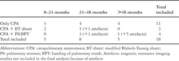
Table 2.
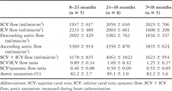
Table 3.
Example of a flow curve obtained using magnetic resonance imaging to image the ascending aorta, the superior caval vein, and the inferior caval vein in a patient after construction of a cavopulmonary anastomosis.
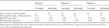
Table 4.

Difference in the mean values of the ratio of pulmonary to systemic flows (Qp/Qs) determined by magnetic resonance imaging (MRI) and the Fick principle.
 You have
Access
- 8
- Cited by