Introduction
Every creature in the biological world has a set of effective survival defense mechanisms to adapt the environment (Boman, Reference Boman1995). Vertebrates have acquired immune system, which relies on B and T lymphocytes to resist the infection of pathogenic microorganisms. On contrary to this, invertebrates like insects lack the acquired immune system and solely rely on the innate immune system comprised of cellular and humoral immunity to produce antimicrobial effectors to resist pathogenic microorganisms (Ferrandon et al., Reference Ferrandon, Imler, Hetru and Hoffmann2007; Lemaitre & Hoffmann, Reference Lemaitre and Hoffmann2007; Vallet et al., Reference Vallet, Lemaitre and Boccard2008; Bao et al., Reference Bao, Chandrasekar, Chuan, Chandrasekar, Tyagi, Gui and Reeck2014).
Antimicrobial peptides (AMPs) are small polypeptides of 20–60 amino acid residues with molecular sizes of 2–7 kDa (Pütsep et al., Reference Pütsep, Brändén, Boman and Normark1999). These active peptides are alkaline and heat stable with a broad spectrum of antibacterial activity. Known insect AMPs are classified into three major groups based on their structures. Linear AMPs with α-helical conformation but without cysteine; ring-type AMPs with cysteine and AMPs have high contents of one or two particular amino acids, most frequently proline and/or glycine residues (Meister et al., Reference Meister, Lemaitre and Hoffmann1997; Bulet & Stöcklin, Reference Bulet and Stöcklin2005; Jin et al., Reference Jin, Sun, Xu, Li, Gao, Xu, Yu and Ren2012). The expression of all the AMPs is activated by microbial infection and regulated by two distinct intracellular signal transduction pathways i.e., the immune deficiency (IMD) and Toll pathways, which also regulate the transcription of hundreds of other genes by controlling the nuclear translocation of the NF-κB transcription factors Relish, Dorsal and Dorsal-related immunity factor (DIF) (Gobert, Reference Gobert2003; Hultmark, Reference Hultmark2003; Ferrandon et al., Reference Ferrandon, Imler, Hetru and Hoffmann2007). Dorsal and DIF are activated by the Toll pathway in response to infection with fungi and Gram-positive bacteria. These factors are localized in the cytoplasm and interact with the mammalian IκB homologue Cactus in unstimulated cells. In response to an infection, DIF and Dorsal translocate into the nucleus by dissociating from Cactus and activate antimicrobial peptide genes to express and release into the hemocoel to defend the hosts against the infection (Lemaitre et al., Reference Lemaitre, Reichhart and Hoffmann1997).
Insect defensins, are in general a family of cationic and inducible AMPs that bear six cysteine residues engaged in three intra-molecular disulfide bridges. These are more active against Gram-positive bacteria and less against Gram-negative bacteria and fungi (Dang et al., Reference Dang, Wang, Huang, Yu and Zhang2010). Defensins have been found in a number of insect orders including Coleoptera (Bulet et al., Reference Bulet, Cociancich, Dimarcq, Lambert, Reichhart, Hoffmann, Hetru and Hoffmann1991; Xu & Faisal, Reference Xu and Faisal2010; Nakamura et al., Reference Nakamura, Iwasaki, Tokino, Asaoka, Yamakawa and Ishibashi2011; Yi et al., Reference Yi, Chowdhury, Huang and Yu2014), Diptera (Richman et al., Reference Richman, Bulet, Hetru, Barillas, Hoffmann and Kafatos1996; Dassanayake et al., Reference Dassanayake, Silva Gunawardene and Tobe2006; Dixit et al., Reference Dixit, Sharma, Patole and Shouche2008; Dang et al., Reference Dang, Wang, Huang, Yu and Zhang2010) and Lepidoptera (Kaneko et al., Reference Kaneko, Tanaka, Ishibashi, Iwasaki and Yamakawa2008) and insect defensin like molecule from mussels and scorpions. In addition to cationic defensins in insects, anionic defensins from Bombyx mori have also been reported (Wen et al., Reference Wen, Lan, Cheng, He, Shiomi, Kajiura, Zhou, Xia, Xiang and Nakagaki2009; Song et al., Reference Song, Park, Kim and Park2010), which indicate their structural dissimilarities. The first two insect defensins were isolated from culture cells of the Sarcophaga peregrina (Matsuyama & Natori, Reference Matsuyama and Natori1988) and by and collaborators from bacteria-challenged larvae of the Phormia terranovae (Lambert et al., Reference Lambert, Keppi, Dimarcq, Wicker, Reichhart, Dunbar, Lepage, Van Dorsselaer, Hoffmann and Fothergill1989). Members of the defensins share a general feature and structure, while they correspond to molecular diversity in amino acid composition and size. The peptide sequences are relatively alike in an insect order but very much different across the orders.
The diamondback moth, Plutella xylostella, is a worldwide pest of cruciferous crops. With the abuse of pesticides, P. xylostella has developed resistance to most of the conventional pesticides and B. thuringiensis in the fields (Tabashnik, Reference Tabashnik1994; Pardo-Lopez & Soberon Bravo, Reference Pardo-Lopez, Soberon and Bravo2013). There is growing recognition that an important element of gut microbiota of both invertebrates and vertebrates is their ability to change the host immune response. In certain conditions this effect can become lethal to the insects. For instance, activation of the immune responses by the bacteria leads to chronic conditions (Bates et al., Reference Bates, Akerlund, Mittge and Guillemin2007; Nenci et al., Reference Nenci, Becker, Wullaert, Gareus, van Loo, Danese, Huth, Nikolaev, Neufert, Madison, Gumucio, Neurath and Pasparakis2007; Maillet et al., Reference Maillet, Bischoff, Vignal, Hoffmann and Royet2008). Current studies have also linked activation of the immune response of Lepidoptera to ingestion of non-lethal doses of B. thuringiensis. The ingestion of B. thuringiensis by greater wax moth, Galleria mellonella larvae increased both oxidative stress in the gut (Dubovskiy et al., Reference Dubovskiy, Martemyanov, Vorontsova, Rantala, Gryzanova and Glupov2008a ) and the phagocytic activity of hemocytes (Dubovskiy et al., Reference Dubovskiy, Krukova and Glupov2008b ), while the exposure of Bt reduced the hemocytes and components of the humoral immune system in cabbage lopper Trichoplusia ni (Ericsson et al., Reference Ericsson, Janmaat, Lowenberger and Myers2009). Insect defensins, a significant group of gene-encoded effectors of innate immunity play a vital part in resisting to the infections caused by different biological agents including bacteria and fungi (Ferrandon et al., Reference Ferrandon, Imler, Hetru and Hoffmann2007; Lemaitre & Hoffmann, Reference Lemaitre and Hoffmann2007; Dang et al., Reference Dang, Wang, Huang, Yu and Zhang2010; Tamura et al., Reference Tamura, Peterson, Peterson, Stecher, Nei and Kumar2011). Up to now, more than 60 defensins have been identified from different species of insect orders including Lepidoptera (Yi et al., Reference Yi, Chowdhury, Huang and Yu2014; Badapanda & Surendra, Reference Badapanda, Surendra, Chandrasekar, Goldsmith and Agunbiade2016). The previous reports depict that majority of the defensins are isolated from the hemolymph, fat body or midgut of bacteria-challenged larvae (Lambert et al., Reference Lambert, Keppi, Dimarcq, Wicker, Reichhart, Dunbar, Lepage, Van Dorsselaer, Hoffmann and Fothergill1989; Boulanger et al., Reference Boulanger, Lowenberger, Volf, Ursic, Sigutova, Sabatier, Svobodova, Beverley, Späth, Brun, Pesson and Bulet2004; Seufi et al., Reference Seufi, Hafez and Galal2011; Cecilia et al., Reference Cecilia, Peter, Débora, Daniele, Cícero, Norman, Garcia and Azambuja2014). Insect defensins are classified according to their activity, while the antimicrobial defensins are recognized to be active mainly against Gram-positive bacteria at different concentrations (Bjellqvist et al., Reference Bjellqvist, Hughes, Pasquali, Paquet, Ravier, Sancez, Frutiger and Hochstrasser 1993 ; Chenna et al., Reference Chenna, Sugawara, Koike, Lopez, Gibson, Higgins and Thompson 2003 ; Boulanger et al., Reference Boulanger, Lowenberger, Volf, Ursic, Sigutova, Sabatier, Svobodova, Beverley, Späth, Brun, Pesson and Bulet 2004 ; Yang et al., Reference Yang, Wang, Liu, Wu, Liu, Gong, Zhao, Hong, Lin, Wang and Lai 2009 ; Bhusan et al., Reference Bhusan, Polamarasetty and Pallu 2010 ; Seufi et al., Reference Seufi, Hafez and Galal2011; Wang et al., Reference Wang, Zhang, Chen, Guang, Li, Yang, Li, You, Yu and Lai 2012 ; Telleria et al., Reference Telleria, Sant'Anna, Alkurbi, Pitaluga, Dillon and Traub-Csekö 2013 ). In the current work, we firstly report the immunological responses of defensins of P. xylostella after challenging with B. thuringiensis. With the analysis of transcriptome sequence of P. xylostella we found only one unique sequence encoding defensin, which has high similarity to the defensin sequence from the P. xylostella genomic sequence and owned the characterization of anionic peptide. In order to elucidate its functions against pathogenic bacterium B. thuringiensis, in the present study, the cloning and characterization of the novel anionic defensin from P. xylostella were reported. The spatial tissue distribution and temporal expression after infection of B. thuringiensis, as well as the antimicrobial activity were also described. In addition, its expression regulated by the Toll-Dorsal pathway as analyzed by RNAi. Our findings may help shed light on immune responses of P. xylostella against B. thuringiensis and have implications for using pathogenic bacteria for the biological control of pests in practice.
Materials and methods
Insect rearing, microorganisms and immunization
The larvae of P. xylostella were collected from the cabbage fields and reared successively on cabbage in the greenhouse of Engineering Research Center of Biological Control, South China Agricultural University, China, at 25 ± 2°C, 70% relative humidity (RH) and a photoperiod of 16:8 h (light : dark).
Gram-negative bacteria, i.e. Escherichia coli DH5a, BL21 (DE3), Bacillus cereus, Bacillus subtilis, Bacillus pumilus; Gram-positive bacteria, i.e. Pseudomonas fluorescent, Salmonella choleraesuis, Staphylococcus aureus Rosenbach; and fungi Botrytis cinerea Klotzsch, Penicillium crustosum Thom, Peronophythora litchi, Colletotrichum orbicular Berk, Fusarium oxysporum Schlecht, Colletotrichum gloeosporioiees Penz. were obtained from the Research Institute of Microbiology, Guangzhou, China, while the insect pathogenic fungus, while the insect pathogenic fungus, M. anisopliae (MaQ 10) was provided by Dr Qiongbo Hu (South China Agricultural University, Guangzhou, China), which was kept in China Center for Type Culture Collection (No. CCTCCM 208173). B. thuringiensis (HD73) was kindly provided by Prof. Jie Zhang (The Chinese Academy of Agricultural Sciences, Institute of Plant Protection, Plant Diseases and Insect Pests, Beijing, China).
The eggs, 1st–4th instar larvae, prepupae, pupae and adults were collected and used in fresh. Each 4th instar larva was injected separately with log phase heat-inactivated B. thuringiensis (2 × 10⁶ CFU ml−1) or live B. thuringiensis (2 × 10⁶ CFU ml−1) in the abdominal region. Every 6 h after injection with B. thuringiensis until 48 h post injection, the fat body, midgut, and epidermis were dissected and collected in ice-cold tubes (Xu et al., Reference Xu, Jin, Yu, Ji, Wang, Cheng, Wang and Zhang2007). The hemolymph from the Bt injected larvae was collected into 1/10 volume of anticoagulant buffer (10% sodium citrate, pH 7.0, and 200 mM phenylthiourea) from the abdominal prolegs of the normal and immune-challenged larvae at 24 h post-injection. The hemocytes were collected by centrifugation at 3000 × g for 10 min at 4°C.
Cloning of defensin from P. xylostella
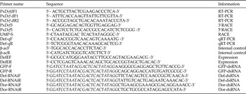
The RNA was extracted from the above dissected tissues using Trizol reagent and was reverse-transcribed into first-strand cDNA using SuperScript Reverse Transcriptase (Invitrogen, Carlsbad, CA, USA) according to the manufacturer's instructions. A UniGene sequence encoding defensin was obtained from P. xylostella transcriptome and was validated by polymerase chain reaction (PCR) using specific primers (table 1).
Primers used for cloning, expression and RNAi analysis.

Primer DefEF, Bold/underlined letter of G represents a designated mutation of letter A to G.
Sequence in Italic (GGATCCTAATACGACTCACTATA) represents T7 promoter.
Gene specific primers (table 1) were designed based on the sequence of the cloned cDNA fragment to amplify cDNA by using SMART™ RACE cDNA amplification kit (Clontech, USA) following the manufacturer's instructions. PCRs were carried out by using the Advantage 2 PCR kit (Clontech, USA) according to the following conditions: 3 min at 94°C, 5 cycles at 94°C for 30 s, 65°C for 30 s and 72°C for 30 s; 3 min at 94°C, 35 cycles at 94°C for 30 s, 60°C for 30 s, 72°C for 30 s and a 8 min elongation at 72°C. The resultant PCR products were subjected to electrophoresis on 1.0% agarose gel to determine the band sizes. The expected sizes bands were excised and purified using an OMEGA gel purification kit (OMEGA, USA). The purified cDNA fragments were cloned into the pMD-18T vector (TaKaRa, Japan) according to the manufacturer's instructions and sequenced. The complete cDNA sequence encoding PxDef was obtained by overlapping the PCR.
Sequence analysis of PxDef
The software Open Reading Frame (ORF) Finder (http://www.ncbi.nlm.nih.gov/projects/gorf/) was used to analyze the ORF of PxDef. The similarity index was analyzed by BLAST algorithm (http://www.ncbi.nlm.nih.gov/blast), while, multiple sequence alignment was carried out by ClustalW (http://www.ebi.ac.ek/clustalw). The signal peptide was analyzed by Signal P4.1 server (http://www.cbs.dtu.dk/services/SignalP/), while, the trans-membrane region was predicted by TMHMM software (http://www.cbs.dtu.dk/services/TMHMM/). The molecular weight and isoelectric point (pI) were determined by using ScanProsite (http://prosite.expasy.org/scanprosite/). MEGA 5.2 software was used to construct the phylogenetic tree on the basis of the Neighbor Joining method (NJ) to assess the relationship of the defensin sequences (Tamura et al., Reference Tamura, Peterson, Peterson, Stecher, Nei and Kumar2011).
Quantitative real-time PCR (qRT-PCR) analysis
The transcription level of PxDef in the developmental stages and dissected tissues after bacterial challenge was measured by qRT-PCR using a CFX96 real-time system. Approximately 1 µg RNA from each sample was reverse-transcribed using the PrimeScriptRT Reagent Kit with a DNA Eraser (TaKaRa, Japan). The first-strand cDNA was subsequently used as a template for PCR with the Def-qF and Def-qR primers (table 1), while β-actin (GenBank accession No. AB282645) was used as the internal control for cDNA normalization and amplification by using the β-actinF and β-actinR primers. The qRT-PCR mixture consisted of 2 µl cDNA sample, 8.5 µl ddH2O, 12.5 µl SYBR Premix Ex Taq™ (TaKaRa, Japan) and 1 µl of each gene-specific primer (10 µM). The PCR conditions consisted of: 95°C for 30 s; 40 cycles of 95°C for 10 s, 58°C for 30 s and 72°C for 20 s, followed by dissociation curve analysis at 95°C for 10 s, 65°C for 5 s and 95°C for 15 s to verify the amplification of a single product. The threshold cycle (CT) values were determined by CFX96 software. The data were exported into a Microsoft Excel spread sheet for subsequent analysis, where the relative expression ratios of the target gene in treated and untreated control groups were calculated using a 2−ΔΔCT method. All assays were performed in three independent replicates.
Recombinant expression and purification of PxDef
The cDNA fragment encoding the mature peptide of PxDef (RIPCQYEDATEDTICQQHCLPKGYSYGICVSYRCSCV) was obtained by RT-PCR with DefEF/DefER primers. In order to obtain mature PxDef with the native N-terminus, the Nco I restriction enzyme was introduced at the cleavage site on the N-terminus of the PxDef nucleotide sequence and mutated the A to G in the primer of DefEF. The products of RT-PCR were purified using agarose gel electrophoresis, digested with Nco I and Xho I enzymes, and then ligated into the Nco I-/Xho I digested expression vector pET-32a(+) (Novagen, USA). The constructed plasmid was transformed into competent BL21 (DE3) cells for expression of the TRX (thioredoxin)-6His-PxDef-fusion proteins. The fusion protein was expressed in BL21 (DE3) cells after induction with 0.5 mM isopropyl-D-thiogalactoside (IPTG) for 6 h at 28°C. The bacterial pellets were harvested by centrifugation and lysed with lysis solution (50 mM Tris–HCl, pH 8.0, 50 mM NaCl, 0.5% Triton X-100, 2 mg ml−1 lysozyme). After sonication, the supernatants were recovered by centrifugation and subjected to Ni2+-NTA chromatography for purification of the recombinant fusion protein, which was dialyzed and dissolved in PBS (pH 7.4) to a final concentration of 1 mg ml−1. In order to release PxDef from fusion proteins, enterokinase was added at a concentration of 8 U mg−1 of protein, and the digestion was performed with the reaction buffer (150 mM Tris–HCl, pH 7.4, 150 mM NaCl, and 2.5 mM CaCl2) at 21°C for 16 h with stirring according to the manufacturer's protocol. The reaction mixture was again subjected to HisTrap HP column to remove thioredoxin and the undigested fusion proteins. The flow-through fractions containing PxDef was collected, concentrated with PEG2000, dialyzed in double distilled water for desalination, and finally lyophilized. The purified recombinant protein PxDef was analyzed by Tricine-SDS–PAGE (Jin et al., Reference Jin, Sun, Xu, Li, Gao, Xu, Yu and Ren2012).
Antibacterial assay
The activity of recombinant protein PxDef against B. thuringiensis was checked by agar diffusion method as described previously (Xu et al., Reference Xu, Jin, Yu, Ji, Wang, Cheng, Wang and Zhang2007). The minimal growth inhibition concentration (MIC) was determined by liquid growth inhibition assay (Kennerdell & Carthew, Reference Kennerdell and Carthew1998) and expressed as the lowest final concentration of the peptide at which no growth was observed (Dhume et al., Reference Dhume, Lu and Horowits2006). Five Gram-positive bacteria, three Gram-negative bacteria and seven fungi were used to test the antimicrobial activity of the purified PxDef. The purified PxDef was serially diluted 10-fold with ultrapure water and aliquots (10 µl) from each dilution were distributed to a 96-well polypropylene microtiter plate, and each well was inoculated with 100 µl mid-log phase bacterial suspension (1 × 10⁵ CFU ml−1) in LB broth.
RNA interference in P. xylostella
Double stranded RNAs (dsRNAs) specific to PxDef and PxDor (GenBank accession number: KF960046) were prepared with gene-specific primers by RT-PCR using T7 RiboMAX™ Express RNAi System (Promega, USA) according to the manufacturer's protocol. The primers (table 1) were designed using the SnapDragon dsRNA design software (http://www.flyrnai.org/cgi-bin/RNAi-find-primers.pl). To minimize potential off-target effects, a dsRNA segment corresponding to the green fluorescent protein (GFP) was also synthesized as a negative control as described previously (Huang et al., Reference Huang, Xu, Freed, Zheng, Wang, Ren and Jin2015). PCR reactions (20 µl) were conducted at an initial denaturation of 94°C for 3 min, followed by 35 cycles of denaturation at 94°C for 30 s, annealing at 51°C for 30 s, and an extension at 72°C for 30 s. The PCR products were purified following the manufacturer's instructions for dsRNA synthesis. The reaction mixture was incubated at 42°C for 16 h, followed by 75°C for 5 min, and 25°C for 30 min. The dsRNA was precipitated with 5 M ammonium acetate and isopropanol, resuspended in ultrapure water and quantified by a spectrophotometer at wave length of 260 nm (Nanodrop 2000, USA).
The 4th instar P. xylostella larvae were injected with 1 µl dsRNA to Dorsal (1 µg µl−1, forty larvae), 1 µl dsRNA to defensin (1 µg µl−1, forty larvae) and 1 µl dsRNA to GFP (1 µg µl−1, forty larvae) and 1 µl DEPC-H2O (1 µl/larvae, forty larvae), respectively. While dsGFP was taken as non-protein injection control, DEPC-H2O was taken as injection control and CK was taken as no-injection control. After 24 h of RNAi treatment, in order to stimulate the immune response, the all 200 larvae were fed on live B. thuringiensis (HD73) by leaf-dipping method (Tabashnik & Cushing, Reference Tabashnik and Cushing1987). The control larvae were treated with phosphate-saline buffer (PBS, pH 7.2). After every 12 h, fat body was extracted from bacterial-challenged larvae and qRT-PCR was performed to analyze the expression of P. xylostella defensin. Each treatment was performed in three independent replicates.
Bioassay on B. thuringiensis pathogenicity
The 4th instar larvae were treated with dsRNA specific to PxDef as described above. After 24 h, the larvae were fed on live B. thuringiensis treated cabbage leaves. While the control larvae were treated with PBS (pH 7.2) only. Each treatment was performed in three independent replicates.
Statistical analysis
The figures were made with the SYSTAT SigmaPlot 12, Excel 2010 and SPSS19 software with one representative set of data. One-way ANOVA tests were performed using SPSS 19.0 to determine significant differences between the tested and control groups at different time in the same tissue (P ≤ 0.001).
Results
cDNA cloning and sequence analysis of PxDef
A 342 bp cDNA fragment was obtained from the transcriptome (Lin et al., Reference Lin, Jin, Hu, Chen, Yin, Li, Dong, Zhang, Ren and Feng2013) and validated by RT-PCR. A complete cDNA sequence encoding defensin (PxDef) was obtained by overlapping the RACE cDNA fragments (GenBank No. KF960050) (fig. S1). The deduced amino acid sequence of PxDef was 96% identical to the defensin from P. xylostella Genome (You et al., Reference You, Yue, He, Yang, Yang, Xie, Zhan, Baxter, Vasseur, Gurr, Douglas, Bai, Wang, Cui, Huang, Li, Zhou, Wu, Chen, Liu, Wang, Li, Xu, Lu, Hu, Davey, Smith, Chen, Xia, Tang, Ke, Zheng, Hu, Song, You, Ma, Peng, Zheng, Liang, Chen, Yu, Zhang, Liu, Li, Fang, Li, Zhou, Luo, Gou, Wang, Wang, Yang and Wang2013), indicating that PxDef is the transcription product from P. xylostella Genome (fig. S2).The phylogenetic analysis showed that PxDef is significantly similar to defensins from other Lepidopteran insects (fig. S3). Sequence analysis showed the presence of potential signal sequence of 22 residues, a propeptide of 60 residues and a mature active peptide of 37 residues with a molecular mass of 4.2 kDa (fig. S1). The mature peptide contains characteristic six-cysteine motif of insect defensins, with a predicted isoelectric point of 5.57, indicated that it is an anionic defensin. In addition, a transmembrane region of 6 to 29 amino acid residues can form an alpha helix, which is consistent with previous reports (Gao & Zhu, Reference Gao and Zhu2013; Lee et al., Reference Lee, Kim, Shin, Jeong, Shin, Lee, Lee, Hwang and Kim2013). A special fragment (Pro64-Pro79) was found in PxDef propeptide, which is longer than that in defensins from other Lepidopteran insect species (fig. S4).
Expression patterns of PxDef
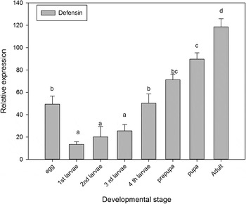
qRT-PCR analysis showed the expression of PxDef in all developmental stages, from eggs to adults. In comparison, the mRNA level in pupae and adults was significantly higher than in the larval stages (fig. 1).
Developmental expression of PxDef in P. xylostella from eggs to adults. The developmental expression of PxDef transcript in P. xylostella was determined by real-time quantitative RT-PCR. Expression of PxDef mRNA is relative to β-actin mRNA. Comparison of PxDef expression levels at different developmental stages was performed by t-test. Data with different letters are significantly different (P < 0.05) (DMRT) among different developmental stages.

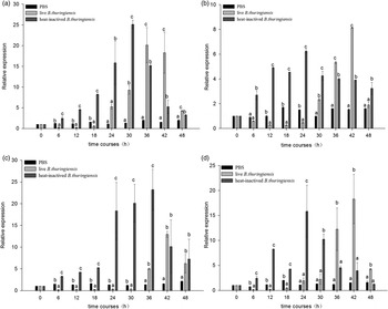
The 4th instar larvae of P. xylostella injected with B. thuringiensis were analyzed for the expression of PxDef in epidermis, hemocytes, midgut, and fat body. PxDef was significantly induced in all four tissues after injection of heat-inactivated B. thuringiensis. Despite the differences in the expression, highest levels of expression were found at 24 h in hemocytes and epidermis, 30 h in midgut, and 36 h in fat body (fig. 2). After injection of live B. thuringiensis, the expression of PxDef was significantly decreased in epidermis, midgut and hemocytes up to 30 h post injection and in the fat boy until 36 h post-injection, then increased rapidly. The results suggest that live B. thuringiensis may release some effectors to inhibit the expression of PxDef (fig. 2).
Induced expression of PxDef in different tissues after B. thuringiensis challenge. The mRNA level of PxDef in midgut (a), epidermis (b), fat body (c), and hemocytes (d) was analyzed by qRT-PCR. Comparison of PxDef mRNA levels was performed by t-test. Data with different letters are significantly different (P < 0.05) (DMRT) among time courses and tissue distribution.

RNA interference (RNAi)
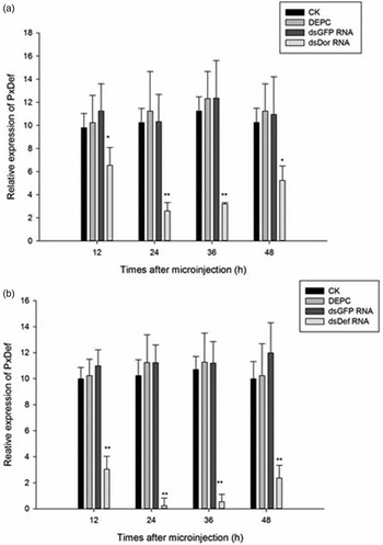
To elucidate the function of PxDef in immunity, DEPC, GFP-dsRNA, PxDef-dsRNA and PxDor-dsRNA were injected into 4th instar larvae of P. xylostella separately. Injection of dsRNA to PxDef significantly down regulated expression of PxDef mRNA at 12 h post- injection, and the RNAi effect sustained until 48 h post-injection (fig 3). Furthermore, RNAi silencing of PxDor also significantly down regulated PxDef transcript from 12 to 48 h post-injection (fig. 3), suggesting that expression of PxDef is regulated by the Toll/Dorsal pathway.
RNAi silencing of PxDor (a) and PxDef (b). P. xylostella larvae were injected with DEPC-treated water (CK), dsGFP RNA, dsPxDor RNA, or dsPxDef RNA, and total RNAs were prepared for qRT-PCR analysis. Actin was used as an internal control. Each bar represents the mean ± S.E. (n=3). Means with an asterisk are statistically different (P<0.05) (Duncan's Multiple Range Test) among treatments; Means with two asterisks are statistically different (P<0.001) (Duncan's Multiple Range Test) among treatments. DEPC: diethyl pyrocarbonate.

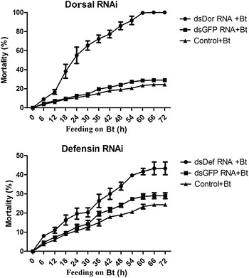
After RNAi silencing of PxDef and PxDor separately, the susceptibility of larval feeding by live B. thuringiensis (2 × 10⁶ CFU ml−1) was analyzed (fig. 4). Control larvae (naïve and GFP-dsRNA injected larvae) showed approximately 20–30% mortality, while larvae treated with dsRNAs to PxDor and PxDef showed significantly higher mortality than the control larvae 3 days after the infection of B. thuringiensis. The results suggest that PxDef may play a role in defense against B. thuringiensis infection in P. xylostella.
Effect of knockdown expression of PxDef and PxDor on susceptibility of P. xylostella larvae to B. thuringiensis (Bt). After dsRNA treatments, the larvae were treated with 200 ppm of Bt. Mortality was recorded every 6 h.

Antibacterial activity of recombinant PxDef
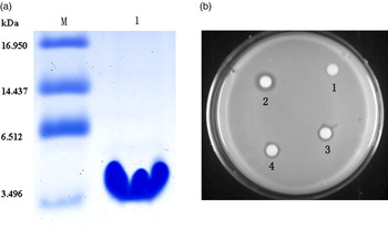
Recombinant PxDef was expressed as a fusion protein to thioredoxin (Ando et al., Reference Ando, Okada and Natori1987). Recombinant PxDef fusion protein was purified by affinity chromatography, thioredoxin was removed by enterokinase and recombinant PxDef was further purified. Analysis of the purified recombinant PxDef by Tricine-SDS–PAGE revealed that PxDef was purified to homogeneity with a molecular mass of approximately 4.2 kDa (fig. 5a).
Activity of recombinant PxDef against B. thuringiensis. (a) Tricine-SDS-PAGE analysis of purified recombinant PxDef. Lane M: polypeptide SDS-PAGE molecular weight standards; lane 1: 2 µg of purified recombinant PxDef. (b) Activity of recombinant PxDef against B. thuringiensis. Recombinant PxDef was tested for antibacterial activity against B. thuringiensis using a radial diffusion assay. The inhibition zone was quantified by measuring distance from the edge of the paper disk to the edge of the clear zone. Each experiment was repeated three times. 1, the inhibition zone of 10 µl PBS; 2, 10 µl (0.5 µg) Ampicillin, the inhibition zone is approximately 0.7 cm; 3 and 4, 10 µl (0.5 µg) recombinant PxDef, the inhibition zone is about 0.5 cm.

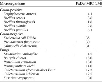
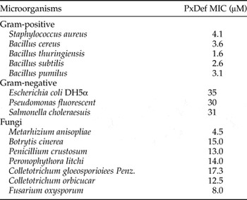
In order to test antimicrobial activity of recombinant PxDef against microorganisms, serial dilutions of purified recombinant PxDef was incubated with bacterial and fungal cultures, and the minimal inhibitory concentrations (MICs) were determined. Recombinant PxDef showed high activity against all Gram-positive bacteria and two fungi (Metarhizium anisopliae and Fusarium oxysporum) tested, showed relatively high activity against other fungi, but low activity against all Gram-negative bacteria tested (table 2).
The activity of recombinant PxDef against microorganisms.
MIC, minimum inhibition concentration.
Recombinant PxDef antibacterial against B. thuringiensis was performed. The inhibition zone was quantified by measuring distance from the edge of the paper disk to the edge of the clear zone (fig. 5b). The results showed that recombinant PxDef at 0.5 µg displayed activity against B. thuringiensis with inhibition zone of approximately 0.5 cm compared with inhibition zone of ~0.7 cm by ampicillin (0.5 µg).
Discussion
Insect defensins, the largest group of insect AMPs, have high activity against Gram-positive bacteria and lower activity against Gram-negative bacteria and fungi (Dang et al., Reference Dang, Wang, Huang, Yu and Zhang2010). To date, most defensins isolated and identified in insects are cationic peptides (Dimarcq et al., Reference Dimarcq, Hoffmann, Meister, Bulet, Lanot, Reichhart and Hoffmann1994; Chernysh et al., Reference Chernysh, Cociancich, Briand, Hetru and Bulet1996; Lauth et al., Reference Lauth, Nesin, Briand, Roussel and Hetru1998; Lee et al., Reference Lee, Yun, Jang, Kim, Lee, Park, Ryu, Seo, Kim and Lee2004; Seufi et al., Reference Seufi, Hafez and Galal2011; El Shazely et al., Reference El Shazely, Veverka, Fučík, Voburka, Žďárek and Čeřovský2013; Porto et al., Reference Porto, Fensterseifer and Franco2014), but little is known about anionic peptides in Lepidopteran insects, especially in P. xylostella. In the present study, PxDef cDNA clone was isolated and sequenced, and the deduced amino acid sequence comprised six cysteine residues in the conserved positions that are typical structural characteristics of invertebrate defensins. Multiple sequence alignment showed that there is a special fragment (Pro64-Pro79) PxDef, which is longer than that in other defensins from Lepidoptera (fig. S4). The special amino acid sequence suggests that PxDef may be different from most defensins reported so far. It is also possible that the longer fragment may account for the anionic pI of PxDef.
Defensins are reported to be cysteine-rich cationic AMPs. In our study, the pI value of mature PxDef is 5.57, indicating that it is an anionic peptide, different to almost all of the known defensins. Recently, an anionic defensin-like gene from silkworm (Kaneko et al., Reference Kaneko, Tanaka, Ishibashi, Iwasaki and Yamakawa2008) and defensin from the tick Amblyomma hebraeum (Lai et al., Reference Lai, Lomas, Jonczy, Turner and Rees2004) have been reported. A. hebraeum defensin (AhDef) contains a net negative charge with a pI value of 4.44, and it exerts antimicrobial activity against Gram-negative bacterium E. coli and Gram-positive bacterium S. aureus (Lai et al., Reference Lai, Lomas, Jonczy, Turner and Rees2004). However, in the present study, recombinant PxDef exhibited higher activity against Gram-positive bacteria and fungi, but lower activity against E. coli and other Gram-negative bacteria. The antibacterial mechanism of AMPs is ascribed to the interaction between the positively charged AMPs and the negatively charged microbial membrane components, which include LPS in Gram-negative bacteria and peptidoglycan in Gram-positive bacteria. But interaction mechanism of anionic defensins is not well understood and future research is needed to elucidate the mechanism.
Insect AMPs have been reported to be expressed predominantly in the fat body, the functional equivalent of the vertebrate liver, and increasing evidence shows that other tissues may also be involved in immune responses (Yi et al., Reference Yi, Chowdhury, Huang and Yu2014; Gambichler & Skrygan, Reference Gambichler and Skrygan2015). In the present study, after challenge with B. thuringiensis, expression of PxDef was significantly increased not only in the fat body, but also in the hemocytes, epidermis, and midgut. These results suggest that PxDef is systematically induced and may play its role in the defense against B. thuringiensis infection. B. thuringiensis is one of the popular pesticides for biological control of Lepidopteran pests including P. xylostella, which has developed a high resistance to B. thuringiensis. In the present paper, the expression of PxDef was analyzed after B. thuringiensis challenge using qRT-PCR method. The transcription levels of PxDef were lower after live B. thuringiensis infection than injection of heat-inactivated B. thuringiensis. In addition RNAi silencing of PxDef caused increase in mortality of P. xylostella larvae after B. thuringiensis infection. These results suggest that defensin may play an important role in P. xylostella against invading B. thuringiensis.
Insect immune related genes are regulated by the Toll and Imd pathways after microbial infections (Hoffmann & Reichhart, Reference Hoffmann and Reichhart2002). RNAi results showed that the expression of PxDef was significantly reduced after RNAi silencing of Dorsal as compared with the control (DEPC H2O and GFP-dsRNA). This result suggests that the expression of PxDef is regulated by the Toll-Dorsal signal pathway. Whether expression of PxDef is also regulated by the Imd pathway or not remains to be determined.
RNAi silence of Dorsal in P. xylostella also down regulates the expression of Cecropins (Ouyang et al., Reference Ouyang, Xu, Freed, Gao, Yu, Wang, Ju, Zhang and Jin2015) and lysozymes and may be many other immune-related genes regulated by the Toll-Dorsal pathway. This may account for almost 100% mortality in P. xylostella larvae treated with dsRNA to Dorsal and after feeding on live B. thuringiensis, since many immune effectors are down regulated after RNAi silencing of Dorsal, resulting in high susceptibility of larvae to live P. xylostella.
Supplementary material
The supplementary material for this article can be found at http://dx.doi.org/10.1017/S0007485316000596.
Acknowledgements
The authors thank Prof. Xiao-qiang Yu (University of Missouri-Kansas City) for correcting the manuscript. This work was supported by grant from The National Natural Science Foundation of China (30800718, 31071734) Science and Technology Program of Guangzhou China (201509010023) and Department of Science and Technology of Guangdong China (2014A020208106).
Disclosure
The authors of this study declare that there is no conflict of interests in this work.