Congenital heart block is a rare conducting system disease with an incidence of about 1/11,000–1/20,000 live births.Reference Kelle, Backer and Tsao1 The aetiologies of congenital heart block include maternal autoimmune antibodies, few structural heart diseases like atrioventricular canal defect with or without left isomerism and congenitally corrected transposition of great arteries, myopathies, genetic disorders, and isolated congenital heart block.Reference Friedman, Duncanson, Glickstein and Buyon2,Reference Sadasivam and Kamupira3 Identification of the cause of congenital heart block is important for effective management.
Delayed pacing therapy or haemodynamic compromise from structural cardiac defects leads to significant morbidity and mortality.Reference Kelle, Backer and Tsao1 This advocates need for immediate diagnosis and aggressive management in case of neonates. In neonates with congenital heart block in structurally normal heart, pacemaker is recommended if the patient develops significant bradycardia (<55 in neonates) and signs of severe congestive heart failure.Reference Li, Zhang and Li4 The recommendation for pacemaker in a child less than 10 kg is the placement of epicardial leads.Reference Li, Zhang and Li4 However, transvenous pacing via umbilical vein can be used as an emergency stabilising procedure in this high vulnerable group of neonates. We report temporary pacemaker implantation in critically ill low birth weight premature neonate via umbilical vein and discuss the technical skills for the same.
Case report
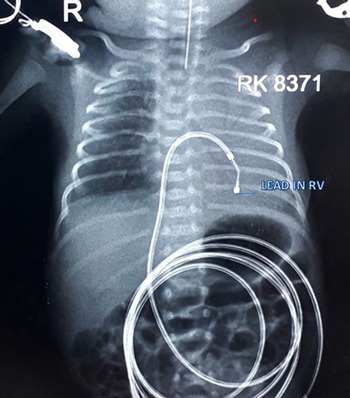
A 4-hour-old male neonate first in birth order, born at preterm at 31 gestational weeks with birth weight of 1.51 kg via non-consanguineous marriage, was delivered at hospital via emergency caesarean section for fetal bradycardia. Baby had cried immediately after birth but developed bradycardia with heart rate of less than 50 beats per minute. The baby was intubated immediately and transferred to NICU and kept under mechanical ventilator support. Baby was managed conservatively with antibiotics (sepsis screen was positive), inotropes (adrenaline 0.5 mcg/kg/min), and IV dexamethasone. ECG revealed complete atrioventricular block. In view of complete heart block with haemodynamic instability, he was referred to our centre for further evaluation and management. Antenatal history of autoimmune disorders was insignificant, and steroids were not given to mother antenatally. On presentation, general physical examination showed saturation of 96% on continuous positive airway pressure with Fi02 of 30% and positive end expiratory pressure: 5 cm of H2O, heart rate – 48 beats per min, BP – 80/60 mm Hg, and respiratory rate – 66 per minute. Cardiovascular examination revealed normal first heart sound, normal splitting of second heart sound, and no murmur. Transthoracic echocardiography showed no structural heart disease. Baby required higher ventilator settings. First dose of surfactant was given via an endotracheal tube. He was shifted to catheterisation lab and temporary pacing was done through umbilical venous route at 4 hours of life (Fig 1). The umbilical vein was first cannulated by 5F paediatric sheath (Arrow Advancer™, Pediatric set). Right ventricle lead could not be placed in spite of repeated attempts, essentially due to number of curves from the umbilicus to the right ventricle. In view of this, the paediatric sheath (7 cm) was replaced by 11 cm sheath (Arrow Percutaneous Introducer sheath) over 0.35 guide wire. 5F introducer could be advanced to inferior vena cava. Temporary bipolar pacing lead (5 French 125 cm BARD® Temporary Pacing Electrode Catheter without screw) was implanted in right ventricle and lead was connected to an external pacemaker (Medtronic single chamber temporary external pacemaker) stimulating heart rate at 140. Long arrow sheath was pulled back leaving pacing catheter in place. Post-procedure heart rate was 140 beats per minute via temporary pacemaker. Baby was kept on inotropic support (dopamine and dobutamine at 10 mcg/kg/min). Due to the requirement of persistent higher ventilator settings, second dose of surfactant was given the next day. Antibiotics were upgraded to meropenem and colistin in milieu of positive repeat septic screen. Mechanical ventilator settings were gradually tapered and baby was extubated to nasal continuous positive pressure airway on 6th day post-operatively. Repeat septic screen was negative and baby was tolerating oral feeds well. Baby was shifted to a surgical unit for permanent pacemaker implantation with epicardial single chamber (CapSure Epi 4968-35; Medtronic Inc., Minnesota, USA) with ventricular lead and pulse generator (Ventricle paced, Ventricle sensed, Rate adaptive, Inhibited response to sensing) on day 22.
Chest X-ray demonstrating the pacing via umbilical vein with lead placed in the right ventricle.

Discussion
Neonates born with atrioventricular block and comorbidities like low birth weight, prematurity, and poor haemodynamic status usually have grave prognosis. This subset of neonates needs meticulous attention and aggressive management. Management strategy for haemodynamically significant atrioventricular block includes medical management followed by staged pacing. Medical management includes administration of chronotropic and inotropic support with isoprenaline, dopamine, and dobutamine for neonatal heart block.Reference Sivarajah, Huggon and Rosenthal5 Staged pacing can be done with either temporary pacing (epicardial or transvenous approach) or permanent pacing. Planned early pacing of high risk neonates with congenital heart block reduces the adverse consequences of bradycardia and asystole.Reference Yildirim, Tunaodlu and Karaadac6 There are several options for pacing ranging from transcutaneous pacing through external defibrillators or temporary pacing catheters via femoral or jugular veins. Temporary pacemaker implantation via umbilical vein can be considered in critically ill neonate as a bridge to a permanent pacemaker. The decision for implanting a device in a neonate is not as straightforward due to many constraints such as patient size, venous access, and haemodynamic instability. Pacing in newborn is generally accomplished with epicardial pacing systems via subxiphoid incision. There have been many reports of implanting epicardial ventricular pacemakers for neonatal congenital heart block since 1973. Epicardial pacing in neonates requires expertise with skilled personnel, and thus in resource-constrained environment, transvenous pacing remains a viable option. However, there are very few studies regarding transvenous approach for temporary pacing in neonates.Reference Kannankeril, Dzurik and Fish7,Reference Doshi and Lokare8 Femoral vein and internal jugular vein access are difficult to get and have their own set of complications in newborns. Smaller patients who are less than 6 years are at risk of subclavian vein occlusion when subclavian vein is used to place temporary pacing leads limiting its use in future permanent pacemaker placement through the same route. In light of these findings, pacing via umbilical vein approach is a promising technique as long-term complication of venous occlusion is not of much concern as umbilical vein gets obliterated.Reference Kannankeril, Dzurik and Fish7 After stabilisation of the neonate and adequate weight gain, temporary leads can be extracted without any complications and permanent epicardial leads can be implanted as was done in our case.
Our case of premature low birth weight neonate having significant bradycardia with heart rate less than 50 beats per minute and ongoing severe sepsis with metabolic acidosis requiring mechanical ventilator support posed challenges on the management. Early temporary pacing was the feasible option as permanent epicardial pacing could not be considered in this patient due to low birth weight, ongoing sepsis, and haemodynamic instability. There was rapid deterioration in the condition of the neonate, and temporary pacing was done as an emergency procedure. Transvenous pacing via umbilical vein was done to avoid potential complications of larger sheath insertion in femoral vein or internal jugular vein leading to vessel injury. The umbilical vein ensured aseptic access without the need for restriction of the baby or restricting limb movements. This allowed for the stabilisation of the baby and weight gain, before a permanent pacemaker implantation was considered at a later date. There are very few reports of umbilical vein approach for pacing in neonates.Reference Li, Zhang and Li4,Reference Kannankeril, Dzurik and Fish7–Reference Hanseus, Sandstrom and Schuller9 Series have been encouraging overall with fewer complications and excellent outcomes.
Conclusion
It is worth re-emphasising that implantation of temporary pacing through umbilical venous route in critically ill neonates with congenital heart block is technically feasible and promising stabilisation procedure prior to permanent pacing. The use of longer sheath in our case allowed for negotiating the curves and hence deploying leads in right ventricle.
Acknowledgements
None.
Financial support
This research received no specific grant from any funding agency, commercial or not-for-profit sectors.
Conflict of interest
None.
Ethical standards
The authors assert that all procedures contributing to this work comply with the ethical standards of the relevant national guidelines on human experimentation and with the Helsinki Declaration of 1975, as revised in 2008, and has been approved by the Max Institute of medical Excellence Institutional committee.