Introduction
The circadian clock maintains intrinsic rhythmical changes of biochemical and physiological processes, which provide optimal adaptation to environmental changes, such as light, temperature, and access to food. In nonmammalian vertebrates, photoreceptors in the brain, pineal gland, and retina serve to entrain the circadian clock to changes in the light–dark cycle (reviewed by Doyle & Menaker, Reference Doyle and Menaker2007; Falcón et al., Reference Falcón, Besseau, Fuentès, Sauzet, Magnanou and Boeuf2009), though evidence from fish suggests that cells and tissues are independently photosensitive and may possess the ability to self entrain to the light–dark cycle (Whitmore et al., Reference Whitmore, Cermakian, Crosio, Foulkes, Pando, Travnickova and Sassone-Corsi2000; Dekens & Whitmore, Reference Dekens and Whitmore2008). In turn, within all tissues, circadian rhythms regulate numerous cellular events. In the retina, these events include retinomotor movements (Pierce & Besharse, Reference Pierce and Besharse1985; Menger et al., Reference Menger, Koke and Cahill2005), disc shedding by photoreceptors (LaVail, Reference LaVail1980), visual sensitivity (Li & Dowling, Reference Li and Dowling1998), dopamine release (Ribelayga et al., Reference Ribelayga, Wang and Mangel2004), the expression of interphotoreceptor retinoid–binding protein (Rajendran et al., Reference Rajendran, Van Niel, Stenkamp, Cunningham, Raymond and Gonzalez-Fernandez1996), and the synaptic output of photoreceptors onto horizontal cells (Wang & Mangel, Reference Wang and Mangel1996; Ribelayga et al., Reference Ribelayga, Wang and Mangel2002, Reference Ribelayga, Cao and Mangel2008).
Midkine (MK) is a member of the family of secreted heparin-binding growth/differentiation factors that also includes pleiotrophin (Obama et al., Reference Obama, Matsubara, Guénet and Muramatsu1994; Kadomatsu & Muramatsu, Reference Kadomatsu and Muramatsu2004). MK was first identified in a screen of retinoic acid–inducible genes in embryonic carcinoma cells (Kadomatsu et al., Reference Kadomatsu, Huang, Suganuma, Murata and Muramatsu1990, Reference Kadomatsu, Tomomura and Muramatsu1998; Muramatsu, Reference Muramatsu2002), and it is now a well-established transcriptional target of retinoic acid signaling. MK is highly conserved throughout the animal kingdom and has numerous biological functions (Winkler et al., Reference Winkler, Schafer, Duschl, Schartl and Volff2003; Kadomatsu & Muramatsu, Reference Kadomatsu and Muramatsu2004). In mammals, MK is expressed during embryonic development, most prominently in the developing neural tube and at epithelial–mesenchymal boundaries (Mitsiadis et al., Reference Mitsiadis, Salmivirta, Muramatsu, Muramatsu, Rauvala, Lehtonen, Jalkanen and Thesleff1995).
The zebrafish genome encodes two distinct midkine genes, midkine-a (mdka) and midkine-b (mdkb), which during early development have distinct cellular patterns of expression and different biological functions (Winkler & Moon, Reference Winkler and Moon2001; Winkler et al., Reference Winkler, Schafer, Duschl, Schartl and Volff2003; Schäfer et al., Reference Schäfer, Rembold, Wittbrodt, Schartl and Winkler2005; Liedtke & Winkler, Reference Liedtke and Winkler2008). In the adult retina, we found that mdka is expressed by horizontal cells and Müller glia, whereas mdkb is expressed by cells throughout the inner nuclear layer (Calinescu et al., Reference Calinescu, Vihtelic, Hyde and Hitchcock2009). In the course of these studies, in situ hybridizations showed that the expression of mdka was high in the morning hours and decreased markedly at the end of the day, suggesting that expression of mdka is regulated either by light or by the circadian clock.
To distinguish between these possibilities, mRNA and protein levels were measured in the retina of fish maintained in complete darkness for 24 h up to 63 h. The data show that over a 24-h dark period, the expression of midkine-a shows a daily temporal rhythm that is not dependent of the presence of light—in the dark, levels of both mRNA and protein increase during the subjective day and decrease at night. This pattern of midkine-a expression is maintained after two complete days in complete darkness, which leads to an interpretation that is consistent with regulation by the circadian clock. Finally, qualitative observations of in situ hybridizations suggest that the expression of mdkb is similarly regulated. The spatial domain of mdkb expression within the inner nuclear layer expands during the night and contracts during the subjective day. Interestingly, within horizontal cells, the two midkine paralogs show asynchronous regulation.
Though the functional significance of the circadian regulation of MK expression in the retina is yet to be determined, this study expands our knowledge of the cellular expression and circadian control of soluble signaling molecules in the vertebrate central nervous system.
Materials and methods
Animals
Wild-type zebrafish (Danio rerio), mixed strains and strain AB, 4.5–7 months old, were purchased from Aquatica Tropicalis (Plant City, FL) and acclimated for at least 2 weeks in aquaria at 28.5°C and a 14/10-h light/dark cycle. Since circadian regulation is indicated by rhythmicity in the absence of environmental cues, a common method to test circadian regulation is by eliminating the light cue and sampling during complete darkness (Menger et al., Reference Menger, Koke and Cahill2005). For experiments described here, either fish were maintained in complete darkness for 24 h and during this period were sacrificed at 4-h intervals corresponding to the Zeitgeber times (ZT): 15, 19, 23, 3, 7, and 11, where ZT0 corresponds to the time of light onset, or fish were maintained in complete darkness for 51 or 63 h. These and the following procedures, except where noted (see below), were repeated in three independent experiments. All procedures were approved by the University Committee for the Use and Care of Animals at the University of Michigan.
Tissue preparation for in situ hybridization and immunohistochemistry
At selected times and using dim red illumination, adult fish were anesthetized in 0.1% 3-aminobenzoic acid-ethyl ester (Sigma-Aldrich, St. Louis, MO) and sacrificed by cervical transection. Eyes were then enucleated, lenses removed, and eyecups fixed by immersion for 4–19 h at 4°C in 4% paraformaldehyde in 0.1 m phosphate buffer containing 5% sucrose. Eyecups were cryoprotected in 20% sucrose, embedded in 2 parts 20% sucrose 1 part Tissue-Tek® O.C.T. Compound (Electron Microscopy Sciences, Hatfield, PA) frozen in Tissue-Tek® O.C.T. Compound, and stored at −80°C. Ten-micron thick cryosections through the dorso-ventral axis of the eyecups were mounted on Superfrost Plus Microscope Slides (Fisher-Scientific, Pittsburgh, PA) and processed for in situ hybridization. Antisense digoxigenin-labeled riboprobes were synthesized using the DIG RNA Labeling Kit (Roche Diagnostics, Indianapolis, IN) from plasmids containing the full-length mdka or mdkb cDNAs (gift from Dr. Christoph Winkler; Winkler et al., Reference Winkler, Schafer, Duschl, Schartl and Volff2003). In situ hybridization was performed as described previously (Hitchcock & Kakuk-Atkins, Reference Hitchcock and Kakuk-Atkins2004) using nitroblue tetrazolium chloride/5-bromo-4-chloro-3-indolyl phosphate as the enzymatic substrate. To compare mdka or mdkb expression across time points, four to five sections from eyes collected at each of the six time points were mounted and processed on the same slide. Therefore, within each replicated experiment, sections were processed identically, and this allowed direct comparison of the levels (intensity of reaction product) and cellular patterns of expression for the two genes.
RNA extraction and quantitative real-time reverse transcription polymerase chain reaction
To isolate retinal RNA, eyecups were removed under dim red illumination and retinas were dissected and carefully separated from the retinal pigment epithelium. Three to four retinas per sample from different zebrafish were pooled for each sample, homogenized with a sterile pestle (Kontes Pellet Pestle; Fisher Scientific) in 200 μl lysis buffer from the Ambion RNAqueous-Micro RNA Isolation Kit (Ambion, Austin, TX), and RNA extraction was performed according to the manufacturer’s instruction. The amount of RNA was quantified using a spectrophotometer, and the quality of the RNA was assessed on ethidium bromide–stained formalin–agarose gels. 0.5 or 1 μg of total RNA was used to synthesize cDNA using the Superscript II First-Strand Synthesis System for RT-PCR (Invitrogen, Carlsbad, CA) following the manufacturer’s protocol with random hexamers. The resulting first-strand reaction was used as a template for the subsequent quantitative real-time reverse transcription polymerase chain reaction (QRT-PCR) using the iQ™ SYBR® Green Supermix (Bio-Rad, Hercules, CA) in an iCycler Real-Time PCR Detection System (Bio-Rad). The following amplification and melt curve analysis protocol was used: 95°C: 3 min, 35× (95°C: 20 s, 57°C: 20 s, and 72°C: 30 s) and 95°C: 1 min, 90× (55°C: 10 s). Gene-specific primers (0.4 μ m) were as follows: for mdka (NM_131070), forward—tgaagttttgttactgagctttgtg, and reverse—agccagtgtacataagtgtgtgtgt; for mdkb (NM_131716), forward—gctgttgtaatttgtagcaggtttt, and reverse—cattcaatctcgttgtcatttacag. Serial dilutions of the first-strand reaction were run for efficiency calculations of each primer using the Pfaffl method (Pfaffl, Reference Pfaffl2001). The threshold cycle (Ct) was determined by the iCycler using the maximum curvature approach and then maintained constant for subsequent runs. Relative gene expression values were determined using the calculated primer efficiencies and threshold cycle with the formula: E^−Ct. Specificity of the amplification products was verified by agarose gel electrophoresis of sample wells. Values obtained were averaged for three to six independent amplification reactions, and statistical significance was calculated by one-way analysis of variance (ANOVA) with Bonferroni correction for multiple comparisons using SPSS software. Samples within each replicate experiment were treated identically. To verify that equal amounts of RNA were used, QRT-PCR was performed with the following primers for ribosomal protein L-19 (rpl-19, accession number: NM_213208): forward—gagtatgctcagacttcagaagagg, and reverse—atcaaaccatccttcaccaacttac. There were no differences in rpl-19 expression across time points.
Midkine-a antibodies and Western blot analysis
Based on antigenicity, hydrophilicity, flexibility, and surface probability (Invitrogen, Camarillo, CA), a C-terminal peptide composed of 16 amino acid residues was chosen as the Mdka immunogen (amino acids 131–145: KVKNKPKGKKGKGKGC; accession number NP_571145). Affinity-purified polyclonal antibodies were generated in rabbits. In Western blots, the resulting antibodies recognized a single band with the correct size in lysates from both transfected 293T cells expressing zebrafish Mdka-MYC and retinas (data not shown). These antibodies did not recognize Mdkb-MYC (data not shown). To assay endogenous Mdka, five retinas were dissected from five different fish at each time point and pooled. The retinal pigment epithelium was carefully removed, and retinas were homogenized with a Kontes Pellet Pestle (Fisher Scientific) in 75 μl of lysis buffer (phosphate-buffered saline with 1% Triton X) and protease inhibitors (Complete-mini EDTA free; Roche, Indianapolis, IN). Lysates were centrifuged for 5 min at 5000 rpm at 4°C to pellet the nuclei and transferred to fresh tubes. The amount of retinal protein was determined with a BCA Protein Assay Kit (Pierce, Rockford, IL). Equal amounts of protein were loaded onto a 12% sodium-dodecyl-sulphate polyacrylamide gel (SDS-PAGE) and processed for immunoblotting. Proteins were separated by electrophoresis on a 12% SDS-PAGE and transferred onto nitrocellulose membranes (Schleicher and Schuell, Keene, NH). Membranes were incubated at 4°C for at least 4 h in blocking buffer [phosphate-buffered saline with 0.05% Tween (PBST) and 5% nonfat dry milk] followed by overnight incubation in Mdka primary antibodies, diluted 1:500. Membranes were washed for 1 h in PBST and then incubated for 1 h in peroxidase-conjugated anti-rabbit IgG secondary antibodies, diluted 1:1000 (Amersham Biosciences, Arlington Heights, IL). Following four to five vigorous washes in PBST, proteins were visualized with the enhanced chemiluminescence ECL detection system (ECL Amersham Biosciences) and radiographic film (Kodak, Rochester, NY). Antibodies against Mdka recognize a single band at the appropriate molecular weight for the native protein (see Results). Equal protein loading was verified by reprobing membranes with a monoclonal antibody recognizing α-, β-, and γ-actin (JLA20; Calbiochem, San Diego, CA).
Data from images
Photographic images were taken with a Nikon DMX 1200 digital camera mounted on a Nikon Eclipse E800 epifluorescent microscope equipped with a differential interference contrast filter. Adobe Photoshop CS2 (Adobe Systems, San Jose, CA) was used to construct the figures. The layer tool was used to generate overlays. If needed, brightness and contrast were adjusted identically in all parts of a given figure.
Digital images of films were taken using a flatbed scanner (Epson expression 1600). The intensity of the midkine and actin bands was measured using the histogram tool in Adobe Photoshop CS2 (Adobe Systems). The intensity of each midkine band was normalized to the intensity of the corresponding actin band. To compare results between replicate experiments, the ZT11 time point was chosen as a reference, and the relative protein levels were normalized. The resulting values were then averaged between experiments and plotted as fold change.
Results
Midkine-a mRNA and protein levels oscillate with a daily rhythm that is independent of light
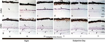
To determine if light itself regulates mdka expression, zebrafish were maintained in total darkness for 24 h, and retinas were collected at 4-h intervals. At each time point, retinas were divided into three groups, and the expression of mdka was evaluated by in situ hybridization and QRT-PCR, and the synthesis of Mdka was evaluated by Western blot analysis. In situ hybridization confirmed that in the adult retina, mdka is expressed at high levels in horizontal cells (Calinescu et al., Reference Calinescu, Vihtelic, Hyde and Hitchcock2009) and showed that this cellular expression is dynamically modulated in the absence of light. Minimum expression was observed in retinas harvested at ZT15 (Fig. 1a). This minimum was observed in retinas harvested at 1 h after the lights were turned off, as well as in retinas kept in total darkness for 25 h. Following this time point, expression increased to reach an apparent maximum at ZT23 (Fig. 1b and 1c), 1 h prior to the normal time of light onset. mdka expression then remained high during the first half of the subjective day (Fig. 1d and 1e, ZT3, ZT7) and gradually decreased during the second half of the subjective day (Fig. 1f, ZT11), approaching the minimum at ZT15 (Fig. 1a).
The expression of mdka and mdkb in the zebrafish retina is independent of light onset. (a–f) are in situ hybridizations that illustrate retinal expression of mdka during the night (a–c) and subjective day (d–f). (g–l) are in situ hybridizations that illustrate retinal expression of mdkb during the night (g–i) and subjective day (j–l). Arrows in (a–f) point to the location of horizontal cells. Arrowheads in (g–l) point to the location of amacrine and ganglion cells, respectively. Sections for all ZT samples for each probe were processed on the same slide. ONL, outer nuclear layer; INL, inner nuclear layer; GCL, ganglion cell layer. Scale bar in (f) equals 50 μm.

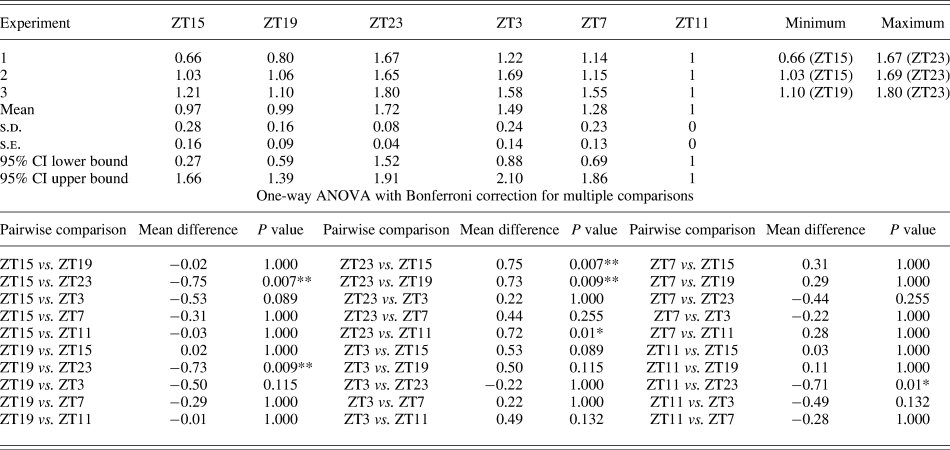
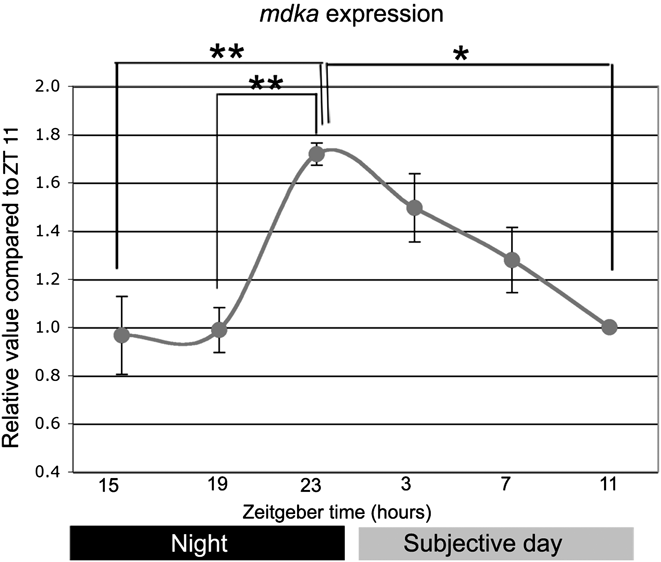
To validate these qualitative observations, QRT-PCR was used to measure mdka mRNA levels. These quantitative data parallel the in situ hybridization data and confirm the temporal pattern of mdka expression. The ANOVA showed that the expression levels of mdka at ZT15 and ZT19 were statistically significantly different from the ZT23 time point, which, in turn, was significantly different from ZT11 (Table 1). Finally, Western blot analysis of retinal lysates showed a pattern of Mdka protein levels that mirror the in situ hybridization and QRT-PCR results (Fig. 3a and 3b). Minimum levels of protein were present in the retinas at ZT15, whereas maximum protein levels were present in the retinas at ZT23. This temporal pattern was observed in two independent experiments. Taken together, these data show that the expression of Mdka in horizontal cells of the zebrafish retina is independent of the presence of light, and both the mRNA and the protein levels follow the same temporal pattern.
Circadian expression of mdka in the zebrafish retina

Evidence for the circadian regulation of the expression of mdka mRNA and the levels of Mdka protein
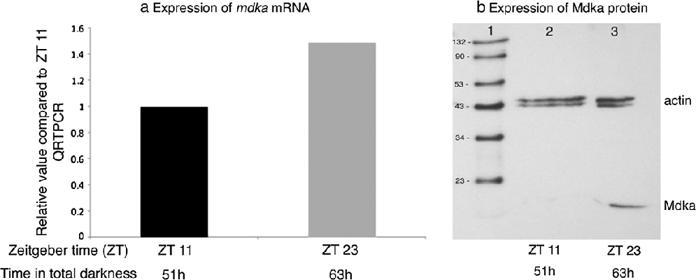
As a minimal test of whether or not the circadian clock regulates the expression of mdka, animals were kept in complete darkness for two complete light cycles, and the mRNA and protein levels were measured at the minimum (ZT11; 51 h in the dark) and maximum (ZT23; 63 h in the dark) expression levels as established in the 24 h experiments (Figs. 2 and 3). In this analysis, mRNA levels at the ZT 23-h time point were 1.5× that at ZT11 (Fig. 4a), which replicates the relative levels of mRNA observed during the initial 24 h of complete darkness. The levels of Mdka protein at ZT11 and ZT23 parallel that shown for the mRNA (Fig. 4b). These data are interpreted to show that mdka is regulated by the circadian clock.
Quantitative analysis of circadian variations of mdka. Gene expression is represented as fold change compared to expression values at ZT11. Data represent the averaged values from three experiments with standard errors represented for each ZT sample. Statistical significance was determined by one-way ANOVA with Bonferroni correction for multiple comparisons using SPSS (**P < 0.01 and *P < 0.05).

Western blot analysis of Mdka expression. (a) Immunoblot of retinal lysates obtained from zebrafish at specified times during 24 h of complete darkness. Lower bands represent the Mdka protein and upper bands represent actin, which was used as a loading control. (b) The quantification of circadian changes in retinal Mdka protein expression, normalized to expression of actin, averaged across two independent experiments. Results are shown as mean fold change compared to the expression at ZT11.

The expression of Mdka may be regulated by the circadian clock. (a) The relative levels of mRNA encoding mdka following 51 and 63 h in complete darkness, normalized to 51 h (ZT11). (b) illustrates a Western blot of retinal lysates obtained from zebrafish at the same ZT as represented in (a). Lower bands are the Mdka protein and upper bands are actin, which was used as a loading control.

The circadian clock may modulate the cellular expression of mdkb
We next asked whether the circadian clock also may regulate the expression of mdkb by evaluating its expression during the initial 24 h of complete darkness. In situ hybridization showed that at ZT3, mdkb is expressed by cells within the innermost (amacrine cell) tier of the inner nuclear layer and in the ganglion cell layer (Fig. 1j; see also Calinescu et al., Reference Calinescu, Vihtelic, Hyde and Hitchcock2009). In contrast, at ZT11, the cellular expression of mdkb expands to include the outer tier of the inner nuclear layer, including horizontal cells (Fig. 1g–1i). This qualitative change in the cellular pattern of expression of mdkb was observed in each of the three replicate experiments. These observations were followed with QRT-PCR using mdkb-specific primers, and though these data showed a trend that followed the qualitative observations of the in situ hybridizations, there were no significant time-dependent differences in the overall levels of mdkb mRNA in the retina (data not shown).
Discussion
In zebrafish, the genes that encode the two MK paralogs are located on different chromosomes have different cellular patterns of expression in the central nervous system (Winkler et al., Reference Winkler, Schafer, Duschl, Schartl and Volff2003; Liedtke & Winkler, Reference Liedtke and Winkler2008) and in the developing hindbrain and are thought to subserve different functions (Winkler & Moon, Reference Winkler and Moon2001; Winkler et al., Reference Winkler, Schafer, Duschl, Schartl and Volff2003; Schäfer et al., Reference Schäfer, Rembold, Wittbrodt, Schartl and Winkler2005; Liedtke & Winkler, Reference Liedtke and Winkler2008). In the zebrafish retina, cellular expression of mdka and mdkb is actively modulated during two neurogenic events: retinal development and photoreceptor regeneration following light-induced photoreceptor death (Calinescu et al., Reference Calinescu, Vihtelic, Hyde and Hitchcock2009). Unexpectedly, a time-course analysis of mdka expression showed high levels of expression during the day and a consistent and substantial decrease 12 h after light onset. This observation suggested that changes in the expression of mdka was either induced by light or regulated by the circadian clock.
To test whether or not the expression of the midkines might be regulated by light itself, the retinal expression of mdka and mdkb was analyzed over a 24-h period in animals kept in complete darkness. This experiment demonstrated that expression of mdka oscillates with a regular daily rhythm that is not dependent on the onset or termination of light. To test whether or not midkine expression may be regulated by the circadian rhythm, animals were kept in constant darkness for an additional 24 h, and the relative levels of mRNA and protein for midkine-a were measured at the predicted peak and trough of the expression levels observed in the 24-h experiments. Results from this experiment replicate the changes observed for animals kept in darkness for 24 h, and the inclusion of an additional subjective light–dark cycle provides for an interpretation consistent with the regulation of midkine-a expression by the circadian clock. Qualitative observations from in situ hybridizations for mdkb indicated that the expression of this gene also may be regulated by the circadian clock. Unlike mdka, which is distinctly expressed in horizontal cells, mdkb is expressed more broadly in the retina and does not show a dramatic temporal change in levels of expression. This may explain why the QRT-PCR analysis of whole retinal mRNA was unable to resolve statistically significant changes in the circadian expression of mdkb. However, the spatial distribution of mdkb mRNA does change, as shown by in situ hybridizations. The distribution of mdkb expression in the outer half of the retina may be regulated by the circadian clock, and if so, the regulation of expression of the two midkine paralogs in horizontal cells is out of phase. This suggests that in the outer retina, these two proteins may exert their biological actions at different times during the circadian cycle.
As for most vertebrates, in teleosts, the retina functions in part (though not alone; Vallone et al., Reference Vallone, Lahiri, Dickmeis and Foulkes2005; Vuilleumier et al., Reference Vuilleumier, Besseau, Boeuf, Piparelli, Gothilf, Gehring, Klein and Falcón2006; Dekens & Whitmore, Reference Dekens and Whitmore2008) to entrain circadian rhythms. Additionally, within the teleost retina, numerous processes are under direct circadian regulation. Among these is the synthesis and release of neurotransmitters and soluble signaling molecules (Ribelayga & Mangel, Reference Ribelayga and Mangel2005; Vuilleumier et al., Reference Vuilleumier, Boeuf, Fuentes, Gehring and Falcón2007), retinomotor movements (Pierce & Besharse, Reference Pierce and Besharse1985; Menger et al., Reference Menger, Koke and Cahill2005), and the synthesis of photoreceptor opsins (Korenbrodt & Fernald, Reference Korenbrodt and Fernald1989; Li et al., Reference Li, Chaurasia, Gao, Carr, Iuvone and Li2008). Whereas the function of retinal midkines is not yet known, our data identify them as components of cyclical signaling events with the vertebrate retina. Furthermore, the demonstration that circadian rhythms regulate the synthesis of these molecules points to important avenues of further inquiry.
Acknowledgments
We thank Dr. Christoph Winkler for the gift of plasmids encoding mdka and mdkb. We also thank Linda Barthel, Dilip Pawar and Laura Kakuk-Atkins for excellent technical assistance. This work was supported by NIH grants R01-EY07060 and P30-EY07003 and a Senior Scientific Investigator Award from the Research to Prevent Blindness (P.F.H.).