Pneumocystis jirovecii pneumonia (PCP) is responsible for significant morbidity and mortality in immunosuppressed patients. Recently, an increasing number of cases of non–AIDS-related PCP has been observed.Reference Thomas and Limper 1 – Reference Mansharamani, Garland, Delaney and Koziel 6 Solid-organ transplant (SOT) recipients are particularly exposed, with historical seriesReference Sepkowitz, Brown and Armstrong 7 – Reference Radisic, Lattes and Chapman 10 showing an overall PCP incidence between 5% and 15%. PCP-related mortality in SOT recipients is between 30% and 50%,Reference Mansharamani, Garland, Delaney and Koziel 6 , Reference Kovacs, Hiemenz and Macher 11 , Reference Sepkowitz 12 and anti-PCP prophylaxis is therefore recommended for SOT recipients, although there is no consensus on the duration of treatment.Reference Roux, Gonzalez and Roux 2 , Reference Gordon, LaRosa and Kalmadi 8 , Reference Martin and Fishman 13 – Reference Chapman, Marriott, Chen and MacDonald 15
Recently, grouped cases of PCP in SOT recipients were described, with epidemiological and molecular data suggesting interhuman transmission.Reference Chapman, Marriott, Chen and MacDonald 15 – Reference Sassi, Ripamonti and Mueller 26 During such episodes, the rapidity with which a grouped characteristic was identified and molecular analyses were performed contributed significantly to limiting the epidemic.Reference Chapman, Marriott, Chen and MacDonald 15
Coordination by infectious diseases specialists (infection control and prevention unit or clinical infectious disease specialist) is recommended for its clinical benefits to healthcare institutions.Reference Kim, Craft and Katzman 27 – Reference Trivedi and Kuper 30 The role played by an infectious disease unit in orchestrating a multidisciplinary response to an unexpected epidemiological situation has not been widely described in the literature.
In this paper, we report grouped cases of PCP among SOT recipients treated at Grenoble University Hospital in 2014, and we highlight the central role played by the infectious diseases unit in coordinating the reactions mounted to deal with these grouped cases.
METHODS
Care Setting
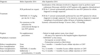
The University Hospital in Grenoble, France, has a total of 2,200 beds. In 2014, 1,899 SOT recipients were followed-up (1,380 kidney, 339 liver, 90 heart, 90 lung). From 2009 to 2015, the numbers of SOTs performed each year were 146, 156, 155, 183, 162, 187, and 180, respectively. SOT patients are cared for in the corresponding organ-centered department, which are located on different floors in our institution. From 2009 to 2014, the incidence of PCP in these patients was <1 case per year. Because of this low incidence, the local recommendations for anti-PCP prophylaxis were (1) absence of prophylaxis for kidney and liver transplant patients and (2) 1 year of prophylaxis after lung and heart transplantation (Table 1).
Main Recommendations for Handling Pulmonary Pneumocystosis in Solid-Organ Transplant Recipients, and Modifications to the Protocol Following the Identification of Grouped Cases in 2014 at Grenoble University Hospital, France

NOTE. BAL, bronchoalveolar lavage; SOT, solid-organ transplant; PCR, polymerase chain reaction; HIV, human immunodeficiency virus; TMP/SMX, trimethoprim/sulfamethoxazole; PaO2, partial pressure of oxygen; CMV, cytomegalovirus.
a Dosage: 40 mg twice daily for 5 days; 40 mg daily for 5 days; then 20 mg daily for 10 days.
b Dosage: 80–400 mg per day or 160–800 mg 3 times per week.
For several years, the Infectious Diseases Department of Grenoble University Hospital has made an infectious diseases hotline (IDH) available, based on a dedicated telephone line available 24 hours a day, 7 days a week. Real-time reports are also sent by the microbiology laboratory to the IDH for all suspected cases of invasive fungal infection, including PCP.
PCP Diagnosis
A diagnosis of PCP was retained (1) if the patient presented with at least the association of cough, dyspnoea, fever, and pulmonary infiltrates on chest computed tomography (CT) scan; (2) if microbiological examination of respiratory specimens (ie, bronchoalveolar lavage [BAL], bronchoscopic aspiration [BA], or induced sputum if a BAL or BA specimen was not available) identified the presence of P. jirovecii upon direct examination or if P. jirovecii DNA was detected by quantitative polymerase chain reaction (qPCR) targeting the gene for P. jirovecii’s major surface glycoprotein (MSG), as previously describedReference Chumpitazi, Flori and Kern 31 (results expressed as the number of MSG copies/mL of respiratory fluid); or (3) if curative treatment with trimethoprim/sulfamethoxazole (TMP/SMX at 15–75 mg/kg body weight per day in 4 intravenous injections initially, for a total of 21 days) was prescribed.
The results of laboratory tests are available on the same day for microscopic tests and after 1–3 days for PCR results. For PCR interpretation, infection was distinguished from colonization as previously described.Reference Maillet, Maubon and Brion 32 The severity of infection was defined based on hypoxemia at <70 mmHg.
Genotyping
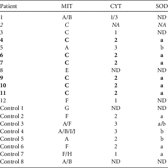
Genotyping by multilocus sequence typing (MLST) was performed on 20 samples (12 from SOT patients, and 8 samples from a control population: 4 HIV patients and 4 patients with malignant haematological diseases). The partial sequences of 2 mitochondrial genes (cytochrome B [CYT] and of the large subunit of ribosomal RNA [MIT]; GenBank accession numbers: AF320344.1 and NC_020331.1, respectively) and of the superoxide dismutase (SOD) gene (GenBank: AF146753.1) were amplified and sequenced as described previously.Reference Maitte, Leterrier, Le Pape, Miegeville and Morio 33 Together, these 3 regions of the P. jirovecii genome have a strong discriminative power, with an H index of 0.987 (corresponding to a 98.7% chance of correctly distinguishing between 2 strainsReference Maitte, Leterrier, Le Pape, Miegeville and Morio 33 ). This combination screening technique is currently recommended by the European Conference on Infections in Leukaemia as the best way to type P. jirovecii strains. 34 To simplify the reporting of the results, different MIT haplotypes were arbitrarily assigned a capital letter; the different CYT sequences were assigned a number; and the different SOD sequences were assigned a lowercase letter. The final genotype is therefore presented in the form A1a.
Clinical Epidemiological Survey
We surveyed all interactions between the patients infected with the same P. jirovecii strain (ie, outpatient clinic, imaging, laboratory, consultation, day care, and inpatient interactions) over the 6 months preceding the onset of the symptoms in the first patient until the diagnosis of PCP in the last patient. This time range was chosen based on reported incubation periods (median, 53 days; range, 1–188 days).Reference Goto and Oka 35 , Reference Yazaki, Goto, Uchida, Kobayashi, Gatana and Oka 36 Because carriers have been shown to transmit the disease,Reference Thomas and Limper 1 , Reference Le Gal, Diamani and Rouillé 18 , Reference Goto and Oka 35 we considered patients to be potentially contagious throughout the entire incubation period (6 months), even in the absence of symptoms, and for 3 days after TMP/SMX treatment was initiated. Transmission was defined as probable when all of the following criteria were met: (1) the 2 patients were present on the same day in the same place; (2) the “source” patient was in the contagious phase; and (3) the duration between the date of potential transmission and the appearance of the first symptoms was compatible with the incubation period for PCP. Transmission was considered possible when only 2 of these criteria were met.
RESULTS
Prior to 2014, the incidence of PCP in SOT recipients in our institution had been <1 case per year since at least 2009 (the time at which PCR was introduced as a routine diagnostic tool). In 2014, the IDH was alerted to 4 cases of PCP diagnosed in SOT recipients from different units (1 lung transplant, 1 kidney transplant, and 2 heart transplant recipients). Faced with these 4 cases, the IDH triggered and coordinated a multidisciplinary reaction.
Case Descriptions
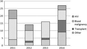
In total, 8 SOT recipients presented with PCP in 2014 and 2 presented in 2015 (Figure 1 and Supplementary Material Figure 1). The analysis presented here includes all cases of PCP in SOT recipients since 2012 (ie, 12 cases).
Number of pulmonary pneumocystosis cases diagnosed at Grenoble University Hospital between 2011 and 2014, classed for various risk factors.

In line with local recommendations, none of the patients received anti-PCP prophylaxis during the episode. In total, 9 patients (75%) presented a severe form of the disease. The median time between transplantation and onset of PCP was 38.5 months (range, 2 months to 18 years). Only 3 patients (25%) had been treated for cytomegalovirus (CMV) in the 3 months preceding the PCP, and 1 patient had developed acute cellular rejection. In total, 10 patients were lymphopenic. CD4+ T-lymphocytes (LTCD4) were counted at the time of diagnosis in 6 patients (median value, 275 mm3; range, 110–700 mm3).
Diagnosis of PCP was based only on a positive PCR result for 6 patients (negative direct examination). Median average fungal burden, determined by qPCR as described by Maillet et al,Reference Maillet, Maubon and Brion 32 was 5.4×10⁶/mL (range, 3×10³ to 1×10¹¹ mL) for SOT patients and 8.7×10⁵ mL (range, 5×10³ to 1.7×10¹⁰/mL) for controls. Three patients were diagnosed with a bacterial pulmonary coinfection. The median time between the first symptoms and the start of anti-PCP treatment was 7.5 days (range, 3–20 days).
All patients were treated with trimethoprim/sulfamethoxazole (TMP/SMX) at curative doses defined above. Systemic steroid therapy (at least 1 mg/kg body weight/day for at least 5 days) was added for 6 of the 9 patients with hypoxia. PCP was directly responsible for the deaths of 5 patients (42%) (Supplementary Material, Table 1).
No correlation was found between results of direct examination, bacterial coinfection, delay in diagnosis or treatment, or treatment regimen (notably use of corticosteroids) and severity or mortality.
Adaptation of PCP Handling in the Institution
As described in Table 1, several modifications were made to our procedure for handling PCP cases in our institution at the end of summer 2014, after the consultation of multidisciplinary working groups set up by the IDH. The modifications related to several key issues in disease management in SOT patients: diagnosis, hygiene measures, anti-PCP prophylaxis, and therapy. Notable therapy modifications included the rapid initiation of curative TMP/SMX therapy before bronchofibroscopy without affecting diagnostic performanceReference Martin and Fishman 13 and the use of systemic steroid therapy in severe cases in line with international recommendationsReference Martin and Fishman 13 , Reference Pareja, Garland and Koziel 37 despite a lack of clinical data.
Epidemiology
Molecular epidemiology
The same genotype (genotype C2a) was identified in 6 SOT recipients (patients 4, 6, 7, 9, 10, and 11) but in none of the control patients. In patient 2, the CYT and SOD sequencing failed; however, the MIT sequence was ‘C,’ which was compatible with the C2a genotype (Table 2). Based on these results, interhuman transmission from a single strain of P. jirovecii was suspected among at least 6 SOT patients who developed PCP in 2014–2015.
Results of Partial Sanger Sequencing of Mitochondrial GenesFootnote a

NOTE. CYT, cytochrome B; MIT, large subunit of ribosomal RNA; SOD, superoxide dismutase; NA, not amplifiable; ND, sequencing not done; PCP, Pneumocystis pneumonia. Italic type indicates possible C2a genotype. Bold type indicates full C2a genotype.
a CYT, MIT, and SOD genes for strains isolated from solid-organ transplant recipients and from 8 control patients presenting with PCP during the same period. The genotypes were arbitrarily named by a capital letter for MIT sequences, a number for CYT sequences, and a lowercase letter for SOD sequences.
Analysis of potential transmission
The epidemiological survey focused on the patients infected with the same strain. A transmission map confirmed probable nosocomial transmission (Figure 2). Indeed, direct transmission was considered probable in the following cases: (1) between patient 4 and patient 7, within the nephrology department; (2) between patient 4 and patient 6, within the heart surgery day clinic; (3) between patient 7 and patient 10 in the nephrology department; and (4) between patient 10 and patient 11, in the nephrology department. Transmission between patient 7 and patient 9 was only considered possible because the patients were in perpendicular corridors and the incubation time for patient 7 was quite short (4 days).
2014 calendar of attendance at the institution for solid-organ transplant recipients with pulmonary pneumocystosis shown to be induced by the same strain (patients 4, 6, 7, 9, 10 and 11). To improve readability, months were divided into 4 distinct periods: from the days 1–8 inclusive, days 9–16 inclusive, days 17–24 inclusive, and day 25 to the last day of each month, inclusive. The grey zones correspond to periods when the patient was an inpatient (in the same building) for at least 1 day during the period; the black zones correspond to the date of diagnosis (start of treatment). The crosses correspond to times when the patients were present for at least 1 day in the institution (emergency services, imaging, laboratory, consultations, outpatient clinic). The black arrows correspond to probable transmission, the dashed arrows correspond to possible transmission.

DISCUSSION
Although the incidence of PCP in SOT recipients in our institution tended to be very low, we observed 10 cases in 1 year. Before the occurrence of these grouped cases, the local recommendations for PCP prophylaxis in SOT recipients had been reduced compared with international recommendations. This reduced treatment protocol is in accordance with meta-analyses indicating that the risk/benefit ratio is only favorable when the incidence of PCP in the target population exceeds 3%.Reference Green, Paul, Vidal and Leibovici 38 , Reference Stern, Green, Paul, Vidal and Leibovici 39
Recently, several series of grouped PCP cases in SOT recipients were reported, particularly among kidney transplant patients.Reference Chapman, Marriott, Chen and MacDonald 15 – Reference Perez-Ordoño, Hojo and Sanclemente 17 , Reference Debourgogne, Favreau, Ladrière, Bourry and Machouart 20 – Reference Chandola, Lall, Sen and Bharadwaj 22 , Reference Goto and Oka 35 Historical series had shown a prevalence between 5% and 40% of PCP in SOT recipients within the first year post transplant.Reference Sepkowitz, Brown and Armstrong 7 , Reference Gordon, LaRosa and Kalmadi 8 , Reference Radisic, Lattes and Chapman 10 , Reference Sepkowitz 12 In contrast, more recent series reported PCP infections occurring long after transplantation,Reference Perez-Ordoño, Hojo and Sanclemente 17 and the phenomenon could be due, at least partially, to the general use of anti-PCP prophylaxis in the months following transplantation. In our series, only 25% of cases occurred in the first year after transplantation, even though kidney and liver transplant patients received no anti-PCP prophylaxis. Cases tended to be very late onset, with a median time between the transplant and the PCP of 38.5 months, and 1 patient had been transplanted 18 years earlier.
As described in other series of PCP cases in SOT recipients,Reference Wang, Partovi, Levy, Shapiro, Yoshida and Greanya 16 , Reference Perez-Ordoño, Hojo and Sanclemente 17 , Reference Borstnar, Lindic and Tomazic 21 additional risk factors for PCP were noted in ~25% of cases, such as reactivation of CMV or acute cellular rejection in the months preceding the infection. We therefore decided to reintroduce anti-PCP prophylaxis for 3 months following CMV reactivation or acute cellular rejection, as suggested in some North American guidelines.Reference Martin and Fishman 13 Interestingly, the median LTCD4 count was 275 mm3. Unlike HIV patients, the association between CD4+ T lymphopenia and the risk of PCP is not well established for non-HIV patients.Reference Roux, Gonzalez and Roux 2 Nevertheless, a series of 56 non-HIV patients diagnosed with PCP had a median LTCD4 count of 120 mm3.Reference Roblot, Le Moal and Kauffmann-Lacroix 5 This apparent discrepancy with our results could be explained by the fact that only 4 of the patients in the cohort described by Roblot et alReference Roblot, Le Moal and Kauffmann-Lacroix 5 were SOT recipients.
Another significant element in our series was the severity of the cases: 9 patients presented with severe PCP and 5 patients (42%) died due to the infection. Among non–HIV-infected patients, mortality rates close to 50% have been reported.Reference Li, Lee, Lee, Lee, Chang and Ko 3 – Reference Roblot, Le Moal and Kauffmann-Lacroix 5 , Reference Ko, Jeon and Park 40 Li et alReference Li, Lee, Lee, Lee, Chang and Ko 3 evoked a link between the time before diagnosis and mortality in patients not infected with HIV compared with HIV-infected patients: they noted a diagnostic delay of 24 days in non-HIV patients versus 7 days in HIV-infected patients, with associated mortality rates of 60% and 9%, respectively. In a prospective French series, Roux et alReference Roux, Canet and Valade 41 also concluded that a delay in initiating treatment in patients not infected with HIV could explain the increased mortality observed. In our patients, the median time between the first symptoms reported by the patient and the start of anti-PCP treatment was 7.5 days (range, 3–20 days). Despite this short delay, mortality remained high (42%), but mortality was not correlated with a delay in initiating treatment.
In our series, for 6 patients, the biological diagnoses of PCP were based only on a positive real-time PCR results from the respiratory specimens (negative direct examinations), but fungal burdens were compatible with possible or probable PCP, as previously defined,Reference Maillet, Maubon and Brion 32 and radioclinical data were also compatible with PCP. Despite the inclusion of these patients, the data regarding severity and mortality remained similar to other series in the literature, the vast majority of which only included patients for whom direct examinations were positive.Reference Fillatre, Decaux and Jouneau 4 , Reference Roblot, Le Moal and Kauffmann-Lacroix 5
The results from the molecular analysis indicated that the same strain of P. jirovecii was responsible for 6 cases of PCP. Various studies have discussed the capacity of certain strains of P. jirovecii to cause epidemics, with arguments in favor not only of interhuman transmission but also of transmission through the environment or from healthy carriers.Reference Le Gal, Diamani and Rouillé 18 , Reference Le Gal, Pougnet and Diamani 19 , Reference Schmoldt, Schuhegger and Wendler 24 , Reference Gits-Muselli, Peraldi and de Castro 25 Genotyping results guided our search for potential dates and places of transmission between the patients infected with the same P. jirovecii strain. This epidemiological survey identified probable transmission among between 5 patients; even though these patients were handled by different medical teams, they could have met in certain public or shared areas of the hospital. In these areas, it would be interesting to screen air samples and/or to screen healthcare workers to identify P. jirovecii carriers.
To our knowledge, previous studies reporting grouped cases of PCP in SOT recipients all described cases occurring in patients grafted with the same organ. The novel aspect of our series is that it reports grouped cases in patients transplanted with different organs. Although these patients were followed-up in distinct hospital departments, the same strain was implicated in several cases. These observations underline the advantage of an infectious diseases unit, which, in collaboration with the microbiology laboratory and the medical hygiene department, have a broader vision of epidemic phenomena within an institution. In our example, multidisciplinary working groups were set up by the IDH to update the protocols for overall management of the risk of PCP in SOT patients. This type of response and coordination are in line with the recommendations made by Chapman et al,Reference Chapman, Marriott, Chen and MacDonald 15 and they probably contributed to the rapid drop-off in the number of new PCP cases detected in SOT patients in our institution (a single case was detected in autumn 2015, caused by a different strain).
In conclusion, to limit PCP in SOT recipients, it is extremely important to follow appropriate prophylaxis and treatment guidelines and to promptly initiate treatment of suspected PCP, even before test results are available. Molecular testing is useful and should trigger a re-assessment of the approach to patient care. Institutional monitoring can be improved by coordination between the infectious diseases unit and the microbiology laboratory, which can facilitate the detection of grouped cases even when patients are treated by different medical teams.
ACKNOWLEDGMENTS
Financial support: No financial support was provided relevant to this article.
Potential conflicts of interest: All authors report no conflicts of interest relevant to this article.
SUPPLEMENTARY MATERIAL
To view supplementary material for this article, please visit https://doi.org/10.1017/ice.2016.274.