




No CrossRef data available.
Article contents
Dilated cardiomyopathy presenting during fetal life
Published online by Cambridge University Press: 13 July 2005
Abstract
Objectives: To describe the echocardiographic features, underlying causes, and outcome of fetuses with dilated cardiomyopathy. Design: A retrospective observational study between 1983 and 2003 at a tertiary centre for fetal cardiology. Patients: Affected fetuses were identified using a computerised database. We included fetuses with dilation and reduced systolic function of either the right ventricle, left ventricle, or both. We excluded fetuses with abnormal cardiac connections, arrhythmias, or stenosis of the aortic or pulmonary valves. In all, we identified 50 fetuses, born to 46 mothers. Of the fetuses, 24 had biventricular cardiomyopathy, 17 had isolated right ventricular cardiomyopathy, and 9 had isolated left ventricular cardiomyopathy. Two-thirds of the fetuses (32) were hydropic at some point during gestation. Main outcomes: A cause of cardiomyopathy was identified in 37 cases (74 per cent). This was genetic or metabolic in 11 fetuses; infective in 11; fetal anaemia, without proven parvovirus infection, in 5; of cardiac origin in 5; and an association with renal disease in 5. In 10 cases (20 per cent), the pregnancy was terminated. Based on an intention to treat, the survival to delivery was 25 of 40 (62.5 per cent, 95 per cent confidence intervals from 46 to 77 per cent), at 28 days was 17 of 40 (42.5 per cent, 95 per cent confidence intervals from 27 to 59 per cent), and at 1 year was 15 of 40 (37.5 per cent, 95 per cent confidence intervals from 23 to 54 per cent). The overall survival of non-hydropic fetuses was 9 of 18 (50 per cent), compared to 6 of 32 (18 per cent) hydropic fetuses. Conclusions: Genetic, metabolic, infective, and cardiac diseases may present with dilated cardiomyopathy during fetal life. There is a high rate of spontaneous intra-uterine and early neonatal death. The prognosis is particularly poor for hydropic fetuses.
Information
- Type
- Original Article
- Information
- Copyright
- © 2005 Cambridge University Press
The annual incidence of cardiomyopathies in childhood is around 1.2 for each 100,000 children.1 More than 40 per cent of affected children ultimately die, or require cardiac transplantation. The scant reports of cardiomyopathy identified during fetal life have either included very few cases or many different types of cardiomyopathy.2–4 We report here a 20-year experience of dilated cardiomyopathy diagnosed during fetal life at a tertiary centre for fetal cardiology.
Methods
Ascertainment of cases
A computerised record is made of all fetuses evaluated at our tertiary unit for fetal cardiology (Filemaker Pro versions 2 to 4; Claris Corporation, Santa Clara, California, United States of America). This database was analysed retrospectively for the period from 1983 to December 2002 to identify all fetuses in whom a diagnosis of “cardiomyopathy” had been made by the attending fetal cardiologist. The videotapes of these fetuses were reviewed by two cardiologists (JMS and SS). This was to identify cases of dilated cardiomyopathy meeting our criterions for inclusion. Retrospective echocardiographic measurements were made by a single fetal cardiologist (JMS).
At the time of presentation during fetal life, the fetal cardiologist makes the diagnosis of “cardiomyopathy” on the basis of the echocardiographic findings. For cases with dilation of the heart and cardiac dysfunction without apparent underlying cause, we used a pragmatic approach for establishing our criterions for inclusion, basing them on the opinion of the attending fetal cardiologist as refined by our echocardiographic findings. We accept that, with the benefit of later investigations, many of which are only possible postnatally, a definite underlying cause will be identified in some fetuses, thus excluding primary myocardial disease. Presentation of such data, nonetheless, may assist those who are faced with evaluating such affected babies during prenatal life.
Echocardiographic criterions for inclusion
Dilated cardiomyopathy was considered to exist when the dimensions as measured in cross-sectional images taken in diastole of either or both right and left ventricles were more than the 95th percentile for the gestational age,5 and the contractile function was considered to be impaired. The left ventricular ejection fraction was quantified as previously described,6 with values from 50 to 80 per cent considered to be normal.7 The errors in observation for such measurements have been published previously.6 Right ventricular function was assessed subjectively. Cases were classified as left ventricular, right ventricular, or biventricular dilated cardiomyopathy.
Other echocardiographic analyses
The thickness of the ventricular septum and the mural ventricular thicknesses were measured by cross- sectional echocardiography and compared to normal reference ranges.5 The cardiothoracic ratio was analysed by ratio of circumferences.8 Regurgitation across atrioventricular and arterial valves, myocardial echogenicity, and Doppler assessment of the outflow tracts were recorded. Hydrops was considered to exist when there were two or more of pleural effusion, pericardial effusion, skin oedema, or ascites.
Criterions for exclusion
We excluded all fetuses with abnormal cardiac connections, aortic valvar stenosis, pulmonary valvar stenosis, and arrhythmias. All but one twin pregnancies were excluded to avoid a potential overlap with cardiac dysfunction related to twin–twin transfusion syndrome. In the only twin pregnancy included, there was an identifiable viral aetiology for the cardiomyopathy.
Echocardiographic equipment
Fetal echocardiograms were performed with systems including the ATL Ultramark 4 ultrasound system, Toshiba SSA 270A ultrasound system (Toshiba Medical Systems, Crawley, United Kingdom), and Hewlett-Packard 1000, 2000 and 5500 ultrasound systems (Philips Inc, Andover, Massachusetts, United States of America).
Results
During the period of study, we evaluated a total of 24,237 pregnancies at our unit considered to be at high risk. We diagnosed 2,743 cardiac abnormalities, excluding arrhythmias, of which 50 cases fulfilled our criterions for dilated cardiomyopathy. Thus, dilated cardiomyopathy accounted for 0.2 per cent of all scans, and 1.82 per cent of fetal cardiac abnormalities. Of the 50 cases, 34 pregnancies had been referred because of suspected congenital heart disease, 17 because of fetal hydrops, 6 because of polyhydramnios, 6 because of a family history of cardiomyopathy, 2 because of oligohydramnios, 1 because of intra-uterine growth retardation, and 1 because of maternal diabetes mellitus. For 15 fetuses, there was more than one indication for referral.
The 50 fetuses with dilated cardiomyopathy were from 46 mothers, with a recurrence of cardiomyopathy in four families. Where the sex could be ascertained in retrospect, there were 29 males and 12 females, giving a ratio of genders of 2.4:1. The median gestational age at presentation was 22 weeks, with a range from 16 to 38 weeks. Seven mothers had frequent miscarriages, and two couples were first cousins.
Echocardiographic features
The right ventricle was involved in isolation in 17 fetuses, the left ventricle alone in 9 (Fig. 1), with the remaining 24 fetuses having biventricular involvement (Fig. 2a,b). Of the fetuses, 26 (52 per cent) were hydropic at presentation, and a further 6 (12 per cent) developed hydrops on follow-up. The echocardiographic features are summarised in Table 1. All fetuses had a cardiothoracic ratio above the normal range. Tricuspid regurgitation was noted in 19 fetuses. Tricuspid regurgitation was noted in 8 of 24 fetuses with biventricular cardiomyopathy, 1 of 9 with left ventricular cardiomyopathy, and 10 of 17 with right ventricular cardiomyopathy. Elevated regurgitant velocity across the tricuspid valve, at 5 metres per second, was noted in 1 fetus with ductal constriction, and in 3 fetuses with renal disease, when velocities ranged from 2.8 to 3.8 metres per second. Mitral regurgitation was noted in 6 fetuses, of whom 4 had biventricular cardiomyopathy, and 2 had left ventricular cardiomyopathy.

Figure 1. Fetal echocardiogram demonstrating a severely dilated left ventricle in the four-chamber view (a) with a non-contractile left ventricle on M-mode (b). LV: left ventricle; RV: right ventricle.

Figure 2. Four-chamber echocardiographic image of a fetus with a dilated cardiomyopathy affecting both ventricles (a) with fetal ascites (b). The cardiothoracic ratio is markedly increased. LV: left ventricle; RV: right ventricle; LA: left atrium; RA: right atrium.
Table 1. Graph of outcome of fetuses with dilated cardiomyopathy in relation to the affected ventricle and presence of fetal hydrops.

Echocardiographic evidence of endocardial fibroelastosis was seen in 12 fetuses. This affected both ventricles in five cases, the left ventricle in two cases, and the right ventricle in three cases. Unusual distributions were seen in two further cases. In one of these, the endocardial fibroelastosis affected the right ventricle and left atrium, with no identifiable cause. In the other, who had calcification of the liver and brain at post-mortem examination, there was marked echogenicity of the right ventricular outflow tract.
Causes or associations of endocardial fibroelastosis included a single case of each of infection with parvovirus, infection with coxsackie virus, toxoplasmosis, renal agenesis, intensely echogenic kidneys, parental consanguinity, calcification of the brain and liver, and extra-medullary haemopoiesis at post-mortem examination suggestive of fetal anaemia.
Causes of dilated cardiomyopathy
The identifiable causes of cardiomyopathy are shown in Table 2.
Table 2. Underlying causes of fetal dilated cardiomyopathy, echocardiographic features and outcome.

Metabolic/genetic.
A possible metabolic or genetic factor was identified in 11 cases. There were four families with recurrent cardiomyopathy. The first family had 2 fetuses with idiopathic medial calcification. The second family had 2 female babies who died in the neonatal period from mitochondrial cytopathy. In the third family, the parents were consanguineous, and biventricular cardiomyopathy was identified in the first pregnancy at 31 weeks associated with fetal hydrops. The subsequent fetus presented at 19 weeks with biventricular dilated cardiomyopathy and hydrops. Both the affected babies died during fetal life. The fourth family had biventricular dilated cardiomyopathy identified in 2 fetuses, one of whom was male diagnosed at 29 weeks and the other female diagnosed at 33 weeks of gestation. The first baby died in the neonatal period and the second died awaiting cardiac transplantation. No cause was identified in spite of extensive postnatal investigation.
Infection.
One mother infected with human immunodeficiency virus had a fetus with dilated cardiomyopathy who was stillborn. Post-mortem examination was declined. Coxsackie viral infection was suspected in 2 babies with left ventricular dilated cardiomyopathy, and confirmed by serological studies. In one of these fetuses, there was increased myocardial echogenicity, and serous effusions, which improved spontaneously, and by term, the cardiac function had normalised and the echogenicity was much reduced. The other case was one of monozygotic twins, with oligohydramnios and features of left ventricular cardiomyopathy, and in whom there was serological evidence for coxsackie viral infection. Laser ablation for twin–twin transfusion was complicated by preterm delivery and death of the twins.
Fetuses with proven parvovirus infection had haemoglobins ranging from 1.5 to 8.2 grams per decilitre in the 5 in whom cordocentesis was done. Of the 7 fetuses, 5 died, despite intra-uterine transfusion in 3. One baby, with endocardial fibroelastosis, had toxoplasmosis confirmed at post-mortem examination.
Fetal anaemia.
We found 5 fetuses to be anaemic other than due to parvovirus infection. In one of these, the cause was anti-C antibodies and in another case a fetomaternal bleed was implicated. In three cases, no cause could be identified.
Cardiac causes.
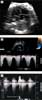
Coarctation of the aorta: Coarctation of the aorta was diagnosed postnatally in 2 babies, which had not been suspected before birth. The first baby with coarctation was born to consanguineous parents, and had very poor left and right ventricular function on both prenatal and postnatal echocardiograms. Preductal coarctation was diagnosed postnatally, and successfully treated with primary balloon dilation at the age of 2 weeks, although it was felt that the impairment of ventricular function was related to cardiomyopathy rather than coarctation of the aorta. Persistence of left ventricular dysfunction necessitated cardiac transplantation at 6 years of age. Histology of the explanted heart showed non-specific myocardial hypertrophy. The second baby had abdominal coarctation of the aorta, with severe hypoplasia of the renal arteries (Fig. 3a,b). This baby underwent balloon dilation of the abdominal aorta, which was technically successful, but there was marked systemic arterial hypertension secondary to renal involvement and the baby died in early infancy. Prenatal constriction of the arterial duct: Ductal constriction was identified in two cases: one of whom was diagnosed prospectively and the other retrospectively. Tricuspid regurgitation of high velocity, at 5 metres per second, aided the recognition of intra-uterine constriction of the arterial duct. This fetus had severe dilation, hypertrophy, and dysfunction of the right ventricle prenatally, and had continuous flow at high velocity across the constricted duct (Fig. 4a–c). The other case had been diagnosed as right ventricular cardiomyopathy early in our series, but retrospective review of the videotape showed clear evidence of ductal constriction. Both babies had an uneventful postnatal course following delivery. Non-compaction of the ventricular myocardium: We diagnosed a single case of isolated non-compaction of ventricular myocardium in a female baby who had biventricular dysfunction prenatally. This child is currently on no medication and is asymptomatic.

Figure 3. Four-chamber echocardiographic image (a) of a fetus who proved to have “middle aortic syndrome” postnatally. Both ventricles are hypertrophied with dilation of the right ventricle. There is reduced systolic function of the right ventricle on M-mode echocardiography (b). LV: left ventricle; RV: right ventricle; LA: left atrium; RA: right atrium; LVW: left ventricle posterior wall; RVW: right ventricular free wall; TV: tricuspid subvalvar apparatus; IVS: interventricular septum.

Figure 4. The four-chamber echocardiographic view demonstrates dilation of the right ventricle (a). There is tricuspid regurgitation with an elevated Doppler flow velocity approaching 5 metres per second confirming elevated right heart pressures (b). The flow pattern in the arterial duct has an increased velocity with diastolic extension of flow consistent with constriction of the arterial duct (c). LV: left ventricle; RV: right ventricle; LA: left atrium; RA: right atrium.
Renal abnormalities.
Renal abnormalities were noted in 5 fetuses. In four of the five cases, there was biventricular hypertrophy. The renal anomalies included bilateral renal agenesis in two cases; and dilated echogenic kidneys, bilateral cystic kidneys, and unilateral hydronephrosis in one case each. These cases were referred because of suspected cardiomyopathy, and renal abnormalities were detected during the subsequent detailed scan for fetal anomalies.
Outcome
Of the 50 cases, 10 pregnancies were terminated (20 per cent), 15 fetuses died during fetal life (30 per cent), 8 died in the neonatal period (16 per cent), and two in infancy (4 per cent). There are 15 survivors (30 per cent), of whom one has undergone cardiac transplantation. Of the 32 hydropic fetuses, only 6 survived (19 per cent), as opposed to 9 of 18 (50 per cent) non-hydropic fetuses (p equal to 0.04, Chi-squared analysis). Based on an intention to treat, the survival to delivery was 25 of 40 fetuses (62.5 per cent, 95 per cent confidence intervals from 46 to 77 per cent), to 28 days was 17 of 40 (42.5 per cent, 95 per cent confidence intervals from 27 to 59 per cent), and to 1 year was 15 of 40 (37.5 per cent, 95 per cent confidence intervals from 23 to 54 per cent). The numbers of cases are not sufficient for a meaningful analysis of mortality within the different subgroups.
Discussion
Dilated cardiomyopathy made up 1.8 per cent of the fetal cardiac abnormalities diagnosed in our centre during our period of study, compared to proportions of from 0.4 to 3.6 per cent given in previous reports.2–4 After birth, the incidence of dilated cardiomyopathy in children peaks during infancy.4, 9, 10 Many babies affected with dilated cardiomyopathy are not represented in the postnatal literature due to the high rate of spontaneous intra-uterine death, which accounted for almost two-fifths of our cases. We observed a clear male predominance. This has also been observed in cardiomyopathies seen during childhood,9 and is attributed to X-linked forms of cardiomyopathy,1 frequently associated with neuromuscular disease. Presentation during fetal life makes this an unlikely explanation for our results, and the reason for the observed sex difference is obscure.
Dilated cardiomyopathy may affect the right ventricle, the left ventricle, or both ventricles during fetal life. In our series, the right ventricle was more commonly affected than the left. For some conditions, such as premature constriction of the arterial duct, the reason why the right ventricle is affected is because of an increase in its afterload. Some causes of fetal dilated cardiomyopathy, in contrast, such as viral infections, may affect differently the performance of the left and right ventricles. Given that left and right heart pressures are normally equal during fetal life,11 it is difficult to explain these findings on a purely haemodynamic basis. Different genes, nonetheless, influence the development of right and left ventricles,12, 13 and differences in contractile properties may also play a role. Postnatally, the pulmonary vascular resistance falls dramatically, perhaps explaining why the right ventricle is less often affected postnatally.
Causes of cardiomyopathy
We observed an extremely diverse range of causes of cardiomyopathy. Myocarditis is a significant cause of dilated cardiomyopathy in children,9 and infective causes accounted for one-fifth of our cases. From postnatal studies, there is a genetic predisposition for the viral infection, as well as the outcome.14 Our data confirm a poor prognosis from intra-uterine viral myocarditis, but spontaneous improvement was also observed. Stem cells are known to play a role in repair of the heart in adults.15 Whether this potential is more in the fetus and neonate is unknown. The low systemic vascular resistance during prenatal life may place less demand on the heart, increasing the potential for spontaneous recovery. Maternal infection with the human immunodeficiency virus was associated with fetal cardiomyopathy in one case, but autopsy confirmation was not available. Human immunodeficiency virus is not known to infect embryonic myocardium, and hence opportunistic infection is considered to account for the high incidence of myocarditis and cardiac failure in association with maternal infection.16 A previous study did not identify impairment of fetal cardiac function in mothers infected with the human immunodeficiency virus.17 The mortality we observed with parvovirus is higher than previously reported.18 It is almost certainly due to referral bias. Even in our referral group, however, 2 of 7 fetuses did survive following intra-uterine transfusion. A variety of modalities for treatment are under trial for viral cardiomyopathy.14, 19 Whether they are useful for treatment of intra-uterine infections is currently not known. Steroids and immunoglobulins have been used in one case of viral myocarditis, where fetal death was imminent, but without success.2
Metabolic and genetic causes accounted for another fifth of the fetuses in our group, with no survivors. Thus, passage of metabolites across the placenta may permit survival to term, but does not completely protect the fetus from dilated cardiomyopathy of this type. A marked deterioration may become evident postnatally, with multi-organ dysfunction, and accumulation of toxic metabolites.20 Given the wide range of possible metabolic diseases affecting the heart, detailed investigation is difficult during fetal life. This makes counselling of affected families extremely difficult. In our study, and in a previous report,4 dilated cardiomyopathy was observed to evolve late in pregnancy, even when initial echocardiograms were normal, emphasising the importance of sequential studies. If pregnancy ends in spontaneous intra-uterine death, then detailed post-mortem examination is essential, and fresh, unfixed tissues and deoxyribonucleic acid should be obtained for further detailed analysis.
It might be argued that some of the cardiac causes of the cases included in our series are not truly dilated cardiomyopathy because there is no intrinsic myocardial disease. Their inclusion can be justified because the echocardiographic features may be very difficult to distinguish from primary dilated cardiomyopathy. One of the main aims of this study was to identify those conditions that should be specifically sought and excluded when fetuses with suspected dilated cardiomyopathy are evaluated echocardiographically. Premature constriction of the arterial duct may be recognised by careful echocardiographic assessment,21 and the parents given a good prognosis following delivery. One fetus had abdominal coarctation, which places an increased afterload on both ventricles, explaining the mode of presentation.22 Non-compaction of the ventricular myocardium was also diagnosed in 1 fetus, and this may manifest during fetal life with restrictive or dilated cardiomyopathy.23, 24 An unexpected association was noted between fetal renal disease and dilated cardiomyopathy. This might be related to renal hypertension causing myocardial dysfunction. The fetal myocardium has a reduced density of contractile elements, and less organised myocytes,25 making the fetus particularly susceptible to conditions causing an increased afterload.
The overall outcome for hydropic fetuses was worse than for non-hydropic fetuses. There was a trend towards a better outcome for fetuses with the right ventricle affected during fetal life. This is likely to be due to reversible conditions such as premature constriction of the arterial duct affecting the right ventricle, and to the fact that demands on the right ventricle are less postnatally due to the low resistance of the pulmonary vasculature. In contrast, hydropic fetuses with impaired left ventricular function had a uniformly poor outcome.
Conclusion
Suspected dilated cardiomyopathy in fetal life can be the consequence of a heterogeneous group of disorders, and has a varied presentation. Detailed and repeated fetal echocardiography is indicated to exclude occult obstructive cardiovascular lesions, and to monitor haemodynamics. This should be combined with appropriate maternal, fetal, and postnatal investigations. Genetic, metabolic, infective, and renal causes should all be considered in the differential diagnosis. The prognosis of affected fetuses is guarded, particularly where there is evidence of intra-uterine cardiac failure.
Acknowledgement
SS was supported by a Commonwealth fellowship grant.
References
Strauss A, Lock JE. Pediatric cardiomyopathy – a long way to go. N Engl J Med 2003; 348: 1703–1705.Google Scholar
Pedra SR, Smallhorn JF, Ryan G, et al. Fetal cardiomyopathies: pathogenic mechanisms, hemodynamic findings, and clinical outcome. Circulation 2002; 106: 585–591.Google Scholar
Webber SA, Sandor GGS, Farquharson D, Taylor GP, Jamieson S. Diagnosis and outcome of dilated cardiomyopathy in the fetus. Cardiol Young 1993; 3: 27–33.Google Scholar
Schmidt KG, Birk E, Silverman NH, Scagnelli SA. Echocardiographic evaluation of dilated cardiomyopathy in the human fetus. Am J Cardiol 1989; 63: 599–605.Google Scholar
Tan J, Silverman NH, Hoffman JI, Villegas M, Schmidt KG. Cardiac dimensions determined by cross-sectional echocardiography in the normal human fetus from 18 weeks to term. Am J Cardiol 1992; 70: 1459–1467.Google Scholar
Simpson JM, Cook A. Repeatability of echocardiographic measurements in the human fetus. Ultrasound Obstet Gynecol 2002; 20: 332–339.Google Scholar
Simpson JM. Echocardiographic evaluation of cardiac function in the human fetus. MD Thesis, University of London, 2000.Google Scholar
Paladini D, Chita SK, Allan LD. Prenatal measurement of cardiothoracic ratio in evaluation of heart disease. Arch Dis Child 1990; 65: 20–23.Google Scholar
Nugent AW, Daubeney PE, Chondros P, et al. The epidemiology of childhood cardiomyopathy in Australia. N Engl J Med 2003; 348: 1639–1646.Google Scholar
Lipshultz SE, Sleeper LA, Towbin JA, et al. The incidence of pediatric cardiomyopathy in two regions of the United States. N Engl J Med 2003; 348: 1647–1655.Google Scholar
Johnson P, Maxwell DJ, Tynan MJ, Allan LD. Intracardiac pressures in the human fetus. Heart 2000; 84: 59–63.Google Scholar
Ross RS. Regulation of gene expression: how can molecules make the right ventricle distinct from the left? Nucl Cardiol 1998; 5: 609–621.Google Scholar
Thomas T, Yamagishi H, Overbeek PA, Olson EN, Srivastava D. The bHLH factors, dHAND and eHAND, specify pulmonary and systemic cardiac ventricles independent of left-right sidedness. Dev Biol 1998; 196: 228–236.Google Scholar
Bowles NE, Kearney DL, Ni J, Perez-Atayde AR. The detection of viral genomes by polymerase chain reaction in the myocardium of pediatric patients with advanced HIV disease. J Am Coll Cardiol 1999; 34: 857–865.Google Scholar
Hornberger LK, Lipshultz SE, Easley KA, et al. Cardiac structure and function in fetuses of mothers infected with HIV: the prospective PCHIV multicenter study. Am Heart J 2000; 140: 575–584.Google Scholar
Levy R, Weissman A, Blomberg G, Hagay ZJ. Infection by parvovirus B 19 during pregnancy: a review. Obstet Gynecol Surv 1997; 52: 254–259.Google Scholar
Kawai C. From myocarditis to cardiomyopathy: mechanisms of inflammation and cell death: learning from the past for the future. Circulation 1999; 99: 1091–1100.Google Scholar
Schwartz ML, Cox GF, Lin AE, et al. Clinical approach to genetic cardiomyopathy in children. Circulation 1996; 94: 2021–2038.Google Scholar
Huhta JC, Moise KJ, Fisher DJ, Sharif DS, Wasserstrum N, Martin C. Detection and quantitation of constriction of the fetal ductus arteriosus by Doppler echocardiography. Circulation 1987; 75: 406–412.Google Scholar
Zeltser I, Parness IA, Ko H, Holzman IR, Kamenir SA. Midaortic syndrome in the fetus and premature newborn: a new etiology of nonimmune hydrops fetalis and reversible fetal cardiomyopathy. Pediatrics 2003; 111: 1437–1442.Google Scholar
Karatza AA, Holder SE, Gardiner HM. Isolated non-compaction of the ventricular myocardium: prenatal diagnosis and natural history. Ultrasound Obstet Gynecol 2003; 21: 75–80.Google Scholar
Guntheroth W, Komarniski C, Atkinson W, Figner CL. Criterion for fetal primary spongiform cardiomyopathy: restrictive pathophysiology. Obstet Gynecol 2002; 99: 882–885.Google Scholar
Brook WH, Connell S, Cannata J, Maloney JE, Walker AM. Ultrastructure of the myocardium during development from early fetal life to adult life in sheep. J Anat 1983; 137: 729–741.Google Scholar

Fetal echocardiogram demonstrating a severely dilated left ventricle in the four-chamber view (a) with a non-contractile left ventricle on M-mode (b). LV: left ventricle; RV: right ventricle.

Four-chamber echocardiographic image of a fetus with a dilated cardiomyopathy affecting both ventricles (a) with fetal ascites (b). The cardiothoracic ratio is markedly increased. LV: left ventricle; RV: right ventricle; LA: left atrium; RA: right atrium.
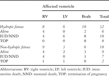
Table 1.
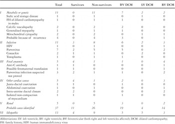
Table 2.

Four-chamber echocardiographic image (a) of a fetus who proved to have “middle aortic syndrome” postnatally. Both ventricles are hypertrophied with dilation of the right ventricle. There is reduced systolic function of the right ventricle on M-mode echocardiography (b). LV: left ventricle; RV: right ventricle; LA: left atrium; RA: right atrium; LVW: left ventricle posterior wall; RVW: right ventricular free wall; TV: tricuspid subvalvar apparatus; IVS: interventricular septum.
The four-chamber echocardiographic view demonstrates dilation of the right ventricle (a). There is tricuspid regurgitation with an elevated Doppler flow velocity approaching 5 metres per second confirming elevated right heart pressures (b). The flow pattern in the arterial duct has an increased velocity with diastolic extension of flow consistent with constriction of the arterial duct (c). LV: left ventricle; RV: right ventricle; LA: left atrium; RA: right atrium.
 You have
Access
- 40
- Cited by