Eliminating healthcare-associated infections (HAI) is a national priority, 1 and accumulating evidence implicates environmental contamination in HAI transmission.Reference Bogusz, Stewart and Hunter 2 – Reference Huslage, Rutala, Gergen, Sickbert-Bennett and Weber 5 Adequate cleaning of environmental surfaces is an important HAI prevention strategy,Reference Septimus, Weinstein, Perl, Goldmann and Yokoe 6 and because cleaning efficacy and/or HAI transmission rates may vary with differences in hospital design, the built environment, with respect to evidence-based design (EBD), is receiving increased attention.Reference Zimring, Denham and Jacob 7 – Reference Hall and Kamerow 10
EBD seeks to improve patient, staff, and organizational outcomes through hospital design. It assumes that such built environmental features as the layout of patient rooms, natural lighting, views of nature, and state-of-the-art technology can have a positive impact.
Our EBD cleaning study is unique in that, unlike any others with which we are familiar, it was conducted in a newly opened EBD hospital, which permitted a post-construction/pre-opening assessment of baseline contamination. Furthermore, it allowed a more reliable reconsideration of potentially underreported and underestimated results. For example, although nosocomial bacteria persist on environmental surfaces for weeks or months,Reference Kramer, Schwebke and Kampf 11 important Gram-negative bacteria, such as Escherichia coli, may be underreported with culture based methods due to their lower viability and environmental bioburden.Reference Guet-Revillet, Le Monnier and Breton 12
DNA on hospital surfaces may also be another underestimated driver of antimicrobial resistance and/or virulence in nosocomial pathogens. Such material, previously considered clinically inconsequential or inert, may be present in viable-yet-uncultivable bacteria, in dead cells, or even in extracellular (ie, naked) form.Reference Overballe-Petersen, Harms and Orlando 13 Recent observations that pathogenic bacteria can integrate short, damaged DNA fragments into their chromosomes expand the potential implications of this contaminant.Reference Overballe-Petersen, Harms and Orlando 13
Our goal was to assess the relationships among environmental DNA, cultivable target organisms (TOs), cleaning, and HAI in a newly opened EBD hospital. DNA included both general (non–species-specific) 16S bacterial DNA, and DNA of species-specific TOs (listed in the Methods section). Because those taxa referred to as the ESKAPE pathogens (ie, Enterococcus faecium, Staphylococcus aureus, Klebsiella pneumoniae, Acinetobacter baumanii, Pseudomonas aeruginosa, and Enterobacter spp.) are among the most nosocomially relevant, we focused on members of that group, as well as E. coli and Clostridium difficile. Furthermore, financial and technical constraints precluded us from studying viral, fungal, and anaerobic organisms. We also sought to prospectively determine: (1) patterns of environmental contamination beginning before the facility opened and after routine terminal cleaning; (2) the genetic relatedness, based on whole-genome sequencing (WGS), of any multidrug-resistant target organism (MDR-TO) isolated from the environment to those isolated from HAIs; and (3) the correlation between environmental TO contamination levels and the frequency of analogous HAIs.
METHODS
Setting
Fort Belvoir Community Hospital (FBCH) is a newly constructed 120-bed, EBD facility in northeastern Virginia. It opened in September 2011 and has 10 operating rooms, a medical-surgical intensive care unit, a telemetry unit, separate medical and surgical wards, and pediatrics and maternity wards. Some bed rail and mattress surfaces (those on the long-term, anti-pressure ulcer beds) in the intensive care unit and telemetry unit are copper impregnated. Although the public restroom faucets are touch-free, all others in the facility are not. FBHC has no ultraviolet units or fogging systems for enhanced room/surface decontamination. FBHC has a beneficiary population exceeding 90,000 and supports one of the busiest emergency departments in the northern capital region. At FBHC in 2014, there were 9,340 inpatient surgeries, 48,793 emergency department patients, 24,976 inpatient bed days, 680,000 visits to ambulatory care clinics, and 1,569 births. The average daily census was 73.
Definitions
We defined thorough cleaning as whether a surface had been wiped enough to remove at least 90% of an invisible marking dye (DAZO fluorescent marking gel, Kleancheck Systems, Hingham, MA) that fluoresces under ultraviolet light. Effective cleaning was defined as surface with no detectable biomaterial. Biomaterial was defined as any microbial growth on blood or MacConkey culture media, or a detectable signal from real-time polymerase chain reaction (PCR) indicating the presence of general or species-specific bacterial DNA.
TOs included any of the following: Acinetobacter baumannii; A. baumannii calcoaceticus complex; E. coli; P. aeruginosa; S. aureus; K. pneumoniae; E. faecium (or faecalis); E. cloacae (or aerogenes); and C. difficile. Multidrug resistance (MDR) was defined according to Magiorakos et al.Reference Magiorakos, Srinivasan and Carey 14 Inpatient infection or acquisition was defined as no evidence of infection or a negative surveillance culture upon admission, followed by a positive clinical or surveillance culture arising no less than 48 hours after admission.
Terminal Cleaning and Surveillance
Terminal cleaning was performed in a standard manner involving collection of trash and linen, high dusting, wet dusting, wiping of surfaces with a quaternary ammonium compound disinfectant, floor cleaning, bathroom cleaning, and room inspection. Surveillance was prospectively conducted for 16 months from October 2011 through January 2013. After patients were discharged, but prior to terminal cleaning of their rooms, 17 high-touch surfaces were sampled for 20 seconds using rayon-tipped swabs premoistened with nutrient transport media. Surfaces were then marked with DAZO gel. After terminal cleaning, the presence or absence of the dye was assessed using the EnCompass monitoring system (Ecolab, St. Paul, MN). Surfaces were then resampled as described above. If the evaluation of the cleaned room could not occur shortly after terminal cleaning, the surveillance episode for that room was aborted. Swabs were used instead of sponges for 2 reasons: (1) swabs are more sensitive than sponges for detecting certain, particularly problematic TOs (eg, Acinetobacter) in the hospital environmentReference Thom, Howard and Sembajwe 15 ; and (2) sponges were too large to fit in tubes used to extract template for PCR. Each surface was sampled with a separate swab.
Sample Processing
Culture method
Swabs were immediately transported to a processing lab and streaked in seriatim onto a blood agar plate (BAP) and a MacConkey agar (MAC) plate. BAP and MAC plates were incubated for 24–48 hours at 35°C. Gram-positive organisms growing on BAP were taxonomically identified using rapid tests (coagulase, catalase, and Staphaurex Remel, Thermo Scientific, Waltham, MA) followed by analysis on the Phoenix automated system (Bekton Dickenson, Franklin Lakes, NJ) using the PMIC/ID-107 panel. Colonies that grew on MAC plates (which were presumably Gram negative) were analyzed on the Phoenix using panel NMIC/ID-133. Any potential TO not definitively identifiable by these methods was further analyzed using a matrix-assisted laser desorption ionization mass spectrometer (MALDI Biotyper, Bruker, Billerica, MA).
PCR methods
After streaking onto BAP and MAC media, swab tips were aseptically removed from rods, submerged in 300 µl of sterile water, and vortexed for 30 seconds. Afterward, 20 µl of the resulting supernatant was added to 40 µl of Lys and Go solution (Pierce Biotechnology, Rockford, IL), and 2 µl of this solution was ultimately used as template for RT-PCR amplification using the protocols of Clifford et al.Reference Clifford, Milillo and Prestwood 16 This 16S rDNA PCR (16S PCR) assay, predicted to detect 94% of all bacterial species,Reference Clifford, Milillo and Prestwood 16 can detect as few as 1×10² copies of purified genomic DNA in a reaction. Laboratory tests showed that the 16S PCR assay could detect bacteria on test surfaces treated with a solution containing as few as 3×10³ organisms per milliliter. Additional species-specific PCR assays detected C. difficile, S. aureus, E. coli, P. aeruginosa, K. pneumoniae, and Acinetobacter spp. To minimize false positives, we defined a positive PCR reaction as 4 standard deviations above the cycle threshold of the negative controls. Lacking specific primers for Enterobacter spp. and Enterococcus spp., those organisms could only be detected through culture-based methods. Finally, as our laboratory does not have equipment needed to grow C. difficile, we could not confirm positive C. difficile PCR results by culturing. Investigators performing the culture and PCR procedures were blinded to each other’s results.
Genetic Relatedness of MDR-TOs and Correlation of Environmental Bio-Burden with Clinical Infections
All methicillin-resistant S. aureus (MRSA) and MDR K. pneumoniae, E. coli, P. aeruginosa, Enterobacter spp. and A. baumannii isolated from the environment or inpatient infections underwent pulsed-field gel electrophoresis (PFGE), and whole-genome sequencing (WGS) was performed as previously described.Reference McGann, Courvalin and Snesrud 17 Logistically, the FBCH microbiology department cannot store bacteria pathogens isolated from routine clinical infections longer than 7 days, so archiving was only feasible for MDR isolates. Therefore, only MDR isolates were available for PFGE and WGS comparison.
Records for all TO-mediated infections occurring during the 16-month observation period were extracted from central electronic medical records and laboratory information systems. The number of TO-positive inpatient infections was compared with the incidence of environmental TO detection.
Additional details of the methods and statistics sections, which were performed using the R software package, are available in the supplemental section. 18
RESULTS
Detection of Species-Nonspecific (General) Biomaterial
Surface, room, and swab totals
A total of 1,273 high-touch surfaces were swabbed before and after terminal cleaning during 77 room visits to 49 unique rooms. Of these, 6 rooms had patients on isolation precautions. Altogether, 2,604 surface samples were obtained; only 2,546 paired swab tips were available for analysis, primarily due to damage during transportation. Of these, 3 swab pairs lacked associated DAZO results. Of the 2,546 paired swabs, 47% (1,197) and 42% (1,069) had cultivable biomaterial and PCR-amplifiable 16S rDNA, respectively. The slightly lower frequency of positive swabs identified by 16S rDNA testing was due to the relative non-selectivity of BAP media. BAP medium accommodates the growth of molds and yeast commonly present in environmental samples; in contrast, the PCR assay is specific to bacteria.
Patterns of contamination and removal
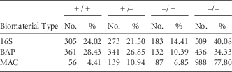
Cleaning was thorough (ie, ≤10% DAZO gel remaining) for 42.6% of surfaces inspected. Cleaning was effective (ie, biomaterial undetected on post-cleaned surface) for 61.6% of surfaces for 16S rDNA, 61.2% for BAP, and 88.7% for MAC (Figure 1). These percentages apply to all room surveys (77 visits to 49 unique rooms). However, cleaning effectiveness varied by assay and surface type; the most and least contaminated surfaces (relative rank order) were very similar, regardless of efficacy measure used. Bathroom sinks were most likely to be contaminated by biomaterial, and side rails were among the least likely to be contaminated. Detection of biomaterial after terminal cleaning differed significantly among the assays used, and as anticipated, PCR assays were more sensitive (16S rDNA vs BAP: P<0.001; 16S rDNA vs MAC: P<0.001; and MAC vs BAP: P<0.001). Terminal cleaning removed cultivable growth on MAC from 71.3% of surfaces that harbored them prior to cleaning. In general, only a limited amount of non–species-specific bacterial 16S rDNA is removed (ie, only 47.2% of PCR-positive precleaned surfaces tested negative after cleaning) (Table 1).
Surface-specific cleaning outcomes per 16S rDNA PCR (Panel A), blood agar (panel B) and MacConkey agar (panel C) measures. “+” indicates positive detection of relevant biomaterial by the cleaning efficacy measure and “–” connotes no detection. Surfaces were assayed for biomaterial presence both before and after terminal cleaning, and histogram legend indicia correspond to the following cleaning event classification types: “+/+” indicates ineffective, “+/–” indicates successful, “–/+” indicates failed (by acquisition), and “–/–” indicates remained clean. Relative frequencies of each event type are presented. Within each biomaterial detection measure–specific chart, stacked bars are rank ordered by their respective surface’s propensity to repulse contaminants.

Detection of Nonspecific Biomaterial Before and After Cleaning

NOTE. 16S, non–species specific; BAP, blood agar plate; MAC, MacConkey agar plate.
Detection of Specific TO Species
Types, ratios, and assay correlation of species-specific TOs
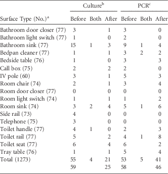
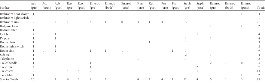
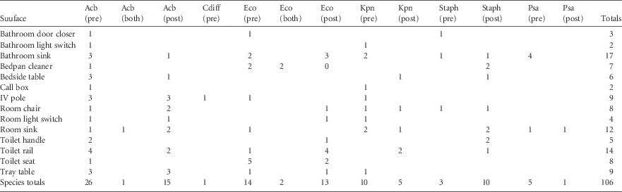
More than 80 different species of cultivable aerobic bacteria were identified, with 10 constituting >55% of all organisms detected (Supplemental Table 1). Coagulase-negative Staphylococcus spp. (30.2%) and Acinetobacter spp. (12.6%) were the 2 most common genera present. MDR-TOs were rarely detected (ie, 3 of 2,546 swabs or 0.1%). The incidence of TO-positive swabs (≥1 TO detected) was 3.3% (84 of 2,546) using the culture method and 4.1% (104 of 2,546) using the PCR method (Table 2). On 1,273 surfaces swabbed, 85 distinct TOs were detected using the culture method and 106 were detected by PCR (Tables 3 and 4). There was no significant discord between TO detection by culture and PCR, suggesting that results from the 2 methods are comparable. On cleaned surfaces, detection of TOs by culturing correlated well with detection using species-specific PCR. The Pearson correlation coefficient for rates of detection by the 2 methods was 0.56 (P=.00947 for a 1-tailed test). The relative order of surfaces, from most to least contaminated, as determined by culturing and PCR was also similar, with a Spearman’s ρ of 0.58 (P=.00688 for a 1-tailed test). Prior to cleaning, however, TO detection by the 2 methods were not strongly correlated (r=0.39; P=.068).
Number of Surfaces That Tested Positive for Any Target Organism

NOTE. PCR, polymerase chain reaction; IV, intravenous therapy.
a For each surface, 2 swabs were obtained, 1 before cleaning and 1 after cleaning.
b Individual surfaces from which any target organism was cultured. Before=tested positive before, but after, room cleaning; After=tested positive only after cleaning; Both=tested positive before and after cleaning. The total number of surfaces that tested positive prior to cleaning is the sum of the sum of the “Before” and “Both” columns. The total number of surfaces that tested positive after cleaning is the sum of the sum of the “After” and “Both” columns.
c Individual surfaces that tested positive for any target organism by PCR.
Recovery of Target Organisms by Culture

NOTE. Acb, Acinetobacter baumanii; Eco, Escherichia coli; Enterob, Enterobacter; Kpn, Klebsiella pneumonia; Psa, Pseudomonas aeruginosa; and Staph, Staphylococcus aureus; Enteroc, Enterococcus.
Recovery of Target Organisms by PCR

NOTE. Acb, Acinetobacter baumanii; Cdiff, Clostridium difficile; Eco, Escherichia coli; Kpn, Klebsiella pneumonia; Psa, Pseudomonas aeruginosa; and Staph, Staphylococcus aureus.
Overall location of TO contamination
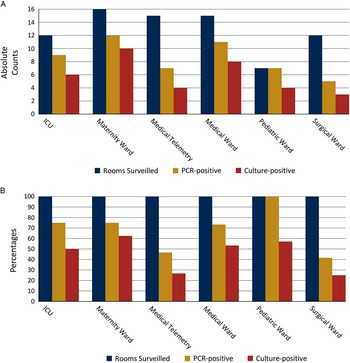
TOs were found in 35 room surveys by culturing and 51 room surveys by PCR (Figure 2). On an absolute basis, the maternity ward harbored the most TOs by both approaches. As a percentage of rooms surveilled, the maternity ward was most likely to have cultivable TOs, and the pediatric ward was most likely to test positive for TO DNA (Figure 2). Certain surfaces were more likely than others to test positive for TOs (Table 2). No TOs were cultured from room door closers, and no TOs were detected on telephones or side rails by PCR (Table 2). A. baumannii and S. aureus were the most common culturable TOs (cTOs) encountered and were typically isolated from sink- and toilet-related fixtures (Table 3). A. baumannii and E. coli were the TOs most commonly detected by PCR (Table 4). A. baumannii was most often found on IV therapy poles, toilet rails, and tray tables, and E. coli was most often found on toilet seats, toilet rails, and bathroom sinks (Tables 3 and 4).
Pathogen presence by hospital unit type. (A) The total count of rooms surveilled is indicated, as well as the frequency with which ≥1 target organism (TO) was detected by culture- or PCR-based methods on ≥1 surface housed in a given hospital unit. (B) As above, but positive detection events are expressed as a percentage of rooms surveilled.

Culture-based recovery of TO after cleaning
TOs were cultured from 80 surfaces. A total of 55 surfaces harbored TOs before, but not after, terminal cleaning; 21 harbored TOs after, but not prior to, cleaning; and 4 surfaces (3 different surface types) had cultivable TOs both before and after cleaning (Table 2). Cleaning resulted in fewer detection incidents on all surfaces but room sinks, on which detection events increased from 5 to 6 after cleaning (Table 2). Among individual surfaces, only bathroom sinks showed a significant reduction in cultivable TOs after cleaning (P=.0004 for a 1-tailed test). Total cTO detection events decreased from 59 to 25 (58% reduction) after cleaning (P=4.88×10⁻⁷ for a 1-tailed test) (Table 2).
Molecular-based detection of TO after cleaning
TOs were detected by species-specific PCR on 99 surfaces (Table 2). In total, 53 surfaces with TOs before room cleaning lacked them afterward, while 41 surfaces without PCR-detectable TOs prior to cleaning tested positive after cleaning. In total, 5 surfaces (of 4 surface types) tested positive for TO DNA both before and after cleaning (Table 2).
Cleaning resulted in more incidents detected on 5 surface types: room chair (3 precleaning vs 4 postcleaning); room light switch (1 vs 2); room sink (6 vs 7); toilet handle (2 vs 3); and toilet rail (5 vs 9) (Table 2). Total detection events by species-specific PCR decreased from 58 to 46 (21% reduction) with cleaning, but this change was not significant (P=.0616 for a 1-tailed test), suggesting that cleaning did not reliably remove all TO DNA (Table 2).
Correlations among environmental contamination with MDR-TOs, TOs, and HAI
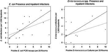
No infections were caused by A. baumannii or C. difficile over the study period. The most common non-MDR TO infection was S. aureus (n=77), followed by E. coli (n=56), K. pneumoniae (n=28), and P. aeruginosa (n=11). The only statistically significant correlation between TO infection and environmental presence of a cultivable TO was for Enterococcus spp. (1-tailed Pearson correlation P=.020; 1-tailed Poisson regression P=.035) (Figure 3). No significant correlations were observed between PCR-detected environmental TO and analogous HAIs except for inpatient E. coli infections and the detection of environmental E. coli DNA (1-tailed Pearson correlation P=.001; 1-tailed Poisson regression P=.002) (Figure 3). The only cultivable environmental MDR-TO was MRSA, which was recovered from 2 terminally cleaned surfaces 7 months after patients and staff arrived at the hospital. A single isolate was collected from a thoroughly cleaned (≤10% DAZO detection) toilet rail, the other was collected from a poorly cleaned (≥10% DAZO detection) call box. Another MRSA was isolated from the respiratory tract of a patient hospitalized on the same ward 5 months later (12 months after opening). According to pulsed-field gel electrophoresis (PFGE), the environmental and infection-derived strains were related (96.3% identical) (Supplemental Figure 1). However, WGS revealed that environmental isolates were genetically identical to each other but were unrelated to the isolates from patient infections because they differed by 126 SNPs, 13 insertions/deletions, and the presence of a prophage.
Environmental pathogen presence and inpatient infections. For each of N=16 months of surveillance conducted, the number of nosocomial infections per 1,000 inpatients was plotted relative to the number of target organism (TO)–positive assays per 20 rooms. (A) A regression plot utilizing all 16 PCR-based E. coli measurements is shown with a dashed line (Y-intercept: 3.21, slope: 0.61). A Pearson’s product–moment correlation coefficient of r=0.48 was computed for these data, which corresponds to P=.0289. (A 1-tailed test was used because it is reasonable to assume that increased environmental presence of a pathogen leads to increased incidence of disease). Because only 2 rooms were surveyed in December 2011, this data point—(10.00, 3.16), plotted with an empty circle—may not be appropriate for analysis. When ignored, the regression plot shown using a solid line (Y-intercept: 2.00, slope: 1.06) is obtained. For this subset of N=15 measurements, Pearson’s r corresponds to 0.71, with P=.00147. (B) A regression plot utilizing all 16 culture-based Enterococcus spp. measurements is shown with a dashed line (Y-intercept: 1.70, slope: 0.30). A Pearson’s product–moment correlation coefficient of r=0.44 was computed for these data, which corresponds to P=.0436 (1-tailed test). The data point for the December 2011 survey, (10.00, 3.16), is represented by an empty circle. When ignored, the regression plot shown using a solid line (Y-intercept: 1.53, slope: 0.58) obtains. For this subset of N=15 measurements, Pearson’s r corresponds to 0.54, P=.0187.

DISCUSSION
Environmental TO peaked early in the surveillance period and then plateaued. Environmental MDR-TOs were rarely isolated and were not implicated in HAIs. The rate of cleaning thoroughness at this facility (43%) was consistent with pre-intervention baselines at other facilities,Reference Carling, Parry and Von Beheren 19 – Reference Rupp, Adler and Schellen 24 and effective cleaning occurred most of the time (62%–88%, depending on assay). Cleaning, however, did not reliably remove bacterial DNA. Further, E. coli DNA was correlated with inpatient infection rates.
To our knowledge, this is the first report of environmental contamination, cleaning outcomes, and HAI in a newly opened EBD facility using culture-based and molecular assays (including WGS). As such, it might raise more questions than answers. First, did EBD contribute to the infrequent isolation of environmental MDR? This cannot be determined without studies conducted at more EBD hospitals. Second, should cleaning assessments be reconsidered or redesigned with DNA in mind, and is environmental DNA, particularly from E. coli, more clinically relevant than previously thought? Cleaning appeared to both remove and introduce biomaterial at this facility. Perhaps more aggressive wiping of surfaces, in an attempt to clean better, led to more surface contamination. Indeed, Manian et alReference Manian, Griesenauer and Senkel 21 found that A. baumannii and MRSA were frequently isolated after as many as 4 rounds of cleaning and disinfection. Based on observations that surfaces cleaned with disinfectants become recontaminated faster than surfaces cleaned with detergents,Reference Stewart, Bogusz and Hunter 25 others have suggested that disinfectants might, through cellular or biofilm disruption, release more bacterial DNA or planktonic forms that lead to a rebound effect, which contributes to the acquisition of resistance genes among environmental bacteria.Reference Bogusz, Stewart and Hunter 2 , Reference Sattar 3 , Reference Stewart, Bogusz and Hunter 25
Third, are current methods of genotyping such as PFGE or spa typing adequate for source attribution or outbreak investigations? Price et alReference Price, Golubchik and Cole 26 recently showed that conventional MRSA genotyping methods can lead to the wrong conclusions regarding relatedness or transmission. Using only PFGE, we might have concluded that environmental MRSA were related to those from infections based on their being >95% similar and from the same ward. However, WGS revealed substantial difference at the genome level.
Limitations of this study include the fact that financial and logistical constraints prevented us from using Petri films, dipped slides, or disinfectant neutralizers. Therefore, the environmental burden of TOs may have been underestimated. However, the PCR assays we used are highly sensitive. Additionally, our findings might not be generalizable to civilian hospitals, but patients of all ages and races are treated at FBCH.
In conclusion, sinks were frequently contaminated at this facility despite EBD; clearly they remain an important potential reservoir for HAI transmission.Reference Hota, Hirji and Stockton 27 – Reference Denham, Kasali, Steinberg, Cowan, Zimring and Jacob 29 We hope that this study spurs similar investigations at other EBD facilities, which are needed to determine how EBD impacts surface contamination, cleaning, and HAI-related outcomes.
ACKNOWLEDGMENTS
We would like to thank Lee Hudson and the late George Nussbaum for their insights into hospital design and construction, and David B. Goldman for critical review of the manuscript.
Financial support: This work was supported by the Military Infectious Disease Research Program; the Congressional War Supplement Fund; the Global Emerging Infections Surveillance System; and the US Army Medical Research and Materiel Command.
Potential conflicts of interest: There are no conflicts of interest and nothing to disclose.
SUPPLEMENTARY MATERIAL
To view supplementary material for this article, please visit http://dx.doi.org/10.1017/ice.2015.151