


Introduction
Mecoptera, also known as scorpionflies, are named from the modern Panorpidae in which upward-tilted male genitalia possess enlarged claspers resembling the stingers of scorpions. The extant Mecoptera is a small relic order that consists of ~600−650 species placed in nine families (Cai et al., Reference Cai, Huang and Hua2008; Bicha, Reference Bicha2010), although the number of families is much smaller than during geological history (Zhang et al., Reference Zhang, Shih, Petrulevičius and Ren2011; Lin et al., Reference Lin, Shih, Li, Ren, Ren, Shih, Gao, Wang and Yao2019b). The earliest fossil record of Mecoptera dates back to the early Permian (Rasnitsyn et al., Reference Rasnitsyn, Aristov, Gorochov, Rowland and Sinitshenkova2004). Mecoptera flourished during the Jurassic (Grimaldi and Engel, Reference Grimaldi and Engel2005; Soszyńska-Maj et al., Reference Soszyńska-Maj, Krzemiński, Kopeć, Cao and Ren2018), but significantly declined since the Cretaceous (e.g., Bittacidae; Kopeć et al., Reference Kopeć, Soszyńska-Maj, Krzemiński and Coram2016).
The suborder Aneuretopsychina Rasnitsyn and Kozlov, Reference Rasnitsyn and Kozlov1990 consists of five extinct families—Mesopsychidae Tillyard, Reference Tillyard1917, Pseudopolycentropodidae Handlirsch, Reference Handlirsch and Schröder1925, Aneuretopsychidae Rasnitsyn and Kozlov, Reference Rasnitsyn and Kozlov1990, Nedubroviidae Bashkuev, Reference Bashkuev2011, and Dualulidae Lin et al., Reference Lin, Shih, Li, Ren, Ren, Shih, Gao, Wang and Yao2019 (Lin et al., Reference Lin, Shih, Li, Ren, Ren, Shih, Gao, Wang and Yao2019b)—which played a dominant role in Mecoptera from the late Permian to the Middle Jurassic (Rasnitsyn and Kozlov, Reference Rasnitsyn and Kozlov1990; Ren et al., Reference Ren, Labandeira, Santiago-Blay, Rasnitsyn, Shih, Bashkuev, Logan, Hotton and Dilcher2009; Bashkuev, Reference Bashkuev2011; Lin et al., Reference Lin, Labandeira, Shih, Hotton and Ren2019a; Zhao et al., Reference Zhao, Wang, Bashkuev, Aria, Zhang, Zhang, Tang and Engel2020). Aneuretopsychina taxa possess a long proboscis probably associated with gymnosperm pollination (Labandeira et al., Reference Labandeira, Kvaček and Mostovski2007; Ren et al., Reference Ren, Labandeira, Santiago-Blay, Rasnitsyn, Shih, Bashkuev, Logan, Hotton and Dilcher2009; Labandeira, Reference Labandeira2010). Ren et al. (Reference Ren, Labandeira, Santiago-Blay, Rasnitsyn, Shih, Bashkuev, Logan, Hotton and Dilcher2009) reported that Aneuretopsychina fed on liquid pollination drops on gymnosperms and co-evolved with early gymnosperm seed plants. Anatomical features of some Aneuretopsychina have been discussed in detail (Grimaldi et al., Reference Grimaldi, Rasnitsyn, Zhang and Fraser2005; Ren et al., Reference Ren, Labandeira, Santiago-Blay, Rasnitsyn, Shih, Bashkuev, Logan, Hotton and Dilcher2009; Grimaldi and Johnston, Reference Grimaldi and Johnston2014; Lin et al., Reference Lin, Labandeira, Shih, Hotton and Ren2019a; Zhao et al., Reference Zhao, Wang, Bashkuev, Aria, Zhang, Zhang, Tang and Engel2020). As for Mesopsychidae, however, very little about body structures has been revealed to date.
Lichnomesopsyche Ren, Labandeira, and Shih, Reference Ren, Labandeira and Shih2010, erected based on fossils from the Daohugou Beds, comprise a total of three described species: L. gloriae Ren, Labandeira, and Shih, Reference Ren, Labandeira and Shih2010, L. daohugouensis Ren, Labandeira, and Shih, Reference Ren, Labandeira and Shih2010, and L. prochorista Lin et al., Reference Lin, Shih, Labandeira and Ren2016 (Ren et al., Reference Ren, Labandeira and Shih2010; Lin et al., Reference Lin, Shih, Labandeira and Ren2016). Lichnomesopsyche spp. bear a distinct long proboscis considered to be adapted for the long pollen catchment funnels of early gymnosperms (Ren et al., Reference Ren, Labandeira, Santiago-Blay, Rasnitsyn, Shih, Bashkuev, Logan, Hotton and Dilcher2009).
The phylogenetic relationship between Mecoptera and Siphonaptera has long been debated (e.g., Whiting et al., Reference Whiting, Carpenter, Wheeler and Wheeler1997; Whiting, Reference Whiting2002; Grimaldi and Engel, Reference Grimaldi and Engel2005; Beutel et al., Reference Beutel, Friedrich and Whiting2008; Friedrich and Beutel, Reference Friedrich and Beutel2010; Zhao et al., Reference Zhao, Wang, Bashkuev, Aria, Zhang, Zhang, Tang and Engel2020), and studies have recently supported that Siphonaptera originated from the long-proboscid-bearing Aneuretopsychina (Huang et al., Reference Huang, Engel, Cai and Nel2012; Gao et al., Reference Gao, Shih, Rasnitsyn, Xu, Wang and Ren2013, Reference Gao, Shih, Rasnitsyn, Xu, Wang and Ren2014) and even that Siphonaptera should be degraded as a suborder of Mecoptera (Tihelka et al., Reference Tihelka, Giacomelli, Huang, Pisani, Donoghue and Cai2020). Herein, we do not intend to settle the phylogenetic problem, but focus on the species-level classification of Lichnomesopsyche.
Material and methods
Five specimens of two species of Lichnomesopsyche were examined: four of L. gloriae and one of L. daohugouensis. All fossils were collected from the Middle−Upper Jurassic Daohugou Beds, near Daohugou Village, Wuhua Township, Ningcheng County, Chifeng City, Inner Mongolia, northeastern China (for a detailed locality map, see Huang, et al., Reference Huang, Cai and Nel2018a, fig. 1c). These specimens were preserved as brownish organic films in the gray-white tuffaceous shale. NIGP 171885 is a male L. gloriae collected from White Hill, eastern Beigou Village that co-occurred with abundant ‘conchostracans’; NIGP 171884 (male), 171886 (female), and 171887 (female), all identified as L. gloriae, were collected from a hill behind Beigou Village. All of these specimens were collected from upper layers of the Daohugou Beds. NIGP 173118, a male L. daohugouensis, was collected from the Chentaizi locality co-occurring with cladocerans, indicating a top-layer production (Huang, Reference Huang2016; Huang et al., Reference Huang, Cai, Fu and Su2018b). The above information reveals that Lichnomesopsyche seems to more frequently appear in upper layers of the Daohugou Beds, which likely suggests a relatively high-altitude environment (Huang, Reference Huang2019).
All specimens were carefully prepared using a sharp knife. Photographs of the general habitus were taken using a Canon EOS 5D Mark II camera with a Canon 100 mm macro lens attached. Photomicrographs of detailed structures were taken using a digital camera attached to a Zeiss Discovery V20 microscope; some were moistened with 70% ethanol to more clearly display fine structures, e.g., setae.
Abbreviations used in diagnosis: CuA = anterior cubitus; M = media; Rs = radial sector; Sc = subcosta. Venational terminologies are based on the nomenclature of Grimaldi et al. (Reference Grimaldi, Rasnitsyn, Zhang and Fraser2005).
Repositories and institutional abbreviations
The fossil materials studied here are deposited in the Nanjing Institute of Geology and Palaeontology, Chinese Academy of Sciences (NIGP). Other repository cited in the text is CNU-M-NN = Capital Normal University (Mecoptera), Ningcheng County, Inner Mongolia.
Systematic paleontology
Order Mecoptera Packard, Reference Packard1886
Family Mesopsychidae Tillyard, Reference Tillyard1917
Genus Lichnomesopsyche Ren, Labandeira, and Shih, Reference Ren, Labandeira and Shih2010
Type species
Lichnomesopsyche gloriae Ren, Labandeira, and Shih, Reference Ren, Labandeira and Shih2010.
Other species
Lichnomesopsyche daohugouensis, Ren, Labandeira, and Shih, Reference Ren, Labandeira and Shih2010; L. prochorista Lin et al., Reference Lin, Shih, Labandeira and Ren2016.
Diagnosis
Forewings and hindwings resembling each other in shape and venation. Sc in forewings with two branches, but with one shorter branch in hindwings; both Rs1+2 and Rs3+4 armed with two branches; M originating from stem of M + CuA; Rs and M forking roughly simultaneously or slightly different. Male terminalia exposed and enlarged, with dististylus approximately as long as basistylus; dististylus varying in shape.
Remarks
Emended from Ren et al. (Reference Ren, Labandeira and Shih2010) and Lin et al. (Reference Lin, Shih, Labandeira and Ren2016).
Lichnomesopsyche gloriae Ren, Labandeira, and Shih, Reference Ren, Labandeira and Shih2010
Figures 1, 2
Holotype
CNU-M-NN 2005020 (1, 2), part and counterpart, a nearly complete specimen with well-preserved body and wings from Daohugou (Ren et al., Reference Ren, Labandeira and Shih2010, pl. 722, pl. 1, fig. 1).
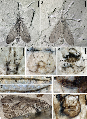
Male Lichnomesopsyche gloriae from Middle−Upper Jurassic Daohugou Beds at Beigou: (1–8) NIGP 171884: (1) part (171884a), general habitus; (2) counterpart (171884b), general habitus; (3) enlargement from (1), indicating details of maxillary palps; (4) enlargement from (2), details of terminalia; (5) enlargement from (1), details of terminalia; (6) tarsus of left foreleg, with T2−T5; (7) tarsus of left midleg, with T1−T5; (8) enlargement from (5), showing comb-like denticles of epandrium (arrow); (9, 10) NIGP 171885: (9) general habitus in lateral view; (10) enlargement from (9), details of terminalia. Bs = basistylus; C = cerci; Cl = claws; Ds = dististylus; Ep = epandrium; Hp = hypandrium; Mp (arrows) = maxillary palps; Pp = paraproct; Tn = nth tarsomere; tX = tenth tergite. Scale bars = 5 mm (1, 2, 9); 0.5 mm (3–7, 10); 0.1 mm (8).

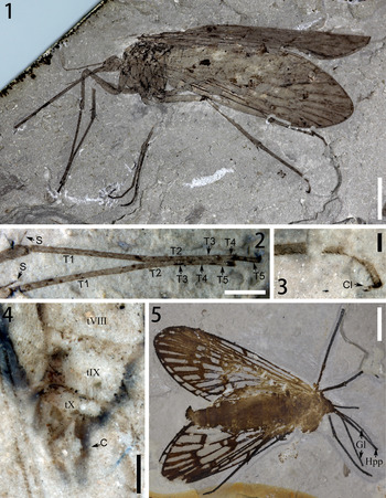
Female Lichnomesopsyche gloriae from Middle−Upper Jurassic Daohugou Beds at Beigou: (1–4) NIGP 171886: (1) general habitus in lateral view; (2) tarsus of forelegs, with T1−T5; (3) claws of left hind leg; (4) terminalia; (5) NIGP 171887, general habitus in dorsal view. C = cerci; Cl = claws; Gl = galeae; Hpp = hypopharynx; S = apical spurs; Tn = nth tarsomere; tN = Nth tergite. Scale bars = 5 mm (1, 5); 2 mm (2); 0.5 mm (3, 4).

Emended diagnosis
Male genitalia trapezoid in dorsal view, covered with small setae; basistylus cylindrical, tapering backward gradually; dististylus enlarged apically, forming a large hook.
Description
Head: proboscis long, sword-shaped; one pair of galeae closed up; hypopharynx visible. Antennae relatively short, filiform, with ~35 flagellomeres; maxillary palps short, with four palpomeres, with two basal palpomeres relatively short, third palpomere elongate, apical palpomere drop-like, pointed distally, shorter than third palpomere (Fig. 1.3).
Wings: with characters resembling description by Ren et al. (Reference Ren, Labandeira and Shih2010).
Legs: slender and long, with length increasing from fore- to hindlegs. Femur moderately thick, longer than tibia; tibia somewhat curving, slightly swelling at apex; two long apical spurs visible in apical tibia (Fig. 2.2); tarsus pentamerous; basitarsus longer than other four tarsomeres together; last three tarsomeres relatively short; fourth tarsomere shortest, with one pair of bifid claws visible (Figs. 1.6, 2.3). Fore tarsus approximately as long as femur and tibia together; hind tarsus slightly longer than femur. Legs covered with dense short setae and some long bristles, with rows of bristles staggered on tibia and tarsus.
Abdomen: 10 segments visible, tapering backward; tergites nearly completely sclerotized; sclerotized section rectangular, with length shorter than width; width gradually decreasing backward; tergite VII small, transverse.
Male terminalia: terminalia large, covered with setae (Fig. 1.4, 1.5, 1.10); hypandrium (sternite IX) near semicircle, slightly hollow posteriorly; epandrium (tergite IX) transverse, bow-tie-shaped, with both anterior and posterior margins concave; two oblique grooves armed laterally with row of comb-like denticles on posterior margin (Fig. 1.8); tergite X small, bearing two oblique lines and one pair of paraprocts visible (Fig. 1.5); cerci clavate (Fig. 1.5). Basistyles and dististyles folding into trapezoid, forming clasping organ; basistylus cylindrical, narrowing gradually to apex; dististylus triangular, distinctly enlarged apically, forming rounded outer angle and large hook apically.
Female terminalia comparatively small with one pair of two-segmented cerci (Fig. 2.4).
Remarks
The wing length/width ratio of Lichnomesopsyche mentioned by Ren et al. (Reference Ren, Labandeira and Shih2010) could reach 3.8. Such a slender wing ratio could be caused by taphonomic deformation (lateral compression).
Lichnomesopsyche daohugouensis Ren, Labandeira, and Shih, Reference Ren, Labandeira and Shih2010
Figure 3
Holotype
CNU-M-NN 2005022 (1, 2), part and counterpart, a nearly complete specimen with well-preserved body and wings from Daohugou (Ren et al., Reference Ren, Labandeira and Shih2010, p. 724, pl. 4).
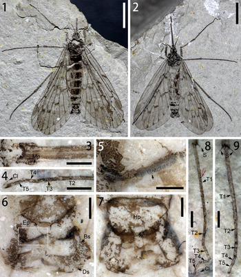
Male Lichnomesopsyche daohugouensis from Middle-Upper Jurassic Daohugou Beds at Chentaizi, NIGP 173118: (1) part (173118a), general habitus; (2) counterpart (173118b), general habitus; (3) enlargement from (2), maxillary palps and long proboscis; (4) tarsus with claws in left midleg; (5) enlargement from (6), showing comb-like denticles (arrow); (6) enlargement from (1), details of terminalia; (7) enlargement from (2), details of terminalia; (8) tarsus of left foreleg, with T1−T5; (9) tarsus of left hindleg, with T1−T5. Bs = basistylus; Cl = claws; Ds = dististylus; Ep = epandrium; Hp = hypandrium; Mp = maxillary palps; S = apical spurs; Tn = nth tarsomere. Scale bars = 5 mm (1, 2); 1 mm (4, 8, 9); 0.5 mm (3, 6, 7); 0.2 mm (5).

Emended diagnosis
Basistylus cylindrical; dististylus dividing a prominent projection on outer margin; apical dististylus rounded, armed with many tiny spines.
Description
General characters of head, legs, and abdomen as in original description of Lichnomesopsyche gloriae described above.
Head: Antennae filiform with > 25 flagellomeres. Maxillary palps short, pointed, with four palpomeres; 1–3 palpomeres relatively short; distal palpomers drop-like, aristiform apically (Fig. 3.3).
Wings: with characters resembling description by Ren et al. (Reference Ren, Labandeira and Shih2010).
Abdomen: tergite VIII triangular; apex with protrusion.
Terminalia: well-exposed, covered with setae (Fig. 3.6, 3.7); hypandrium oval; epandrium (tergite IX) bow-tie-shaped, with both anterior and posterior margins concave, armed with one row of comb-like denticles on posterior margin (Fig. 3.5); basistyles and dististyles folding into trapezoid, forming clasping organ; basistylus cylindrical; dististylus dividing a prominent projection in outer part, discriminated by tuft of hairs; apical dististylus rounded, with inner portion armed with many tiny spines.
Remarks
The circular, pale spots on the holotype of Lichnomesopsyche daohugouensis could have been produced by pathological factors.
Discussion
Shih et al. (Reference Shih, Qiao, Labandeira and Ren2013) summarized the wing parameters of 13 specimens of Mesopsychidae; based on the high frequency of broader left wings, they concluded that mesopsychids possess asymmetrical wings. However, such characters could be attributed to taphonomic factors that would clearly deform some specimens. The wing ratios of the newly illustrated specimens display variation in wing appearances under different postures, or the left and right wings are simply symmetrical (Table 1). Thus, mesopsychids would have symmetrical wings.
Parameters for left and right forewings of Lichnomesopsyche. L = length (mm); L/W = length/width ratio; W = width (mm); − = not available.

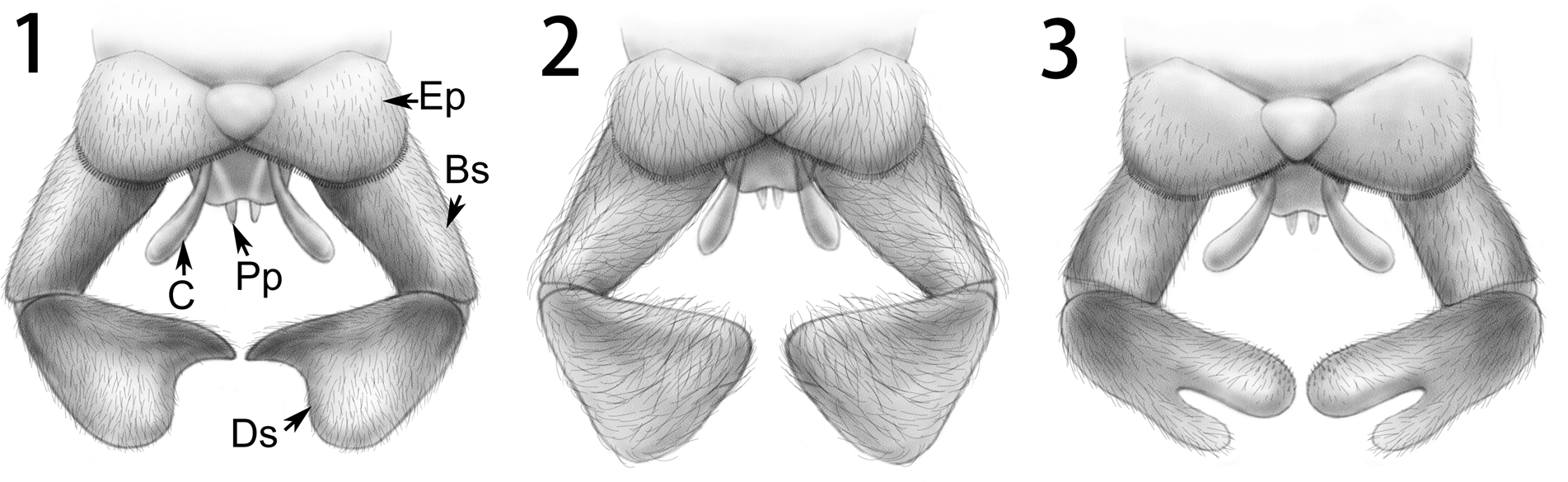
Lichnomesopsyche, reconstructed male terminalia in dorsal view: (1) L. gloriae; (2) L. prochorista (based on Lin et al., Reference Lin, Shih, Labandeira and Ren2016, fig. 6c); (3) L. daohugouensis. Bs = basistylus; C = cerci; Ds = dististylus; Ep = epandrium; Pp = paraproct.

Species of Lichnomesopsyche are very similar to one another in wing characters, and previous classifications focused on very minor differences, e.g., the relative bifurcation between Rs1+2 and Rs3+4, and circular and pale spots on the wings, which are not necessarily preserved in all specimens. The present study indicates that the male genitalia of the three species of Lichnomesopsyche are clearly different from each other (Fig. 4). The male genitalia of L. daohugouensis distinctly differs from those of both L. gloriae and L. prochorista by a series of characters, including the basistylus not tapering backward and the dististylus not enlarged apically, but with a remarkable projection presented at the outer margin. The male genitalia of L. gloriae and L. prochorista closely resemble each other, but they differ based on the following characters: the dististylus of both species are somewhat triangular, but the apical dististylus of L. gloriae has a large hook instead of rounded angles as in L. prochorista (see Lin et al., Reference Lin, Shih, Labandeira and Ren2016, fig. 6c). In addition, comparatively long hairs are developed on the surface of the genitalia of L. prochorista, whereas the genitalia of L. daohugouensis and L. gloriae are covered with shorter setae. A key to the species of Lichnomesopsyche is presented below. There are no alternative characters that can be clearly used for separating the female species of Lichnomesopsyche, although the body sizes of L. prochorista and L. gloriae are generally larger than that of L. daohugouensis. According to all known specimens, the forewing length of L. daohugouensis varies from 18.0−22.0 mm, of L. gloriae from 20.5−27.9 mm, and of L. prochorista from 22.0−25.9 mm.
Key to the species of Lichnomesopsyche based on characters of the male terminalia.—
1 Basistylus not tapering; dististylus not enlarged apically; apical dististylus with outer projection ……………. L. daohugouensis
Basistylus tapering; dististylus enlarged apically………………………………………………………………………………. 2
2 Apical dististylus with a large hook…………………………………………………………………………………… L. gloriae
Apical dististylus with rounded angles …………………………………………………………………………… L. prochorista
The male terminalia of Lichnomesopsyche are extraordinary in Mecoptera. The posterior margin of the epandrium is armed with comb-like denticles (Figs. 1.8, 3.5) resembling those of Parapolycentropus paraburmiticus Grimaldi and Rasnitsyn in Grimaldi et al., Reference Grimaldi, Rasnitsyn, Zhang and Fraser2005 from mid-Cretaceous Burmese amber (see Grimaldi and Johnston, Reference Grimaldi and Johnston2014, fig. 8B). This character represents a synapomorphy of Aneuretopsychina.
The species of Lichnomesopsyche from Daohugou biota are rare cases of preserved male genitalia structure that are enabled as diagnostic characters. Meanwhile, little differentiation is found in venation, so it is indisputable to regard genital structures as diagnostic characters within this genus. Our study provides a new perspective on the classification of Lichnomesopsyche.
Acknowledgments
Financial support was provided by the National Key Research and Development Program of China (grant no. 2016YFC0600406), Strategic Priority Research Program of the Chinese Academy of Sciences (XDB18000000 and XDB26000000), and the National Natural Science Foundation of China (41925008 and 41688103).