Introduction
Gravity's role in plant growth and development has been studied by re-orienting plants in relation to the vector, removing gravity's influence by putting plants in free-fall (in orbiting spacecraft), and imposing radial acceleration by means of on-board rotors to discover detection thresholds in the ‘microgravity’ ( < 10− 3 g) to 1 g range (for a review, see Musgrave, Reference Musgrave2002). How acceleration forces in excess of Earth's gravity might affect plants was studied by growing them for extended periods on large-diameter centrifuges (Brown et al., Reference Brown, Dahl and Chapman1976), and Fitzelle and Kiss (Reference Fitzelle and Kiss2001) made elegant use of hypergravity treatments to elucidate gravitropism mechanisms in starch-deficient mutants. However, beyond these phenomenological approaches lie questions to be answered about gravity's role in sculpting plant life processes. Seed development from a fertilized egg is an ideal system for investigating these fundamental questions.
Rapid-cycling Brassica rapa L. has been a convenient model for these investigations (Musgrave, Reference Musgrave2000). Because of the self-incompatibility system of its flowers, the start of seed development is initiated by manual transfer of pollen between plants, and thus the timing is readily controlled. Seeds develop within a silique, which contains a unique internal atmosphere that both controls and is controlled by the metabolism of the developing embryos (Porterfield et al., Reference Porterfield, Kuang, Smith, Crispi and Musgrave1999; Vigeolas et al., Reference Vigeolas, van Dongen, Waldeck, Huhn and Geigenberger2003; Borisjuk et al., Reference Borisjuk, Rolletschek, Radchuk, Weschke, Wobus and Weber2004; Goffman et al., 2004; Musgrave et al., Reference Musgrave, Allen, Blasiak, Tuominen and Kuang2008).
Gases within the siliques of B. rapa can be measured using a variety of methods (Porterfield et al., Reference Porterfield, Kuang, Smith, Crispi and Musgrave1999; Vigeolas et al., Reference Vigeolas, van Dongen, Waldeck, Huhn and Geigenberger2003; Goffman et al., 2004; Musgrave et al., Reference Musgrave, Allen, Blasiak, Tuominen and Kuang2008). We developed an in vitro silique maturation system (Musgrave et al., Reference Musgrave, Allen, Blasiak, Tuominen and Kuang2008) to provide a large cohort of standard siliques, and refined a gas extraction technique to give good reproducibility in sampling these small, closed environments. The relationship between internal gas composition of siliques and seed storage reserves was investigated using this system (Musgrave et al., Reference Musgrave, Allen, Blasiak, Tuominen and Kuang2008) prior to putting it to work in assessing gravity's role in modulating seed development.
A series of experiments in our laboratory has elucidated the tight co-regulation of seed development and the composition of the gaseous microenvironment surrounding the seeds. Removing seeds from this microenvironment disrupts this co-regulation, so procedures that would directly measure respiration or photosynthesis rates by the immature seeds are not practical because they disturb this equilibrium. Non-invasive experiments on the seed microenvironment performed with a range of model species have led us to conclude that seed development in green pepper (Blasiak and Musgrave, Reference Blasiak and Musgrave2002; Blasiak et al., Reference Blasiak, Kuang, Farhangi and Musgrave2006), Arabidopsis (Kuang et al., Reference Kuang, Crispi and Musgrave1998) and Brassica (Porterfield et al., Reference Porterfield, Kuang, Smith, Crispi and Musgrave1999; Musgrave et al., Reference Musgrave, Allen, Blasiak, Tuominen and Kuang2008) is oxygen limited. When plants were grown in a range of sub-ambient oxygen concentrations, the growth of their developing embryos was proportional to the amount of oxygen in the surrounding atmosphere (Kuang et al., Reference Kuang, Crispi and Musgrave1998). Similarly, pepper fruits that developed with synthetic internal gases passing through their locular spaces would mature their seeds normally if the gas composition mimicked what occurs normally inside the fruit (Blasiak et al., Reference Blasiak, Kuang, Farhangi and Musgrave2006). Raising the O2 concentration in the flow-through mixture by even a few per cent resulted in larger seeds with more advanced storage reserve deposition (Blasiak et al., Reference Blasiak, Kuang, Farhangi and Musgrave2006). Comparison of storage reserve deposition in cotyledons of Brassica seeds developing in vitro (shown to be a more oxygen-limited environment than in vivo) revealed that the loss of starch and formation of lipids are retarded by oxygen restriction to the developing seeds (Musgrave et al., Reference Musgrave, Allen, Blasiak, Tuominen and Kuang2008).
The impetus for the experiments reported here arose from our prior findings that Brassica seed development is altered in the absence of gravity (Kuang et al., Reference Kuang, Popova, Xiao and Musgrave2000, Reference Kuang, Popova, McClure and Musgrave2005; Musgrave et al., Reference Musgrave, Kuang, Xiao, Stout, Bingham, Briarty, Levinskikh, Sychev and Podolski2000, Reference Musgrave, Kuang, Tuominen, Levine and Morrow2005). Seeds matured in microgravity were smaller and contained different storage reserves than those developing in 1 g (Musgrave et al., Reference Musgrave, Kuang, Xiao, Stout, Bingham, Briarty, Levinskikh, Sychev and Podolski2000). Because the changes in seed maturation and storage reserve composition were similar to those occurring in low oxygen environments (Kuang et al., Reference Kuang, Crispi and Musgrave1998), we hypothesized that the lack of convection in microgravity (Musgrave et al., Reference Musgrave, Kuang and Matthews1997) results in a different gaseous microenvironment for the developing seed than that maintained at 1 g.
To test this hypothesis regarding the mechanism behind gravity's effect on seed development, we conducted a series of experiments in hypergravity on a large-diameter centrifuge. We sought data from centrifuge experiments at 2 g and 4 g to compare with our existing data on seed development in microgravity and at 1 g. We have hypothesized that the changes in seed development occurring in microgravity may be caused by lack of convective air movement within the developing seed pod. Because convection is gravity-dependent, we expected to enhance internal gas mixing by subjecting plants to hypergravity treatments. We predicted that growing the parent plant in hypergravity would lead to an accelerated rate of seed development.
Materials and methods
Hypergravity treatments
A 24-ft diameter centrifuge at NASA Ames Research Center, Moffett Field, California, USA, was used to provide hypergravity (2 g and 4 g) exposures over a 16-d experimental period. Protocols and hardware previously used by our laboratory for experiments in microgravity (Kuang et al., Reference Kuang, Popova, Xiao and Musgrave2000) were used for these experiments. Briefly, B. rapa L. cv. ‘Astroplants’ plants were pre-grown between two flat faces of phenolic foam (RootCube; Smithers Oasis, Kent, Ohio, USA) half cylinders moistened with modified Hoagland's solution, with two plants spaced 1 cm apart in each foam-containing plastic sleeve. When the plants were 13 d old, they were loaded into plant growth chambers (PGCs) and inserted into NASA's Plant Growth Facility (PGF) hardware. Six plants were loaded into each PGC. Each PGF held three of these plant-containing PGCs in addition to three PGCs fitted with racks to hold the tubes for in vitro silique culture (described below). Average (SE) light intensity 10 cm below the PGC lids measured in three positions across the length of the chambers was 137 (10) and 130 (8) μmol m− 2 s− 1, photosynthetically active radiation (PAR) for the two PGF units, which were assigned randomly to treatments in the three different hypergravity experiments. One PGF was mounted in a cabinet attached to the rotor and the other was kept stationary in a cabinet in the same area. Because conditions were being matched to those of the earlier spaceflight experiments, carbon dioxide was fed into each PGF to maintain chamber levels at 1000 ppm. Environmental data (temperature, relative humidity and carbon dioxide concentration) were collected every 5 min by the PGF for each PGC and acceleration data for the centrifuge (torque, gz and rpm) were logged every 5 min. For the three experiments, average temperatures in the chambers ranged between 23 and 26°C, RH ranged between 57 and 82%, and CO2 ranged between 700 and 1300 ppm.
As the plants began to flower, the rotor was stopped daily to allow for a period of manual pollination. Open flowers were marked with colour-coded tape prior to pollination, to allow identification of pollination dates for the resultant siliques at the end of the experiment. Rotor stops for these pollinations, watering of the plants, and hardware maintenance typically took an hour or less. Over the three 383-h run periods (one 2-g experiment and two 4-g experiments) that were performed using the large-diameter centrifuge, the plants experienced hypergravity (2 g or 4 g) for at least 370 h (>97% of the total time).
Watering intervals conformed to the schedule previously used for the microgravity experiment and occurred 5, 9 and 13 d into the experiment. The plenum air supply system in the PGF provides a steady flow of air through the PGCs. The PGCs containing plants growing in phenolic foam were weighed, and nutrient solution was added to replace water that had been lost. Chambers lost water at equivalent rates (~15 ml d− 1) whether they were in the PGF mounted on the rotor or in the stationary PGF.
In addition to the experiments with whole plants and cultured siliques on the large-diameter rotor, experiments were performed using a laboratory centrifuge (Eppendorf 5702) that was modified to provide light and temperature regulation comparable to the main experiments. In vitro siliques (Musgrave et al., Reference Musgrave, Allen, Blasiak, Tuominen and Kuang2008; see below) were matured at 4 g (200 rpm) using this system, primarily to generate additional data on internal silique gas composition for comparison to stationary control material.
Production of in vitro siliques
B. rapa plants were interpollinated 16 d after planting, to produce a large number of high-quality, same-aged siliques for in vitro culture. Siliques were cut from the plants 10 d after pollination and sterilized in a solution of 10% (v/v) Clorox and 0.1% (w/v) Tween 20 (Sigma-Aldrich, St. Louis, Missouri, USA) for 10 min, then rinsed in sterile water three times for 5 min each. The cut ends of the siliques were removed using a sterile scalpel and siliques were placed with cut end down in Murashige Skoog (MS) medium (Sigma-Aldrich) containing 30 g l− 1 sucrose, 5.0 g l− 1 agar, 0.25 mg l− 1 benzylaminopurine, 0.025 mg l− 1 naphthalene acetic acid, at a pH of 5.8. Siliques were cultured individually in 17 × 100 mm sterile polystyrene culture test tubes (Fisher, 14-956-6A), that were placed into stainless-steel holders within three PGCs or directly in the Eppendorf 5702 rotor. Siliques were harvested from their respective treatments at 4–6 d intervals using a randomized sampling scheme, with final harvest occurring 29 d after pollination.
Determination of internal silique gases
Approximately 1 min elapsed between stopping the laboratory centrifuge rotor and collection of internal gas samples for analysis. Sampling of gases in the developing siliques was accomplished by rupturing individual siliques in a pure helium atmosphere and then using gas chromatography (GC) to determine CO2 and O2 concentrations in the silique's internal atmosphere. A system of gas-tight Hamilton syringes (2.5 and 5 ml), connected by a three-way Popper valve, (Popper & Sons, Inc., New Hyde Park, New York, USA) was purged with ultrapure helium prior to introduction of the silique in the 5-ml crushing syringe. After an additional 20 s helium rinse, the syringe was sealed with the plunger. The syringe plunger was used to rupture the silique and expel its gases into the helium surrounding it as the aggregate gases were transferred into the 2.5-ml sample syringe for injection onto the GC column, as described in Blasiak et al. (Reference Blasiak, Kuang, Farhangi and Musgrave2006). Gas samples were analysed on a series 6890 gas chromatograph (Agilent Technologies, Wilmington, Delaware, USA). Oxygen and carbon dioxide were measured simultaneously by injection onto a 6-ft CTR1 column (Alltech, Deerfield, Illinois, USA) at 35°C, utilizing helium as the carrier gas at 28 ml min− 1, linked to a thermal conductivity detector. Output from the detector was fed to a computer and integrated using EZChrom 6.8 software (Scientific Software Inc., Pleasanton, California, USA).
Cytochemical localization of reserves
Material for light microscopy was dissected from in vitro or in vivo siliques and fixed overnight at 4°C in 2.5% (v/v) glutaraldehyde and 1% (w/v) formaldehyde in 0.1 M phosphate buffer, pH 7.0. Tissues were then washed in the same buffer, dehydrated in an ethyl alcohol series, and infiltrated and embedded in LR White resin. Embedded tissues were sectioned with a Leica microtome. Semi-thin sections 1 μm thick were stained with toluidine blue in a 2% sodium borate solution for general tissue staining. Cytochemical reaction with periodic acid–Schiff reagent (PAS) was used to localize polysaccharide, and 1% Aniline Blue Black in 7% acetic acid was used as a stain for gross protein.
Morphometric analysis
Stained sections were examined with an Olympus compound microscope (BX40) and photographed with an Olympus digital camera (Olympus PM10SP). The size of cotyledon cells was measured using Image-Pro Plus (Version 4.5; Media Cybernetics, Bethesda, Maryland, USA), and starch grains and protein bodies in the cells were counted directly on the digitized images.
Biochemical quantification of storage reserves
Seeds were dissected from in vitro siliques that had been used for internal gas samples or from in vivo siliques and immediately frozen for subsequent analyses. Frozen seeds were lyophilized overnight and then weighed into appropriate sample aliquots. Analyses for starch, soluble carbohydrates and protein followed the procedures used previously (Musgrave et al., Reference Musgrave, Kuang, Tuominen, Levine and Morrow2005), using dry samples ranging from 2 to 12 mg. Lipid was quantified using the charring method as described by Mansfield and Briarty (Reference Mansfield and Briarty1996).
Embryo rescue
In vivo siliques from the 14 d after pollination (DAP) cohorts (4-g centrifuge treatments and controls) were surface sterilized and opened to extract the immature seeds within. The embryos were removed from the seed coats under sterile conditions and then transferred first to embryo rescue medium and then to rooting medium, which encouraged root development. At the time of transferring these plants to soil, plants were scored for developmental stage for comparison with those generated from the stationary-control embryos. Flowering plants from each treatment were then interpollinated to create the seed for an E2 generation from the 4-g centrifuge and control treatments for subsequent comparisons (Kuang et al., Reference Kuang, Popova, Xiao and Musgrave2000).
Results
In vitro
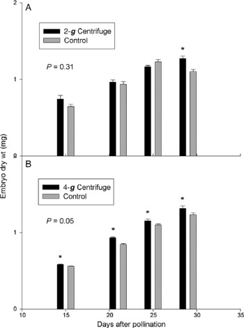
To test the role of gravity in modifying the seed microenvironment, we conducted a series of experiments with in vitro siliques placed in culture at 10 DAP. These siliques were exposed to treatment conditions from 12–29 DAP and so represent maturation events during late stages of seed development. Figure 1 shows the time-line for seed weight gain during this period. Significant differences were not found in the 2-g seed weights uniformly across the time-line (Fig. 1A), but the 4-g seeds were significantly heavier than the 1-g controls (Fig. 1B; P = 0.05).
Weights of lyophilized Brassica embryos dissected from in vitro siliques maturing at (A) 2-g, (B) 4-g or their respective 1-g controls (grey bars). The 4-g results are from two independent experiments. P-values refer to the results of t-tests for paired comparisons for the time course. Mean values with SE bars are shown. Asterisks indicate that mean values for the centrifuge treatment were significantly greater than controls (P ≤ 0.06) as determined by t-tests.

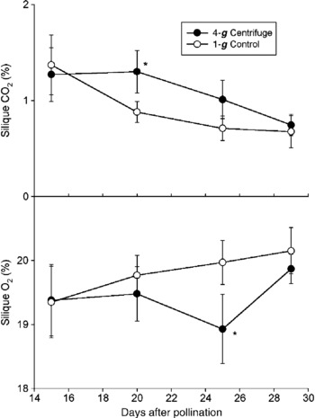
Sampling of gases from in vitro siliques from the stationary (1-g) treatment (Fig. 2) showed the expected decline in CO2 over the course of the maturation period (Fig. 2A), and the expected increase in O2 (Fig. 2B), based on our prior experimentation (Musgrave et al., Reference Musgrave, Allen, Blasiak, Tuominen and Kuang2008). CO2 in the 4-g siliques was significantly higher than the 1-g controls at the intermediate sampling dates (20 DAP). In contrast, O2 concentration in the 4-g treatment differed from the 1-g controls at 25 DAP.
Carbon dioxide (A) and oxygen (B) concentrations in gas samples recovered from in vitro Brassica siliques maturing at 4-g or in the stationary control. Data are from two independent experiments and represent the mean values for n = 12; bars denote SE. Asterisks indicate statistical differences from the control (P ≤ 0.03 for O2; P ≤ 0.09 for CO2).

Whole plant
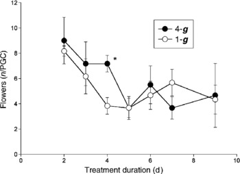
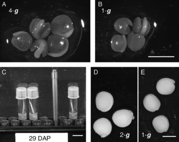
Plant exposure to hypergravity (2-g and 4-g) during the reproductive stage had no adverse effects on flowering, fertilization or embryogenesis. As shown in Fig. 3 for the two 4-g experiments, the number of flowers in anthesis was equivalent to the 1-g control with the exception of 4 d into the treatment period, when the 4 g treatment had significantly more flowers than the controls. Sequential pollinations over an 8-d period gave rise to siliques bearing embryos of different ages. The embryos from the most mature (14 DAP) of these compared favourably to the control materials, both at 2 g and 4 g (4 g experimental material shown in Fig. 4A, B). Similarly, embryos from in vitro siliques (Fig. 4C) that experienced hypergravity for the 12–29 DAP interval were comparable to the control materials (2-g experiment material shown in Fig. 4D, E). In both experimental systems, the hypergravity embryos appeared to be larger than the 1-g controls, as was confirmed statistically using dry weight data (Fig. 1).
Number of B. rapa flowers in anthesis after 2–9 d of exposure to hypergravity (4 g), compared to 1-g controls. Six plants were present in each PGC. Mean values for two independent experiments, n = 36; bars denote SE. Plants were 14 d old at time of transfer to the different treatments. The value marked by an asterisk was significantly different from the control (P ≤ 0.005).

Dissecting microscope images of embryos from 1 g, 2 g and 4 g. Embryos produced by Brassica plants grown in hypergravity ranged in age from 8 to 15 d after pollination (about halfway to maturity). The embryos shown (A, B; 14 DAP; scale bar = 2 mm) were rescued and grown into plants using tissue culture (Fig. 5). Hypergravity effects on the later stages of seed development (Reference Allen, Bisbee, Darnell, Kuang, Levine, Musgrave and van LoonFigs 1 and Reference Blasiak and Musgrave2) were studied in Brassica siliques put into culture at 10 DAP and kept in culture up to 29 DAP (C; scale bar = 2 cm). The embryos shown (D, E) were harvested from tissue culture at 29 DAP (scale bar = 0.77 mm).

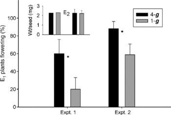
Some of the oldest embryos (Fig. 4A, B) from these plants were also used in a functional test of their maturity. Since the immature seeds are too young at this stage to survive being dried, we used an embryo rescue technique (Kuang et al., Reference Kuang, Popova, Xiao and Musgrave2000) to enable growth of the embryos into plants, while bypassing the drying step. As shown in Fig. 5, plants from these hypergravity embryos showed developmental advancement compared to the 1-g controls, with more plants entering the flowering stage earlier. Progeny (E2) from these 4-g embryos were indistinguishable in developmental time-line and seed weight from the 1-g controls (Fig. 5, inset).
Comparison of plants (E1) produced by B. rapa embryos rescued from siliques formed in 4 g and 1 g indicates accelerated development in the 4-g treatment. In both experiments, more plants from the 4-g embryos had entered the flower stage (as indicated by presence of buds) at time of transfer to soil than from the stationary control group. Progeny (E2) from these plants were indistinguishable in their seed weights (inset) and developmental time-line (data not shown). Asterisks indicate significant differences from control (P ≤ 0.07).

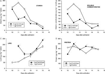
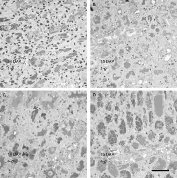
Embryos of different ages frozen after their development on the centrifuge or in the stationary controls were analysed for storage reserve components. Figure 6 shows representative results for one of the 4-g experiments. Declines in starch (Fig. 6A) and soluble carbohydrates (Fig. 6B) occurred at an earlier stage in the 4-g plants than in the corresponding 1-g controls. Further, lipid accumulation (Fig. 6C) was enhanced at 4 g, compared to 1 g. The rapid decline in starch reserves during development could also be detected through cytochemical localization (Fig. 7). As with the biochemical analyses, starch grains visible in cotyledon thin sections were more numerous in the 1-g control than in the 4-g centrifuge material, indicating accelerated development in hypergravity.
Biochemical quantification of reserves (starch, soluble carbohydrates, lipid and protein) stored in B. rapa seeds during early development (10–15 DAP) at 4 g compared to 1-g controls. Mean values with SE bars are shown (symbols appearing without error bars are larger than the SE). Values marked by asterisks are significantly different from control (P ≤ 0.03) as determined by t-test.

Cytochemical localization of storage reserves in embryos of B. rapa developing at 1 g (A and B) or 4 g (C and D). As the embryos age, starch, shown here as magenta-coloured grains because of staining with periodic acid–Schiff (PAS) reagent, disappears from the cotyledon tissue. Starch begins disappearing at an earlier time point in hypergravity than in 1 g, indicating developmental acceleration. Protein bodies are stained blue. Bar = 20 μm.

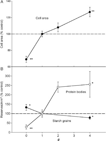
These experiments were conducted with the same procedures and hardware that had been used during our prior spaceflight experiment on STS-87 (Kuang et al., Reference Kuang, Popova, Xiao and Musgrave2000, Reference Kuang, Popova, McClure and Musgrave2005) so that we would be able to compare our results across a range of g-values. Although biochemical analyses (Fig. 6) were not available for the STS-87 experiment, we were able to compare storage reserve deposition using cytochemical localization techniques on the oldest embryos produced on the plants (15 DAP). In Fig. 8, microscopy data for cell size, starch grains per area and protein bodies per area were normalized against the control values for each experiment and then plotted against g. Cell cross-sectional area increased with increasing g, as did number of protein bodies per unit area of cotyledon, while starch grains decreased per unit area of cotyledon.
Cell size (A) and storage reserves (B) across the g-continuum. Data shown are from cotyledons from 15 DAP Brassica embryos that developed on plants growing in microgravity, 1 g, 2 g or 4 g. For purposes of comparison among experiments, data have been normalized against their respective 1-g controls. Mean values ( ± SE); n = 8–13. Asterisks indicate values significantly different from controls (*, P ≤ 0.07; **, P ≤ 0.02).

Discussion
Prior long-term plant growth experiments in hypergravity had not investigated seed production (Brown et al., Reference Brown, Dahl and Chapman1976), leaving in question whether seed production could occur normally in hypergravity. We found that B. rapa successfully formed seeds at both 2 g and 4 g (Fig. 4). Furthermore, the seeds formed in hypergravity were more advanced developmentally than those formed in 1 g, as judged by storage reserve composition (Fig. 6) and performance of rescued embryos (Fig. 5). This is the converse of what had been found previously when the same model system was subjected to microgravity conditions on the space shuttle (Kuang et al., Reference Kuang, Popova, Xiao and Musgrave2000, Reference Kuang, Popova, McClure and Musgrave2005), Mir space station (Musgrave et al., Reference Musgrave, Kuang, Xiao, Stout, Bingham, Briarty, Levinskikh, Sychev and Podolski2000), and International Space Station (Musgrave et al., Reference Musgrave, Kuang, Tuominen, Levine and Morrow2005). In each of these microgravity settings, seed development had been delayed. Embryos rescued from immature siliques produced plants that were developmentally delayed compared to the 1 g controls, flowering 12 d later (Kuang et al., Reference Kuang, Popova, Xiao and Musgrave2000). As in our hypergravity experiments (Fig. 5, inset), these effects were not carried on into the E2 generation. In the one experiment that produced mature seeds in microgravity (Musgrave et al., Reference Musgrave, Kuang, Tuominen, Levine and Morrow2005), seed weights were depressed by 25%. Plants grown from these seeds were smaller than those grown from 1-g seeds, but the F2 seeds and plants were normal sized as long as the seeds were matured at 1 g. Collectively these data all suggest that gravity modulates seed development by controlling the maternal environment of the seed.
How the maternal environment is modulated by gravity during late stages of seed development was studied using our in vitro system (Musgrave et al., Reference Musgrave, Allen, Blasiak, Tuominen and Kuang2008). Embryos were significantly heavier when raised at 4 g, while there was no statistically significant difference between 2-g and control embryos (Fig. 1). The in vitro system provided a means to determine changes in the captive atmosphere around the developing seeds, since large numbers of same-aged siliques could be rapidly sampled after rotor stops. The finding that CO2 accumulates to higher concentrations in the 4-g siliques than in those maturing at 1 g bears out our hypothesis that oxygen becomes more available to embryos in the hypergravity environment, supporting higher metabolic rates that allow the 4-g embryos to reach a greater size and a more advanced developmental stage. Subsequent to the significant elevation in CO2, oxygen in the 4-g siliques became significantly lower than in controls, consistent with the accelerated embryo growth (Fig. 1B) transiently overshooting the oxygen resupply capacity within the siliques. This interesting pattern was observed in both of the 4-g time course experiments.
Although there is no literature on long-term hypergravity effects on plant reproduction and embryo development, there is some pertinent information on animal reproduction in hypergravity, and in plant models subjected to related stresses. In sea urchin, hyper-g (2 g or 5 g) caused a 50% reduction in both the rate of sperm–egg binding and the rate of fertilization (Tash et al., Reference Tash, Kim, Schuber, Seibt and Kinsey2001). This contrasts with our results in Brassica, in which seed set was not affected by hypergravity (up to 4 g), but is consistent with results in Arabidopsis thaliana, where we found seed set to be normal at 2 g but close to zero at 4 g (Musgrave et al., Reference Musgrave, Kuang, Allen, Darnell, Wagers-Hughes and Blasiak2006). The effects of another thigmo-stress, vibration, on reproductive fitness were investigated in a closely related plant, Capsella bursa-pastoris, by Niklas (Reference Niklas1998). He found that mechanically disturbed plants produced 43% fewer seeds (indicating disruption of seed set), but the seeds that were produced had no difference in weight from those of the control plant. In agreement with Niklas' (Reference Niklas1998) finding that mechanical disturbance delayed anthesis of the last flower by 5 d, we observed that Brassica plants flowering at 4 g had significantly more flowers in anthesis than the stationary controls after 4 d of exposure to the treatments, although total number of flowers produced by the plants was not influenced by gravity (Fig. 3).
Reviewing animal metabolic responses to gravity, Wade (Reference Wade and Sonnenfeld2005) rejected earlier notions that gravitational effects operated in an all-or-none mode, and concluded that gravity acts as a continuum. Increasingly, evidence points to changes in gas exchange, through alteration of convective air movement, as a prime candidate for how gravity is affecting metabolic processes (Musgrave et al., Reference Musgrave, Kuang and Matthews1997; Porterfield, Reference Porterfield2002; Briarty and Maher, Reference Briarty and Maher2004; Kitaya et al., Reference Kitaya, Kawai, Takahashi, Tani, Goto, Saito, Shibuya and Kiyota2006). Using artificial sensors that consume oxygen to model the respiratory activity of a plant root, Liao et al. (Reference Liao, Liu, Monje, Stutte and Porterfield2004) elegantly demonstrated that oxygen is less available in microgravity than at 1 g, and conversely becomes more available at hypergravity (2 g). Consistent with enhanced availability of oxygen in hypergravity, we observed more CO2 accumulation inside Brassica siliques maturing at 4 g compared to the 1-g controls. Similarly, starch and soluble carbohydrates in the immature embryos were lost earlier in the developmental sequence at 4 g than at 1 g (Fig. 6A, B), and lipid accumulation was enhanced (Fig. 6C), as we have found in better-oxygenated environments (Kuang et al., Reference Kuang, Crispi and Musgrave1998; Musgrave et al., Reference Musgrave, Allen, Blasiak, Tuominen and Kuang2008). Working with rats raised on the same centrifuge that we used for our experiments, Plaut et al. (Reference Plaut, Maple, Wade, Baer and Ronca2003) concluded that gravitational load influences mammary tissue metabolism. The oxidation of glucose to CO2 showed a linear decrease with g-load in the micro- to 2-g range, while incorporation of glucose into lipid decreased in a curvilinear manner with increased g. For these free-moving animals, hypergravity caused a decrease in body weight for both the dam and the fetus, as they had to expend more energy to move around. In contrast, developing plant embryos had enhanced weight when forming in a hypergravity environment and lipid deposition in the seed was enhanced. Hypergravity also affected the growth of the maternal plant, causing it to assume a prostrate growth form with thickened stems and heavier root systems (Allen et al., Reference Allen, Bisbee, Darnell, Kuang, Levine, Musgrave and van Loon2009). Interestingly, cell size increases were responsible for stem thickening at 4 g (Allen et al., Reference Allen, Bisbee, Darnell, Kuang, Levine, Musgrave and van Loon2009), consistent with our cell size results for developing embryos across the micro- to 4-g range (Fig. 8).
These findings underscore the importance of previous work, which demonstrated that embryos are oxygen limited (Kuang et al., Reference Kuang, Crispi and Musgrave1998; Rolletschek et al., Reference Rolletschek, Weschke, Weber, Wobus and Borisjuk2004; Blasiak et al., Reference Blasiak, Kuang, Farhangi and Musgrave2006). Tipping the balance on this limitation, be it through directly changing oxygen in the exterior atmosphere (Quebedeaux and Hardy, Reference Quebedeaux, Hardy, Burris and Black1976; Kuang et al., Reference Kuang, Crispi and Musgrave1998; Blasiak et al., Reference Blasiak, Kuang, Farhangi and Musgrave2006) or by changing gas resupply processes (Tuominen and Musgrave, Reference Tuominen and Musgrave2006) by altering gravity, results in a change in seed size, storage reserve composition and vigour. B. rapa embryo development is slowed in microgravity and accelerated in hypergravity, due to this delicate balance. Our results (Fig. 8) have significance to NASA because they allow us to predict seed size and storage reserve composition for a model plant developing in habitats on the moon (1/6 g) or Mars (3/8 g). Furthermore, the notion that gravity plays a fundamental role in plant gas relations is an important consideration for scientists interested in geo-centric aspects of evolution (Volkmann and Baluska, Reference Volkmann and Baluska2006).
Acknowledgements
Supported by NASA grant NAG-10-329 and NNX07AT77G to Mary E. Musgrave, and by NOW-ALW-SRON grant MG-057 to Jack J.W.A. van Loon. We thank Matt Regan, April Spinale, Bill Wells, Howard Levine, Daniel Shultz and Doug Gruendel from Kennedy Space Center, Florida, USA, and Tom Luzod, Tianna Shaw and Jon Rask at Ames Research Center, Moffett Field, California, USA, for supporting the experiments.