Non-technical Summary
Oligochaete cocoons, previously interpreted as water fleas, are described from upper Permian freshwater strata of eastern Kazakhstan. The ovoid wrinkled cocoons contain eggs and have a prominent neck and a long stalk at opposite poles. By their size range and distinct morphological features, the cocoons are identical to those of some extant freshwater lumbriculid annelids and, thus, represent the oldest undoubted record of clitellates.
Introduction
Despite the important role of clitellate annelids in Recent terrestrial faunas and their influence on sediment bioturbation, pedogenesis, mineral dissolution and clay mineral precipitation, soil fertility, and feeding basis for a number of animals, their fossil record is obscure, controversial, and mostly restricted to occasional finds of single incomplete specimens in Mesozoic and Cenozoic strata. The record of clitellate body fossils was recently revised and summarized by Shcherbakov et al. (Reference Shcherbakov, Timm, Tzetlin, Vinn and Zhuravlev2020).
The earliest body fossil clitellates have been discovered in the Olenekian (Lower Triassic) lacustrine siliciclastics of the southern Cis-Urals of Russia (Shcherbakov et al., Reference Shcherbakov, Timm, Tzetlin, Vinn and Zhuravlev2020) followed by the Lower Cretaceous and later finds only, namely from the terrestrial Koonwarra Fossil Bed of Victoria, Australia (Jell and Duncan, Reference Jell and Duncan1986; Dettmann et al., Reference Dettmann, Molnar, Douglas, Burger and Fielding1992), the Helvetiafjellet Formation of Spitsbergen (Manum et al., Reference Manum, Bose and Sawyer1991), the Las Hoyas Lagerstätte of Spain (Timm et al., Reference Timm, Vinn and Buscalioni2016), and the Yixian Formation of northeastern China (Hethke et al., Reference Hethke, Fürsich, Jiang, Wang, Chellouche and Weeks2019). Several putative leech impressions, characterized by dense transverse ribbing and possible suction discs, were reported from the Upper Jurassic Solnhofen lithographic slates of Bavaria (Ehlers, Reference Ehlers1869: pl. 36; Kozur, Reference Kozur1970).
The existence of oligochaetes in the Early Triassic is also inferred by the presence of some microburrows, although the identification of exact producers is disputable. Among rare reports of more convincing oligochaete burrows in paleosols, the oldest are of the Lower Triassic age and are represented by fine, open, deeply folded tubules filled with elliptical faecal pellets and pelletoid masses; the burrow fill commonly differs in quantitative mineral grain composition from the host sediment (Retallack, Reference Retallack1976, Reference Retallack1997). These microburrows can be attributed to aquatic oligochaetes because they feed in a conveyor-belt mode, more or less in a vertical position, leaving small faecal mounds at the sediment surface (e.g., Tevesz et al., Reference Tevesz, Soster and McCall1980; White and Miller, Reference White and Miller2008).
In addition, abundant fossil clitellate cocoons, which are resilient toward thermal, chemical, and proteolytic decay, commonly occur in lacustrine and purely terrestrial palynological samples from the Upper Triassic onward (Fritsch, Reference Fritsch1910, pl. 3, fig. 5; Manum et al., Reference Manum, Bose and Sawyer1991; Jansson et al., Reference Jansson, McLoughlin and Vajda2008; Tosolini and Pole, Reference Tosolini and Pole2010; Steinthorsdottir et al., Reference Steinthorsdottir, Tosolini and McElwain2015; McLoughlin et al., Reference McLoughlin, Bomfleur, Mörs and Reguero2016). The structure of such sac-like organic, acid-resistant, chambered, two-layered, mostly reticulate mesofossils allows interpretation as hard-shelled cocoons of leeches. A more precise taxonomic attribution of the majority of these fossils is difficult, even though they can preserve such remarkable anatomical content as fossilized spermatozoa with a conspicuous helical ‘drill-bit’ structure as in the branchiobdellid acrosome (Bomfleur et al., Reference Bomfleur, Mörs, Ferraguti, Reguero and McLoughlin2015). It should be emphasized that the preservation of such cocoons can show unusual taphonomic features, because they are hardened and often covered with a spongy outer cuticle resistant to desiccation due to the terrestrial lifestyle of the worms that deposited them (Sawyer, Reference Sawyer1970; Coleman et al., Reference Coleman, Marotta and Shain2008).
In general, clitellate cocoons provide a microenvironment for embryonic development, preventing desiccation and reducing predation and microbial invasion. Clitellate annelids have a series of midbody segments, the clitellum, specialized to secrete the sheath of the cocoon; their glandular epidermal cells become hypertrophied and filled with proteinaceous granules prior to egg laying (Sayers et al., Reference Sayers, Coleman and Shain2009). The process starts with the secretion of a thin external mucus layer into which a fibrous proteinaceous matrix is deposited to form a multilayered sheath surrounding the clitellum (Hirao, Reference Hirao1965; Westheide and Müller, Reference Westheide and Müller1996). As the granules are released, a tubular sheath develops around the clitellum, into which the eggs are released from the female pores and subsequently fertilized by spermatozoa that had previously been stored in spermathecae during copulation. After egg deposition, the tubular sheath with its content swells and is moved by muscle contractions anteriorly to the worm’s head. Then, the cocoon tube sticks to the substratum and remains attached to it while the worm withdraws. Immediately after deposition, both ends of the tube contract and the cocoon acquires an ovoid shape (Hirao, Reference Hirao1965). In a population of Tubifex tubifex (Müller, Reference Müller1774), the number of eggs per cocoon varied from 1–18 (Hirao, Reference Hirao1965). More eggs, up to 35–40, are enclosed in one cocoon by some enchytraeids (Chekanovskaya, Reference Chekanovskaya1962). Some worms camouflage the cocoon by covering it with substratum particles, thus, demonstrating parental behavior (Timm, Reference Timm1970, Reference Timm2020; Westheide and Müller, Reference Westheide and Müller1996). Following deposition, the wall of the cocoon gradually hardens to form a protective case, which is evident from the formation of a dense zone of concentric and longitudinal protuberances on its poles resulted from a tension gradient along the cocoon surface (Sawyer, Reference Sawyer1970; Coleman et al., Reference Coleman, Marotta and Shain2008). Once development is completed, the hatchlings leave the cocoon either through a self-made hole in its shell or through one of its polar tufts/extensions, or by pushing open a part of the shell like an operculum (Evans and Guild, Reference Evans and Guild1947; Chekanovskaya, Reference Chekanovskaya1962; Sawyer, Reference Sawyer1970; Arkhipova, Reference Arkhipova and Shilova1989).
For a long time, Mesozoic and Cenozoic acid-resistant, two-layered, reticulate cocoons have variously been interpreted by paleobotanists and palynologists as megaspores or seeds, or lichen, fungal, algal, or insect remains (Manum et al., Reference Manum, Bose and Sawyer1991; Steinthorsdottir et al., Reference Steinthorsdottir, Tosolini and McElwain2015). Similarly, the oligochaete cocoons from the upper Permian lacustrine Karaungir Lagerstätte (eastern Kazakhstan) were erroneously ascribed to water fleas (Crustacea: Cladocera) and described as five different species belonging to two genera, namely, Archedaphnia kazakhstanica Smirnov, Reference Smirnov1970 (type species), Archedaphnia novojilovi Smirnov, Reference Smirnov1970, Archedaphnia borutzkyi Smirnov, Reference Smirnov1970, Archedaphnia strelkovi Smirnov, Reference Smirnov1970, and Propleuroxus freyi Smirnov, Reference Smirnov1970. The author based his attribution on the presence of two valves with an ephippium in the rear part, an eye, a head helmet (although poorly defined) and a caudal needle, but noted the absence of either antennae or any other jointed appendages. The attribution was further confused because, later, the same author described a genuine cladoceran possessing a bivalved carapace, setigerous antennae, and other limbs, which he referred to the same genus, Archedaphnia (Archedaphnia testacea Smirnov, Reference Smirnov1971), from the Middle Jurassic of Transbaikalia, Russia (on its age, see Arzhannikova et al., Reference Arzhannikova, Frolov, Arzhannikov, Demonterova, Ivanov, Jolivet, Rubtsova and Dorozhko2018). These fossils have widely been cited as the oldest cladocerans (e.g., Novojilov, Reference Novojilov1970; Negrea et al., Reference Negrea, Botnauriuc and Dumont1999; Chiambeng and Dumont, Reference Chiambeng and Dumont2005; Korovchinsky, Reference Korovchinsky2006) until a revision of the type material undertaken by Kotov (Reference Kotov2007; Van Damme and Kotov, Reference Van Damme and Kotov2016). The latter author established a new genus Smirnovidaphnia Kotov, Reference Kotov2007 for the Jurassic species, whereas the alleged upper Permian ‘cladocerans’ were recognized as problematic fossils lacking any arthropod features.
Here, based on a comparison with Recent clitellate cocoons, we reinterpret these enigmatic fossils from the Lopingian (upper Permian) siliciclastic lacustrine Karaungir Lagerstätte in eastern Kazakhstan as the oldest oligochaete cocoons and thus as the earliest undoubted record of oligochaete annelids and the Clitellata in general. A description of cocoons of an extant freshwater lumbriculid worm is added for comparison, because no detailed descriptions of such cocoons have been made before.
Geological setting
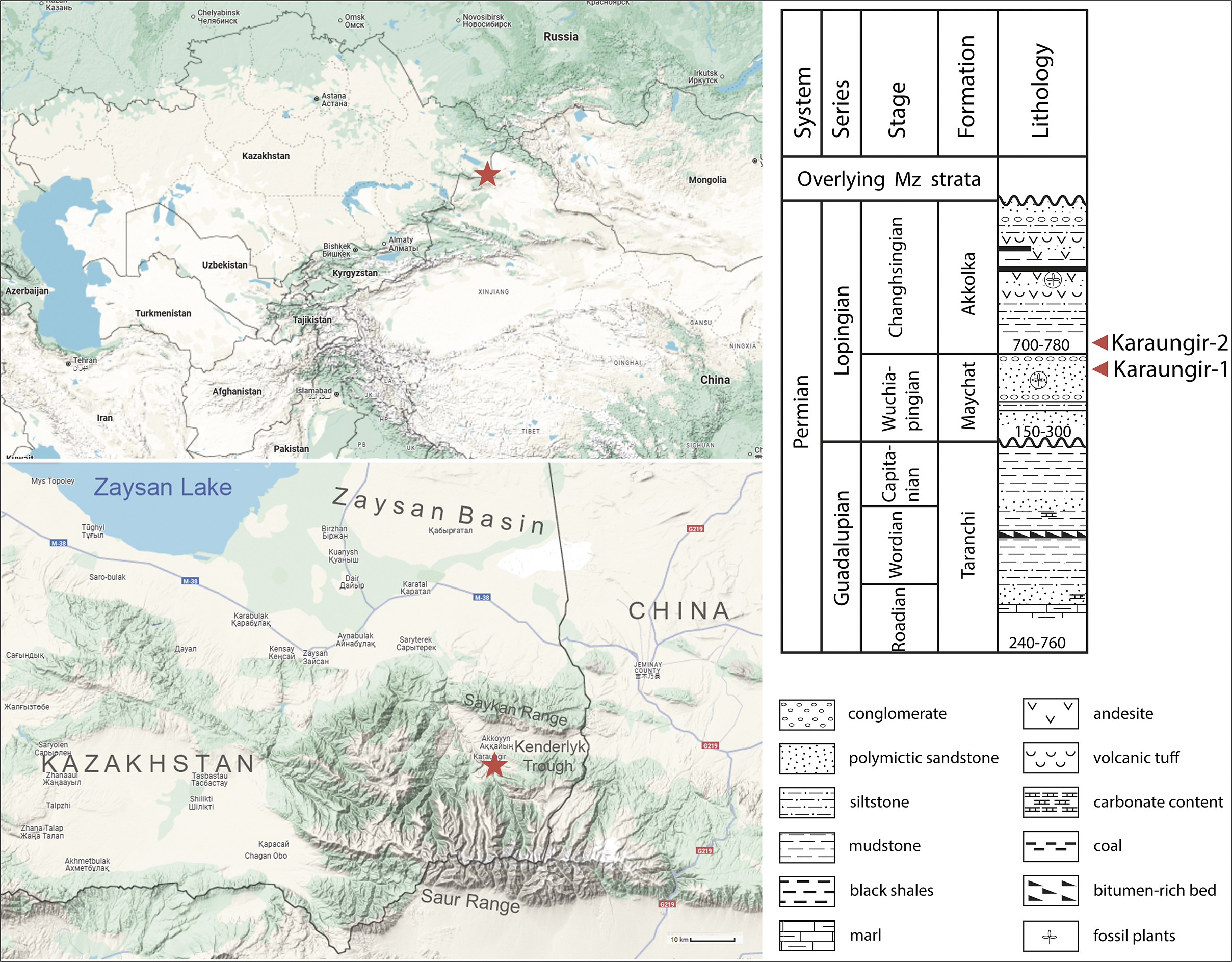
The fossils originate from the right bank of the Karaungir River, the Saur Range, eastern Kazakhstan, where the river valley cuts through upper Permian strata (Fig. 1). There, two localities, named Karaungir-1 and -2, yielded numerous oligochaete cocoons. The latter are preserved as organic compressions on bedding planes of the upper mudstone-siltstone member of the Maychat Formation (Karaungir-1) and the lower member of the Akkolka Formation of the same composition (Karaungir-2) separated by 20 m of sediment (Smirnov, Reference Smirnov1970). These coal-bearing formations were dated as upper Permian (Lopingian, ~254 Ma) based on regional stratigraphy and the assemblage of fossil plants, spinicaudatans, and insects (Ponomarenko, Reference Ponomarenko1969; Novojilov, Reference Novojilov1970; Vakhrameev et al., Reference Vakhrameev, Dobruskina, Zaklinskaja and Meyen1970; Krassilov, Reference Krassilov1999; Shcherbakov, Reference Shcherbakov2000, Reference Shcherbakov2024; Nurbekova et al., Reference Nurbekova, Smirnova, Goncharev, Sachsenhofer, Hazlett, Smirnov, Yensepbayev, Mametov and Fustic2023). In some publications, a middle Permian age was suggested for the paleoentomofauna of Karaungir based on the relatively high proportion of ancient insect families (Rasnitsyn et al., Reference Rasnitsyn, Aristov and Rasnitsyn2013). This suggestion is untenable because primitive forms persist commonly as relicts in refugia with milder climates, and this effect was especially strong during intervals of pronounced climatic zonation, e.g., the Permian (Krassilov, Reference Krassilov1999; Shcherbakov, Reference Shcherbakov2008).
Geographic (star) and stratigraphic (arrows) locations of the Karaungir Lagerstätte, Kenderlyk Trough, Zaysan Basin, eastern Kazakhstan (modified: basemaps after Google Maps, section after Korobkin et al., Reference Korobkin, Chaklikov, Ismailov and Tulemissova2023).

Figure 1. Long description
The graphic is divided into three main sections.
1. Top-Left Map: A regional map of Central Asia showing Kazakhstan and neighboring countries. A red star marks the study site in eastern Kazakhstan, near the borders of Russia, Mongolia, and China.
2. Bottom-Left Map: A detailed topographic map of the Zaysan Basin. Zaysan Lake is in the Northwest. The Saykan Range and Saur Range are in the Southeast. A red star marks the Karaungir site within the Kenderlyk Trough, located between these mountain ranges near the Chinese border.
3. Right Panel: A stratigraphic column and lithology legend.
- The column is organized by System (Permian), Series (Guadalupian and Lopingian), Stage, Formation, and Lithology.
- The Lopingian series includes the Changhsingian stage (Akkolka Formation) and Wuchiapingian stage (Maychat Formation).
- The Guadalupian series includes the Capitanian, Wordian, and Roadian stages (Taranchi Formation).
- Two red arrows point to specific fossil horizons: Karaungir-2 is located in the lower Akkolka Formation (700-780 meters), and Karaungir-1 is located in the upper Maychat Formation (150-300 meters).
- The lithology column shows a sequence of conglomerate, sandstone, siltstone, and mudstone with specific markers for coal, volcanic tuff, and fossil plants.
- The legend at the bottom right defines symbols for conglomerate, polymictic sandstone, siltstone, mudstone, black shales, marl, andesite, volcanic tuff, carbonate content, coal, bitumen-rich bed, and fossil plants.
The 2 km thick Carboniferous-Permian succession, including the Maychat and the lower Akkolka formations, were accumulated in the Kenderlyk Trough of the Zaysan Basin bordered by the Rudny Altay and Zharma-Saur volcanic arcs to the northwest and southeast, respectively (Nurbekova et al., Reference Nurbekova, Smirnova, Goncharev, Sachsenhofer, Hazlett, Smirnov, Yensepbayev, Mametov and Fustic2023). This continental basin was formed during the Pennsylvanian-Permian rotational collision of the Kazakh microcontinent and Siberia (Buslov et al., Reference Buslov, Watanabe, Fujiwara, Iwata, Smirnova, Safonova, Semakov and Kiryanova2004; Liu et al., Reference Liu, Chen, Ma, Liu, Gong, Shi and Han2018). During this interval, a transition occurred from shallow marine to brackish-lagoonal and finally to freshwater-brackish-saline lacustrine and fluviodeltaic facies of the Maychat and the lower Akkolka formations (Nurbekova et al., Reference Nurbekova, Smirnova, Goncharev, Sachsenhofer, Hazlett, Smirnov, Yensepbayev, Mametov and Fustic2023). The Maychat Formation comprises 150–300 m of organic-rich mudstone interbedded with coal in its lower part and a coarsening upward succession of siltstone-sandstone-conglomerate cycles overlain by thin light gray to black shale, coal, and volcanic ash beds in its upper part (Novojilov, Reference Novojilov1970; Smirnov et al., Reference Smirnov, Nurbekova, Fustic, Mathews, Hazlett, Mametov and Yensepbayev2022; Nurbekova et al., Reference Nurbekova, Smirnova, Goncharev, Sachsenhofer, Hazlett, Smirnov, Yensepbayev, Mametov and Fustic2023). Variable pristane/phytane ratios (<1–>3) and abundant β-carotane suggest a frequent alternation of redox conditions, from anoxic to oxic, and changes of the salinity regime from freshwater to brackish (Zhu et al., Reference Zhu, Shen, Zhao, Hu, Ge and Wang2022; Nurbekova et al., Reference Nurbekova, Smirnova, Goncharev, Sachsenhofer, Hazlett, Smirnov, Yensepbayev, Mametov and Fustic2023). The Maychat Formation is overlain by the Akkolka Formation (700–780 m thick) consisting in its lower part of a rhythmic alternation of siltstone and calcareous mudstone grading into conglomerates and further into coal-bearing strata (Kumpan et al., Reference Kumpan, Dobretsov and Mitrofanova1969; Korobkin et al., Reference Korobkin, Chaklikov, Ismailov and Tulemissova2023). The brackish rather than entirely freshwater condition is inferred for the depositional environments of this formation by the depauperated fauna of freshwater bivalves—only Abiella subovata (Jones, Reference Jones1901)—and by the presence of calcareous mudstone (Betekhtina, Reference Betekhtina1966; Kumpan et al., Reference Kumpan, Dobretsov and Mitrofanova1969).
The cocoons were accompanied by numerous insects as well as by clam shrimp carapaces (spinicaudatans) and bivalve mollusk shells (Smirnov, Reference Smirnov1970). The assemblage comprises 14 insect orders, including beetles, cicadas, scorpionflies, stoneflies, caddisflies, lacewings, protorthopterans, and miomopterans (Shcherbakov, Reference Shcherbakov2024).
Due to the occurrence of a number of complete specimens preserving fine morphological details and soft tissues (e.g., clitellate cocoons containing eggs), the Karaungir-1 and -2 localities can be classified as Konservat-Lagerstätten (e.g., Kimmig and Schiffbauer, Reference Kimmig and Schiffbauer2024). In summary, the cocoons under study were laid under fresh- to brackish, calm lacustrine conditions in the eastern Kazakhstan part of northern Pangaea by the end of the Permian.
Materials and methods
Numerous fossil cocoons (~90 specimens) were collected in the 1960s by field parties of the Arthropoda Laboratory, Paleontological Institute, USSR Academy of Sciences, headed by Nestor I. Novojilov (1907–1992) in the upper Permian (Lopingian) Maychat and Akkolka formations. They occurred in the localities Karaungir-1 and 2, on the right bank of the Karaungir River, 0.4–0.6 km downstream from the Maychat Creek, 50 km east-southeast of the town of Zaysan, northern slope of the Saur Range, eastern Kazakhstan. The fossils are preserved on a bedding surface of a brownish-green, polymictic, microwavy-laminated siltstone.
The specimens were photographed using a Leica M165C stereomicroscope equipped with a Leica DFC425 digital camera. High-resolution images of both gold-coated and uncoated specimens were taken with a Tescan VEGA scanning electron microscope (SEM), using back-scattered electron (BSE) and secondary electron (SE) detectors at the Borissiak Paleontological Institute, Russian Academy of Sciences. Elemental analysis of an uncoated, unpolished specimen, containing both the fossil and the adjacent matrix, was performed with a quantitative energy dispersive X-ray (EDX) INCA microanalyser coupled to the same SEM, at an accelerating voltage of 15 keV.
For comparison, two cocoons of the extant freshwater microdrile oligochaete Rhynchelmis (Rhynchelmis) tetratheca Michaelsen, Reference Michaelsen1920 (Lumbriculida: Lumbriculidae) were collected at Aravete Spring and Lake Peipsi (Chudskoe), Estonia, and fixed in 70% ethanol. One of them was critical point dried with CO2, covered with a palladium-gold alloy and examined in the same SEM.
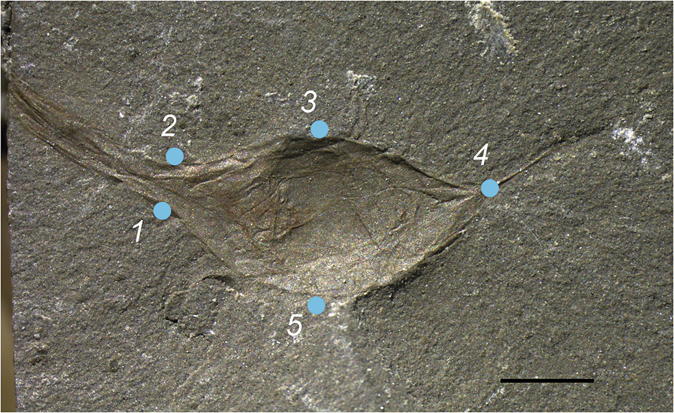
Landmark-based geometric morphometrics was applied to determine whether all of the Karaungir cocoon compressions represented a continuous morphospace occupation or were subdivided into clusters. We compared the capsule variations in shape for a total of 60 Permian compressions and six cocoons of three extant microdrile species—two above-mentioned cocoons of Rhynchelmis tetratheca and four cocoons of Rhynchelmis limosella Hoffmeister, Reference Hoffmeister1843, Rhynchelmis tetratheca, and Tatriella slovenica Hrabĕ, Reference Hrabĕ1939 figured earlier (Chekanovskaya, Reference Chekanovskaya1962; Timm, Reference Timm1970; Timm and Vvedenskaya, Reference Timm and Vvedenskaya2006). tpsUtil ver. 1.82 program (Rohlf, Reference Rohlf2015) was used to compile the database. The sliding semilandmarks (SSL) technique was applied to analyze the shape of the cocoons because this technique is suitable for describing the shape with a continuous morphospace occupation, although, instead of the semilandmarks, conventional landmarks were used (Zelditch et al., Reference Zelditch, Swiderski, Sheets and Fink2004). To each specimen image, five landmarks were digitally added using the program tpsDig2 ver. 2.32 (Rohlf, Reference Rohlf2015; Fig. 2). All compressions and cocoons were uniformly oriented (the stalk to the right, the more convex side downward) and outlined clockwise from left to right. Landmarks 1, 2, 3, and 5 were attributed to type 2 landmarks; landmark 4 was defined as type 1 (Weber and Bookstein, Reference Weber and Bookstein2011; Smith and Wilson, Reference Smith and Wilson2017). To fix coordinates of each landmark for the subsequent Procrustes analysis, an auxiliary file ‘Link file.tps’ defining the number of semilandmarks to be analyzed was created (Voyta et al., Reference Voyta, Golenishchev and Tiunov2019). Then, the program tpsRelw ver. 1.53 (Rohlf, Reference Rohlf2003) was used to perform Procrustes analysis.
Cocoon of the late Permian microdrile worm Archedaphnia kazakhstanica Smirnov, Reference Smirnov1970 sensu lato from the Karaungir Lagerstätte, Zaysan Basin, eastern Kazakhstan, PIN 2500/571, with five landmarks added (1, 2, 3, 5 = type 2 landmarks; 4 = type 1 landmark). Scale bar = 1 mm.

All specimen data were compiled into a single ‘*.tps’ database and analyzed in tpsRelw ver. 1.53 (Rohlf, Reference Rohlf2003). The Procrustes-aligned landmark data were then subjected to principal component analysis (PCA) and canonical variate analysis (CVA) using the cross-validation jackknife method performed in the program PAST ver. 3.15 (Hammer et al., Reference Hammer, Harper and Ryan2001). The morphospace for cocoon compressions and extant cocoons was constructed in the program PAST ver. 3.15, and images of morphotypes (wireframes on transformation grids) for specific specimens were constructed in the program tpsRelw ver. 1.53.
The terminology used follows Evans and Guild (Reference Evans and Guild1947), Christensen (Reference Christensen1956), Piearce et al. (Reference Piearce, Oates and Carruthers1992), Dimitriu et al. (Reference Dimitriu, Sayers, Coleman and Shain2006), Coleman et al. (Reference Coleman, Marotta and Shain2008), and Romdhane et al. (Reference Romdhane, Quilichini and Tekaya2017).
Repository and institutional abbreviation
The examined specimens of Archedaphnia kazakhstanica are deposited in collections of the Borissiak Paleontological Institute, Russian Academy of Sciences (PIN), Moscow, Russia, nos. 2499 (Maychat Formation) and 2500 (Akkolka Formation).
Systematic paleontology
Phylum Annelida Lamarck, Reference Lamarck1802
Class Clitellata Michaelsen, Reference Michaelsen1919
Subclass Oligochaeta Grube, Reference Grube1850
Genus Archedaphnia Smirnov, Reference Smirnov1970
Reference Smirnov1970 Archedaphnia Smirnov, p. 96, pl. 7, fig. 1, text-fig. 1.
Reference Smirnov1970 Propleuroxus Smirnov, p. 100, pl. 7, fig. 4, text-fig. 5; n. syn.
Type species
Archedaphnia kazakhstanica Smirnov, Reference Smirnov1970.
Diagnosis (emended)
Small ovoid attached membranous cocoons with two long, striated, polar extensions, shorter narrow stalk, and longer, broader neck with tattered edge; monotypic.
Remarks
In the description of these fossils by Smirnov (Reference Smirnov1970), a bivalved carapace was recognized, although in fact each fossil is a single, hollow, variably wrinkled object. The alleged ‘caudal needles’ are stalks, the ‘helmets’ are necks, and the ‘ephippia’ are eggs. The appearance of the cocoons, similar to that of spinicaudatan carapaces and insect wings and bodies found in the same layers, suggests their original rigid structure rather than a gelatinous or membranous one.
Archedaphnia kazakhstanica Smirnov, Reference Smirnov1970
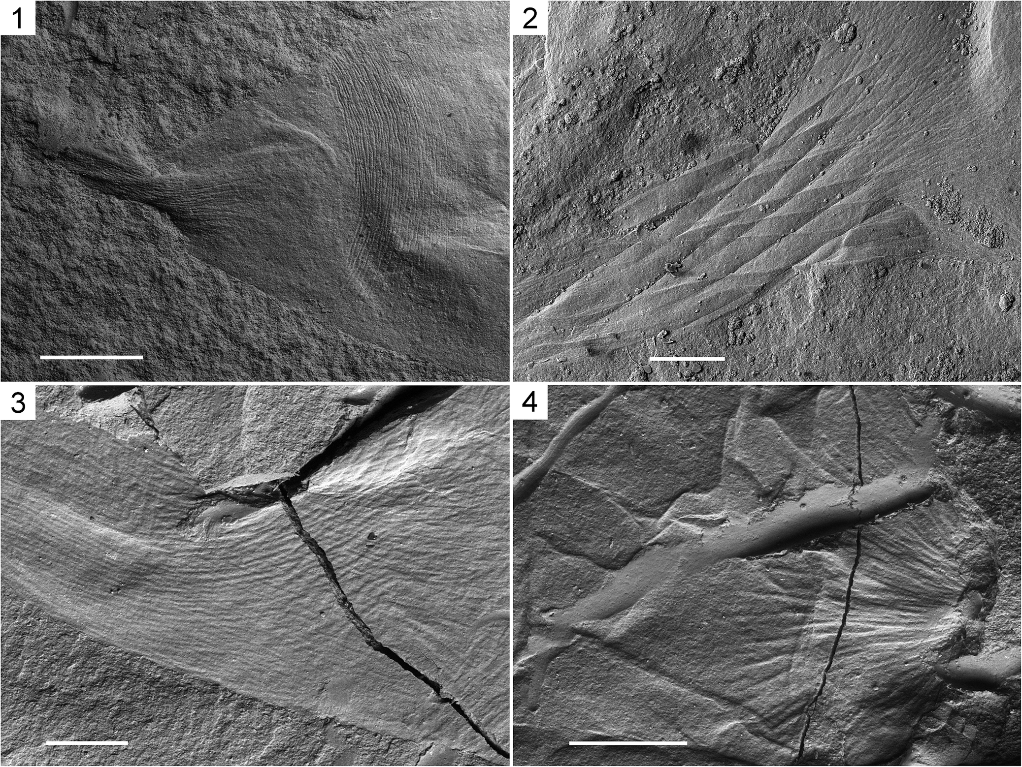
Cocoons of the late Permian microdrile worm Archedaphnia kazakhstanica Smirnov, Reference Smirnov1970 sensu lato from the Karaungir Lagerstätte, Zaysan Basin, eastern Kazakhstan, SEM, BSE, uncoated (1, 3, 4) or gold-coated (2): (1) PIN 2500/279, Archedaphnia borutzkyi Smirnov, Reference Smirnov1970, holotype, twisted neck with longitudinal ridges and the adjacent part of the capsule with transverse striation; (2) PIN 2500/556, specimen figured by Van Damme and Kotov (Reference Van Damme and Kotov2016, fig. 3A–D), strongly twisted neck; (3, 4) PIN 2500/567: (3) neck with longitudinal ridges, twisted at the base; (4) stalk base with convergent ridges. Scale bars = 0.5 mm (1); 0.2 mm (2–4).

Figure 3. Long description
A four-panel set of S E M micrographs.
Panel 1, top-left: A B S E micrograph of the holotype P I N 2500 forward slash 279. It shows a twisted neck featuring distinct longitudinal ridges that transition into an adjacent capsule area marked by fine transverse striations. A scale bar representing 0.5 mm is at the bottom-left.
Panel 2, top-right: A gold-coated specimen P I N 2500 forward slash 556. This view highlights a strongly twisted neck structure with overlapping, elongated folds. A scale bar representing 0.2 mm is at the bottom-center.
Panel 3, bottom-left: Specimen P I N 2500 forward slash 567 showing a neck with prominent longitudinal ridges that are twisted at the base. A large diagonal crack runs through the fossilized material. A scale bar representing 0.2 mm is at the bottom-left.
Panel 4, bottom-right: Another view of specimen P I N 2500 forward slash 567, focusing on the stalk base. The surface features convergent ridges that meet at a central point. A scale bar representing 0.2 mm is at the bottom-left.
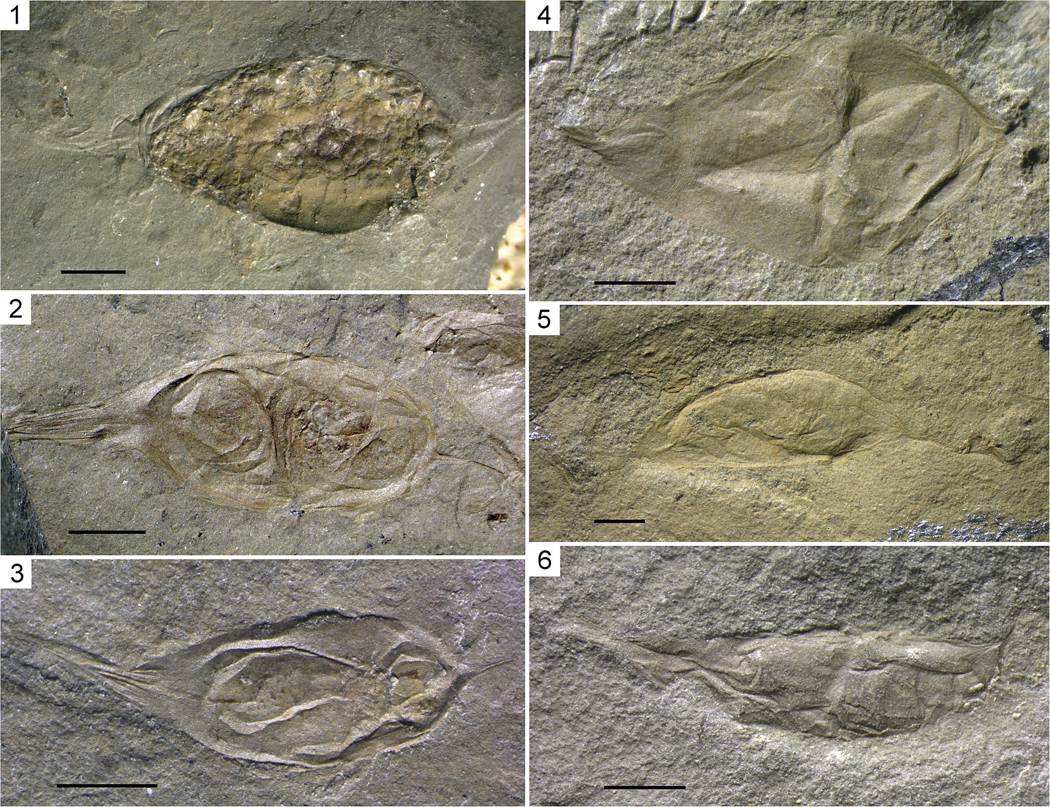
Cocoons of the late Permian microdrile worm Archedaphnia kazakhstanica Smirnov, Reference Smirnov1970 sensu lato from the Karaungir Lagerstätte, Zaysan Basin, eastern Kazakhstan, type specimens of four species described by Smirnov: (1, 2) Archedaphnia kazakhstanica: (1) holotype, PIN 2500/282; (2) paratype, PIN 2500/542; (3) Archedaphnia novojilovi Smirnov, Reference Smirnov1970, paratype, PIN 2499/206; (4) Archedaphnia borutzkyi Smirnov, Reference Smirnov1970, holotype, PIN 2500/279; (5, 6) Archedaphnia strelkovi Smirnov, Reference Smirnov1970: (5) holotype, PIN 2500/543; (6) paratype, PIN 2499/205. Necks on the left, stalks on the right. Scale bars = 1 mm.

Figure 4. Long description
The six panels show fossilized cocoons of Archedaphnia species embedded in grey and tan rock matrix. Each panel includes a black scale bar representing 1 mm in the bottom-left corner.
* Panel 1: Holotype of Archedaphnia kazakhstanica, P I N 2500 forward slash 282. It has a bumpy, textured surface and a tapered neck on the left.
* Panel 2: Paratype of Archedaphnia kazakhstanica, P I N 2500 forward slash 542. The cocoon appears more translucent with visible internal folds and a long, striated neck.
* Panel 3: Paratype of Archedaphnia novojilovi, P I N 2499 forward slash 206. This specimen shows a distinct inner layer or wall within the main cocoon body.
* Panel 4: Holotype of Archedaphnia borutzkyi, P I N 2500 forward slash 279. It has a broad, diamond-shaped body with sharp creases and a short neck.
* Panel 5: Holotype of Archedaphnia strelkovi, P I N 2500 forward slash 543. This cocoon is more elongated and smooth, appearing in a lighter tan matrix.
* Panel 6: Paratype of Archedaphnia strelkovi, P I N 2499 forward slash 205. The specimen is highly elongated with a wrinkled texture and a thin, trailing stalk on the right.
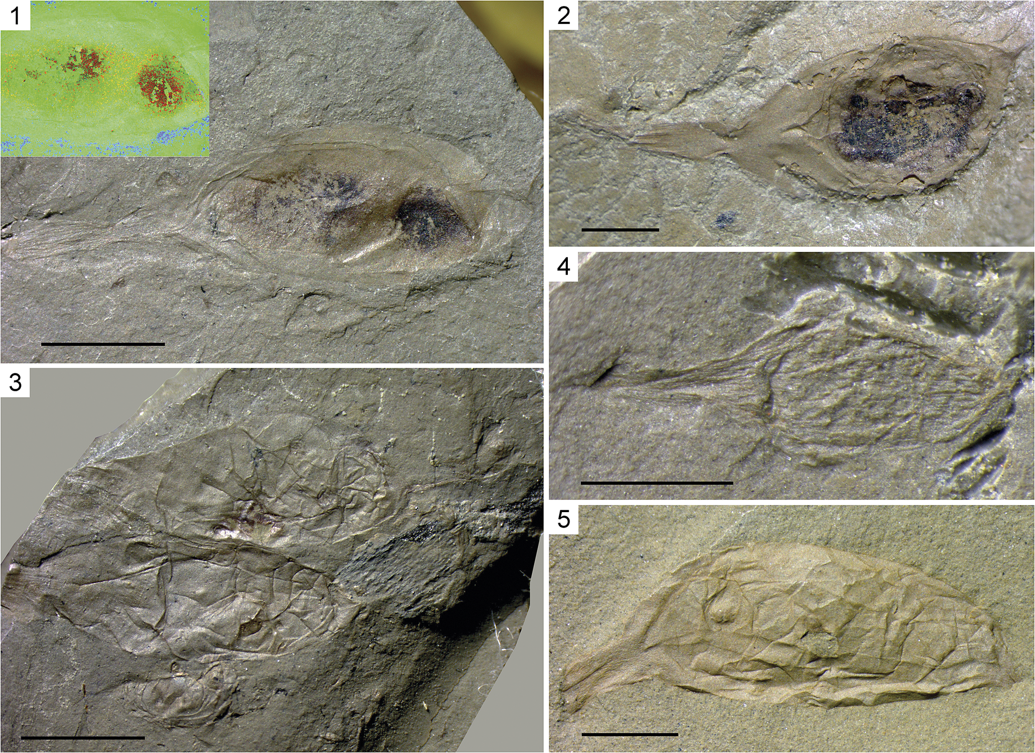
Cocoons of the late Permian microdrile worm Archedaphnia kazakhstanica Smirnov, Reference Smirnov1970 sensu lato from the Karaungir Lagerstätte, Zaysan Basin, eastern Kazakhstan: (1) PIN 2500/552a, cocoon with two eggs; inset, capsule in artificial colors (red = carbon, green = iron, blue = calcium), EDX; (2) PIN 2500/557, cocoon with one egg; (3) PIN 2500/541a and b, paratypes of Archedaphnia kazakhstanica, two cocoons of similar size and shape, each with a small dark object inside, preserved closely parallel, stalks directed toward a plant fragment; (4) PIN 2500/561, very small abortive cocoon; (5) PIN 2500/566, pale empty cocoon. Necks on the left, stalks on the right. Scale bars = 2 mm (1, 3); 1 mm (2, 5); 0.5 mm (4).

Figure 5. Long description
A collection of five macro photos of fossilized microdrile worm cocoons preserved in rock. All specimens are oriented with the neck on the left and the stalk on the right.
* Panel 1: A large cocoon containing two distinct dark oval eggs. An inset in the top-left shows an E D X map where red indicates carbon, green indicates iron, and blue indicates calcium. Scale bar is 2 mm.
* Panel 2: A cocoon containing a single large dark egg. Scale bar is 1 mm.
* Panel 3: Two paratype cocoons of similar size and shape preserved parallel to each other. Each contains a small dark object. Their stalks point toward a plant fragment on the right. Scale bar is 2 mm.
* Panel 4: A significantly smaller, abortive cocoon with a wrinkled texture. Scale bar is 0.5 mm.
* Panel 5: A pale, empty cocoon with a highly textured, folded surface. Scale bar is 1 mm.
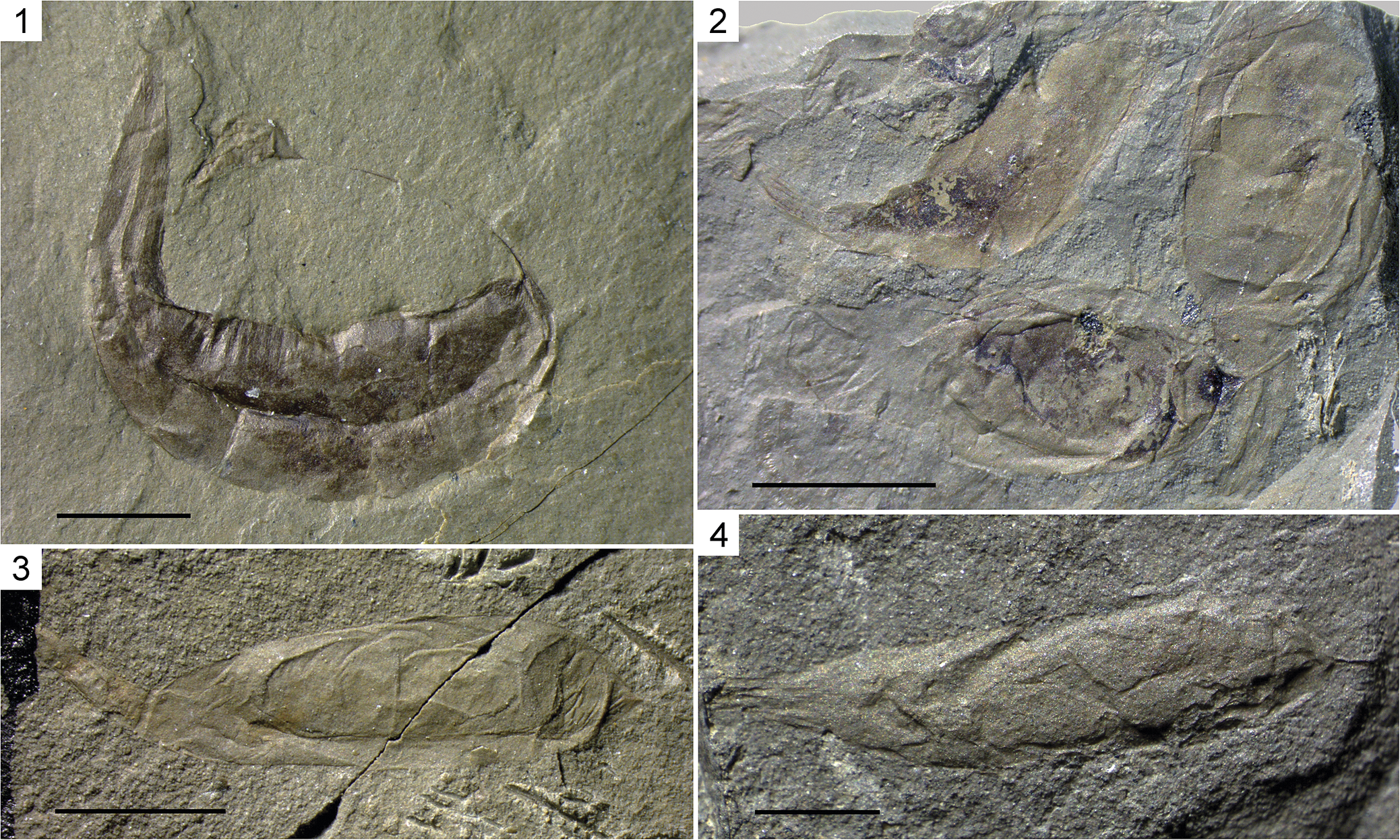
Cocoons of the late Permian microdrile worm Archedaphnia kazakhstanica Smirnov, Reference Smirnov1970 sensu lato from the Karaungir Lagerstätte, Zaysan Basin, eastern Kazakhstan: (1) PIN 2500/563, holotype of Propleuroxus freyi Smirnov, Reference Smirnov1970, strongly curved cocoon with a widened neck; (2) PIN 2500/550, three cocoons, the upper left one curved, with a dark object inside the neck, the lower left one with a very wide neck; (3) PIN 2500/278, paratype of Archedaphnia kazakhstanica; (4) PIN 2500/573, one of the most elongated cocoons. Necks on the left, stalks on the right. Scale bars = 2 mm (2, 3); 1 mm (1, 4).

Figure 6. Long description
A four-panel photo collage of fossilized microdrile worm cocoons embedded in grey rock matrix.
* Panel 1 (top-left): P I N 2500/563, the holotype of Propleuroxus freyi. It shows a single, strongly curved, dark brown cocoon. The neck on the left is widened, and the body curves downward and back up toward the stalk on the right. A 1 mm scale bar is at the bottom left.
* Panel 2 (top-right): P I N 2500/550, containing three cocoons. The upper left specimen is curved with a dark object visible inside its neck. The lower left specimen features a notably wide neck. A 2 mm scale bar is at the bottom left.
* Panel 3 (bottom-left): P I N 2500/278, a paratype of Archedaphnia kazakhstanica. The cocoon is more elongated and horizontal, with a visible crack in the rock matrix running diagonally through the right side of the specimen. A 2 mm scale bar is at the bottom left.
* Panel 4 (bottom-right): P I N 2500/573, showing a highly elongated, slender cocoon with a textured surface. The neck is on the left and the stalk on the right. A 1 mm scale bar is at the bottom left.
Reference Smirnov1970 Archedaphnia kazakhstanica Smirnov, p. 96, pl. 7, fig. 1, text-fig. 1.
Reference Smirnov1970 Archedaphnia novojilovi Smirnov, p. 98, pl. 7, fig. 2, text-fig. 2; n. syn.
Reference Smirnov1970 Archedaphnia borutzkyi Smirnov, p. 98, pl. 7, fig. 3, text-fig. 3; n. syn.
Reference Smirnov1970 Archedaphnia strelkovi Smirnov, p. 98, text-fig. 4; n. syn.
Reference Smirnov1970 Propleuroxus freyi Smirnov, p. 100, pl. 7, fig. 4, text-fig. 5; n. syn.
Reference Van Damme and Kotov2016 Archedaphnia sp. indet., Van Damme and Kotov, fig. 3A–D.
Type specimens
Holotype, PIN 2500/282 (Fig. 4.1), Akkolka Formation, eastern Kazakhstan, Saur Range, right bank of the Karaungir River, 0.4–0.5 km downstream from Maychat Creek, Karaungir-2, 47.260°N, 85.410°E. Paratypes, PIN 2499/204, 2500/278 (Fig. 6.3), 2500/541a and b (Fig. 5.3), 2500/542 (Fig. 4.2), 2500/549, Maychat and Akkolka formations, eastern Kazakhstan, Saur Range, right bank of the Karaungir River, 0.4–0.6 km downstream from Maychat Creek, Karaungir-1 and -2.
Occurrence
Karaungir-1, 47.261°N, 85.409°E and Karaungir -2, 47.260°N, 85.410°E, right bank of the Karaungir River, 0.4–0.6 km downstream of Maychat Creek, Saur Range, eastern Kazakhstan; Maychat and Akkolka formations, Lopingian.
Description
Ovoid, slightly elongated, wrinkled membranous cocoons with prominently striated neck and stalk at the ends (Figs. 3, 4). The capsule length between the neck and the stalk bases varies from 1.6–8.5 mm; its diameter 0.5–2 mm. The neck is long (to 3.6 mm), constricted at the base or not, bearing a terminal opening with a distinct, irregular margin; the neck is usually fluted and sometimes twisted, with diamond-shaped reticulation (Fig. 4.1–4.3). The stalk is very narrow, to 2 mm long, rarely preserved completely, with a terminal expansion attached to the substratum in life (Figs. 4.5, 6.1). Both neck and stalk bear fine longitudinal ridges (~10 μm in width) oriented along the long axis of the cocoon (Fig. 3.3, 3.4). In well-preserved specimens, the stalk is separated from rest of the capsule by a beltlike area of fine transverse ridges (Fig. 3.1). Toward the attachment point, these ridges become thickened and more widely spaced. On some cocoons, both transverse and longitudinal striae are visible on the entire capsule, although not as distinct as in the polar areas.
The number of eggs in a cocoon varies from one to two, and one cocoon appears to contain a worm ready to hatch (Figs. 5.1, 5.2, 6.2 top left). In some other cocoons, clutch-like masses of small, rounded objects are visible (Fig. 4.1).
Remarks
The cocoons were preserved as compressions on the siltstone bedding planes, without distinct orientation. EDX analysis revealed that their organic membranes are either destroyed or replicated by clay minerals, containing silica, aluminium, and iron; magnesium and potassium are less common. In contrast, the eggs are preserved as carbonaceous films due to kerogenization of organic precursors (Fig. 5.1, inset).
In this regard, these fossilized cocoons are similar to the carbonaceous/aluminosilicified fossils of the Burgess Shale-type that were formed by replication of organic matter in clay minerals due to the presence of organic adhesion molecules, which enhance ion binding and the recalcitrance of relatively labile organic substrata (e.g., Orr et al., Reference Orr, Briggs and Kearns1998; Wilson and Butterfield, Reference Wilson and Butterfield2014; Naimark et al., Reference Naimark, Kirpotin, Boeva, Gmoshinskiy and Kalinina2020). In the Karaungir localities, the factors facilitating this process could be bottom water stagnation and adhesive ash layers (Smirnov et al., Reference Smirnov, Nurbekova, Fustic, Mathews, Hazlett, Mametov and Yensepbayev2022; Nurbekova et al., Reference Nurbekova, Smirnova, Goncharev, Sachsenhofer, Hazlett, Smirnov, Yensepbayev, Mametov and Fustic2023). These factors provided conditions suitable for a rapid, organic-matter mineralization and early-diagenetic sealing of sediments similar to some famous Konservat-Lagerstätten, e.g., the Lower Cretaceous Jehol Biota (Jiang et al., Reference Jiang, Harlow, Wohletz, Zhou and Meng2014). A fine undisturbed lamination of the host rocks is indicative of the absence of animal scavenging, bioturbation, and sediment processing, which further facilitated the exceptional preservation of the cocoons.
Smirnov (Reference Smirnov1970) described from Karaungir four species of Archedaphnia, differing in length/width ratio, shape of the neck base (constricted or gradually tapering), and symmetry (extensions axial or displaced to one side), as well as the monotypic genus Propleuroxus, based on one elongate, strongly bent cocoon. Oligochaete cocoons assume their definitive shapes only after deposition and their variation remains poorly studied. Therefore, we synonymize all five cocoon species from Karaungir, although we admit that these might belong to more than one oligochaete species.
Comparative description of cocoons produced by extant Rhynchelmis (Rhynchelmis) tetratheca
Description
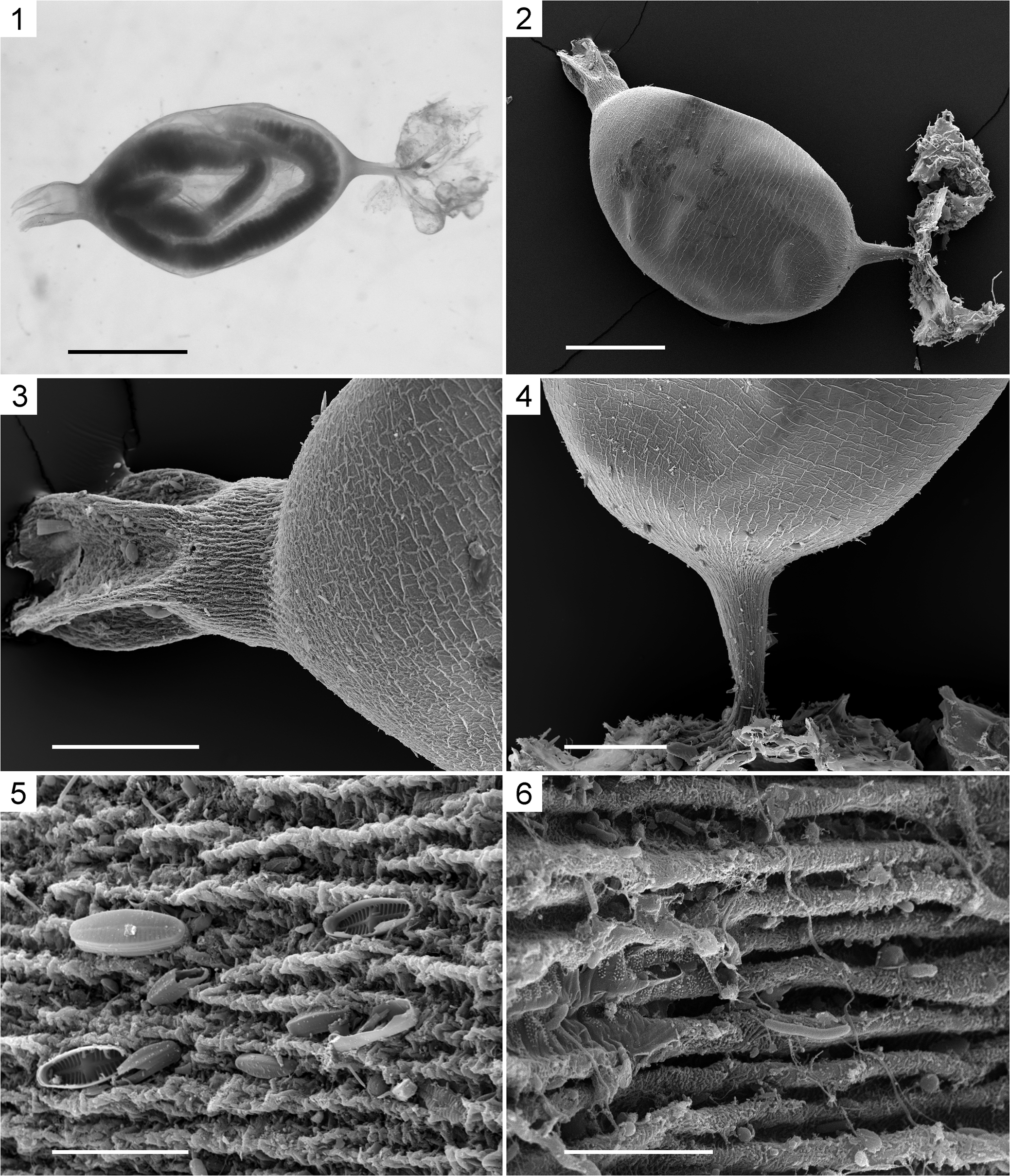
Golden-green, translucent, ovoid, slightly elongated, wrinkled membranous cocoons with prominent polar tufts/extensions (Fig. 7.1, 7.2). The capsule varies from 1.4–2.1 mm in length between the bases of extensions and from 0.9–1.2 mm in diameter. The external surface of the capsule membrane is wrinkled mostly transversely in the middle and longitudinally toward the poles imparting to these areas an irregular reticulate pattern (Fig. 7.3, 7.4). The wrinkles are loosely spaced, discontinuous, ridge-like (150–1000 μm in length and ~2 μm in height). The anterior extension (stalk), attached to the substratum, is narrow (0.12 mm in diameter at the base), tapering toward the attachment point (Fig. 7.4). The opposite, posterior extension (neck), 0.2 mm in diameter, is constricted at the base and fluted (Fig. 7.3). Both extensions are sculptured by fine longitudinal protuberances. The stalk and neck protuberances are parallel to the long axis of the cocoon, converging toward the apex on the stalk (Fig. 7.3, 7.4). On the neck, the protuberances (2–4 μm in width) are continuous, loosely spaced (4–6 μm apart), consisting of dense, equidistantly arranged, subtriangular thickenings, which are gently inclined toward the apex (Fig. 7.5). In turn, these subtriangular thickenings are covered with fine papillae, ~1 μm in diameter. The stalk protuberances are densely spaced (0.5–0.7 μm apart), wavy, continuous, rope-like strands (~1 μm in width) consisting of tiny fibrils (< 0.1 μm in diameter) (Fig. 7.6). Toward the attachment point, these strands are thickened and the spaces between them are widened and filled with fine transverse lintels. At the attachment point, they form thick root-like tendons (~10 μm in width) spreading over the substratum and anchored to it.
Cocoons of the Recent freshwater lumbriculid microdrile worm Rhynchelmis tetratheca Michaelsen, Reference Michaelsen1920 from Estonia: (1) cocoon with two young worms inside, light photograph; (2–6) cocoon with two eggs, SEM, SE, gold-coated: (2) general view; (3) neck; (4) stalk; (5, 6) longitudinal protuberances on the neck (5) and stalk (6) with adherent frustules of pennate diatoms. Scale bars = 1 mm (1); 0.5 mm (2); 0.2 mm (3, 4); 20 μm (5); 5 μm (6).

Figure 7. Long description
A multi-panel figure containing one light photograph and five S E M micrographs.
* Panel 1: A light photograph of a translucent, oval-shaped cocoon. Inside, two dark, coiled young worms are visible. The cocoon has a short neck on the left and a thin stalk on the right. Scale bar is 1 millimeter.
* Panel 2: An S E M micrograph showing the general external view of a cocoon. The surface appears textured. It features a distinct neck at the top-left and a stalk at the bottom-right attached to debris. Scale bar is 0.5 millimeters.
* Panel 3: A high-magnification S E M view of the cocoon neck. The surface transitions from a cracked, tile-like pattern on the main body to dense, longitudinal ridges on the neck. Scale bar is 0.2 millimeters.
* Panel 4: A high-magnification S E M view of the cocoon stalk. The stalk is thin and tapers from the main body, showing similar longitudinal ribbing. Scale bar is 0.2 millimeters.
* Panel 5: A close-up S E M view of the longitudinal protuberances on the neck. Several oval-shaped pennate diatom frustules are attached to the ridged surface. Scale bar is 20 micrometers.
* Panel 6: An extreme close-up S E M view of the stalk surface. The parallel longitudinal ridges are highly detailed, with small organic particles and diatom fragments caught between the folds. Scale bar is 5 micrometers.
Materials
Two specimens from the Aravete Spring, 59.149°N, 25.750°E (dried and gold-coated) and Lake Peipsi (Chudskoe), station 58 near the town of Mustvee, 58.814°N, 27.033°E, Estonia; Recent.
Remarks
One cocoon contained two fully developed segmented young worms, with a pharyngeal bulb, ready to hatch (Fig. 7.1), and the other contained two embryos at different developmental stages. Numerous diatoms and other freshwater organisms adhering to the cocoons prove that these had once been sticky.
The protuberances covering the neck and stalk are similar, overall and in detail, to those of some leech cocoons (Dimitriu et al., Reference Dimitriu, Sayers, Coleman and Shain2006). Thus, a proteinaceous composition and twisted helicoid fibrillar ultrastructure are inferred for the cocoon wall of Rhynchelmis (Rhynchelmis) tetratheca.
Variation of Karaungir cocoons
All of the fossil cocoons from Karaungir are similar to those of the extant genus Rhynchelmis (Lumbriculidae) in having an ovoid to fusiform capsule, a long thin stalk, and a long wider neck, tattered at the apex. In another genus of this family, Stylodrilus Claparède, Reference Claparède1862, both extensions are long and rather wide. In all other microdrile genera for which cocoons have been described, these are ovoid to fusiform with rudimentary extensions; however, cocoons are not yet known for most oligochaete taxa.
The Karaungir cocoons were misinterpreted as Cladocera and classified according to their shape into four species of Archedaphnia (Daphniidae) and a monotypic genus Propleuroxus (Chydoridae) by Smirnov (Reference Smirnov1970). The 14 specimens, assigned by him to Archedaphnia kazakhstanica, Archedaphnia novojilovi, and Archedaphnia borutzkyi (Fig. 4.1–4.4), have a pale, ovoid, more or less elongated capsule (2.6–6.1 mm long, aspect ratio 1.6–3.3:1); sometimes such cocoons contain one large, dark, rounded egg or, rarely, two eggs (Fig. 5.1, 5.2). Usually, the capsule has its maximum width near the midlength, but sometimes closer to the stalk or the neck (varies in extant species, e.g., Rhynchelmis tetratheca). These three species were distinguished by the length/width ratio and the presence of a constriction at the base of the neck (the neck was misinterpreted as a cladoceran helmet and the egg as an ephippium; Smirnov, Reference Smirnov1970). However, the aspect ratio shows considerable variability in Rhynchelmis limosella (cf. Chekanovskaya, Reference Chekanovskaya1962, fig. 57; Timm, Reference Timm1970, fig. 20) and Rhynchelmis tetratheca (3.20–4.90 x 0.61–1.70 mm; Timm, Reference Timm1970), the number of eggs per cocoon varies in most microdrile species, and the capsule length varies more than twofold in some species (variation studied in only a few species; Arkhipova, Reference Arkhipova and Shilova1989). Moreover, one of the numerous cocoons from Karaungir that was not assigned to any species by Smirnov (Reference Smirnov1970) has an elongated, slightly curved capsule with a large dark object partially inserted into the wide neck base (PIN 2500/550[590]; Fig. 6.2, top left), indicating that the shape of microdrile cocoons can change after hatching. Another nontype specimen (PIN 2500/561; Fig. 5.4) is very small, with a densely wrinkled capsule < 1 mm long, and possibly represents an abortive cocoon.
The two cocoons assigned to the fourth Archedaphnia species, Archedaphnia strelkovi, have a pale, elongate (aspect ratio 2.6–3.5:1), slightly asymmetrical capsule with one weakly convex or nearly straight side (Fig. 4.5, 4.6). Cocoon capsules of a similar shape (but with rudimentary extensions) have been recorded only in extant Bothrioneurum vejdovskyanum Štolc, Reference Štolc1886 (Naididae; Timm, Reference Timm2020, fig. 19.4). The single fossil cocoon described as Propleuroxus freyi is dark and bent into a C-shape; its elongate capsule (aspect ratio ~3:1) has one side concave and transversely wrinkled, a deep groove along the midline, the other side convex, with transverse cracks, the neck and stalk continuing the curve of the convex side, and the expanded base of the very long stalk almost touching the neck (Fig. 6.1). This shape appears unnatural, caused by deformation during or after burial. This entire curve was mistaken for the outline of an oval cladoceran carapace by Smirnov (Reference Smirnov1970). Several other, nontype specimens are slightly curved or have extensions bent to one side. None of the asymmetrical or curved cocoons show distinct dark eggs; some are completely darkened (sometimes unevenly), some have a small dark object inside, and others are pale, only slightly darker than the rock (Figs. 4.5, 4.6, 5.5). Probably, these are cocoons without eggs: empty after hatching or attacked by parasites or pathogens. Among extant oligochaetes, a curved cocoon is reported only in Tatriella slovenica Hrabĕ, Reference Hrabĕ1939 (Lumbriculidae). The only known cocoon of this species had a very thin, soft, transparent, apparently flexible wall and rudimentary extensions and contained young worms almost ready to hatch (Timm and Vvedenskaya, Reference Timm and Vvedenskaya2006, fig. 5e), so its curvature might have been accidental.
Therefore, all or almost all of the observed morphological differences between Karaungir cocoons can be explained by intraspecific variability and taphonomic deformations, as well as differences between cocoons with eggs, cocoons in which eggs failed to develop, and empty cocoons after hatching. Consequently, all of these cocoons can be assigned to a single form/species, Archedaphnia kazakhstanica sensu lato. However, they might have belonged to more than one worm species and even genus, because the cocoons with ovoid capsules and short extensions, typical of extant microdriles, show little differences between species and genera. It is possible that in the geological past, cocoons with long necks and long thin stalks, now known only in Rhynchelmis, were characteristic of various microdrile genera. The names Archedaphnia (= Propleuroxus) freyi and probably also Archedaphnia strelkovi reflect different types of preservation rather than different taxa.
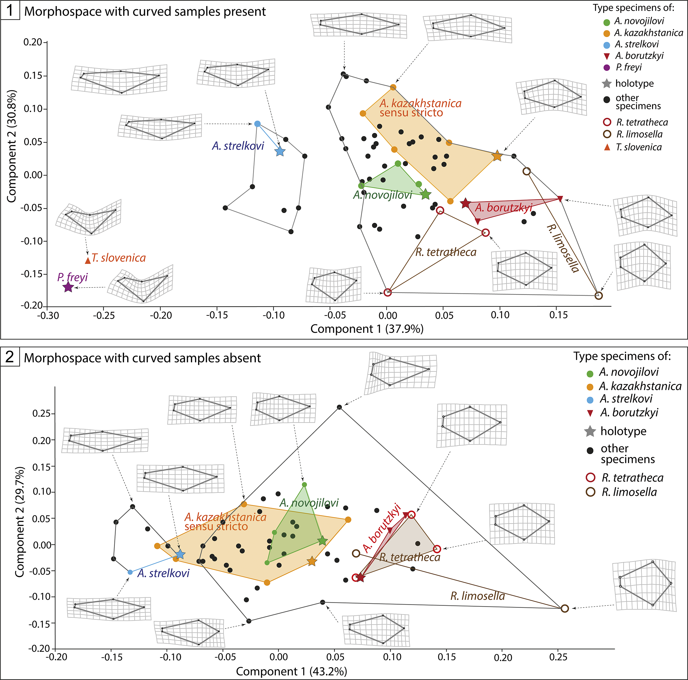
Using geometric morphometrics, we compared capsule shapes of 60 Permian cocoon compressions from Karaungir (among the type specimens, one deformed Archedaphnia kazakhstanica paratype was not included) and six extant microdrile cocoons. When all 66 cocoons were included in the analysis (scenario 1), most of the fossils clustered together abutting three closely spaced but not overlapping subclusters for the type specimens of Archedaphnia kazakhstanica, Archedaphnia novojilovi, and Archedaphnia borutzkyi (Fig. 8.1). Somewhat separated was a small cluster condensed around the holotype and paratype of Archedaphnia strelkovi. Propleuroxus freyi occupied a morphospace farther away from the principal cluster. The extant Rhynchelmis tetratheca and Rhynchelmis limosella presented ranges of variation that partially overlapped with the larger fossil cluster, whereas Propleuroxus freyi was closer to the extant Tatriella slovenica (in capsule shape; its extensions are rudimentary).
Results of geometric morphometric analyses: (1) scenario 1, with curved specimens included; dark gray outline with no fill surrounds clusters with 83.85% of specimens correctly identified; (2) scenario 2, with curved specimens excluded and a more homogeneous cloud of specimens; dark gray outline with no fill surrounds clusters with 78.33% of specimens correctly identified.

Figure 8. Long description
Panel 1, titled Morphospace with curved samples present, features an X-axis for Component 1 (37.9%) ranging from -0.30 to 0.20 and a Y-axis for Component 2 (30.8%) ranging from -0.20 to 0.20. A large dark gray outline contains most specimens. Within this space, colored polygons represent species clusters: A. kazakhstanica sensu stricto (orange), A. novojilovi (green), and A. borutzkyi (red). Individual points for A. strelkovi (blue), T. slovenica (orange triangle), and P. freyi (purple star) are located on the left. R. tetratheca and R. limosella are indicated by open circles on the right. Deformation grids showing shape variation are linked to specific points by dashed arrows.
Panel 2, titled Morphospace with curved samples absent, features an X-axis for Component 1 (43.2%) ranging from -0.20 to 0.25 and a Y-axis for Component 2 (29.7%) ranging from -0.20 to 0.25. The specimen cloud is more homogeneous. The A. kazakhstanica sensu stricto (orange) and A. novojilovi (green) clusters overlap significantly in the center. A. strelkovi (blue) is positioned to the left, while A. borutzkyi (red) and R. tetratheca (open red circle) form a cluster to the right. R. limosella (open brown circle) is an outlier on the far right.
Legends on the right of both panels identify type specimens: A. novojilovi (green dot), A. kazakhstanica (orange dot), A. strelkovi (blue dot), A. borutzkyi (red inverted triangle), P. freyi (purple dot), holotypes (stars), other specimens (black dots), R. tetratheca (red open circle), R. limosella (brown open circle), and T. slovenica (orange triangle).
The cross-validation jackknife test supports correctly identified groups (Archedaphnia kazakhstanica + Archedaphnia novojilovi + Archedaphnia borutzkyi, Archedaphnia strelkovi, Propleuroxus freyi, Rhynchelmis limosella, Rhynchelmis tetratheca, Tatriella slovenica) with 53.03% variation. It is noteworthy that these estimates are affected by a restricted morphospace array of all of the Permian and extant species, which precludes a more precise identification of the Permian cocoons. If the groups were considered solely by morphology and all specimens were combined into three morphological clusters, namely, Archedaphnia strelkovi, curved cocoons (Propleuroxus freyi and Tatriella slovenica), and Archedaphnia kazakhstanica + Archedaphnia novojilovi + Archedaphnia borutzkyi (extant Rhynchelmis species belonged to the same cluster), the proportion of correctly identified specimens increased to 83.85%. Such clusters eliminated the inexact superposition of Permian and extant cocoons in the morphospace and provided a basis for their morphological subdivision, although these clusters did not allow us a firm species-specific interpretation (Fig. 8.1, including curved specimens). In summary, only three distinct clusters, separated by Principal Component 1 (PC1), reflected the most important differences in the shapes of fossil cocoons.
In the second scenario, we assumed that cocoon curvature was an artifact of fossil preservation or an individual anomaly, and excluded the five curved compressions and the similarly curved cocoon of Tatriella slovenica from further analysis. In this case, the distribution of cocoons appeared as a more homogeneous figure-eight-shaped cloud; the holotype and two of three paratypes of Archedaphnia novojilovi and the holotype of Archedaphnia strelkovi fell within the cluster of Archedaphnia kazakhstanica type specimens, whereas the Archedaphnia borutzkyi cluster merged with the Rhynchelmis spp. cluster (Fig. 8.2). Among several ways to split this figure-eight-shaped cloud into two or three, either at its waist or at the gaps between point aggregations, only one topology yielded subclusters that correlated with the described fossil species—a very small Archedaphnia strelkovi subcluster and a much larger subcluster combining all other fossil and Recent species. With this clustering, the percentage of correctly identified specimens was 78.33%. Other cloud splittings yielded unnamed subclusters, with the percentage of correctly identified specimens ranging from 86.67% (at the waist) to 66.67%.
Thus, geometric morphometrics failed to subdivide the morphological continuum of the Karaungir cocoons into discrete groups corresponding to Smirnov’s species, except for Propleuroxus freyi, Archedaphnia strelkovi, and Archedaphnia kazakhstanica + Archedaphnia novojilovi + Archedaphnia borutzkyi. However, the analysis confirmed the affinities of Archedaphnia fossils to clitellate cocoons.
Discussion
The Permian Archedaphnia-type cocoons are similar to membranous cocoons of the modern microdrile oligochaete genus Rhynchelmis Hoffmeister, Reference Hoffmeister1843 in their ovoid shape, long narrow stalk attached to the substratum, and even longer, wider neck with a tattered edge (Chekanovskaya, Reference Chekanovskaya1962; Timm, Reference Timm1970, Reference Timm2020). Similarly to the cocoons of Rhynchelmis tetratheca examined by us, the cocoon wall of Archedaphnia is finely wrinkled over most of the surface, whereas the stalk and neck bear ridges oriented parallel to the long axis of the cocoon (cf. Figs. 3, 4, 7). These ridges are especially well expressed on the neck and, probably, correspond to the protuberances of extant clitellate cocoons (Figs. 3.1–3.3, 7.3, 7.5). The Permian cocoons agree in all principal features to extant Rhynchelmis cocoons, and differ only by their coarser microsculpture. Taking into account that cocoons of many oligochaete genera remain unknown, we interpret Archedaphnia as a genus for fossil microdrile cocoons similar to those of Rhynchelmis.
The cocoons of the Clitellata are diverse and usually specific for a genus or even species (in contrast, polychaetes secrete protective dwellings and/or brood tubes, but not egg cases). In leeches (Euhirudinea and Acanthobdellida), the cocoons are free-lying or laterally attached, hard-shelled, and thick-walled (except for Glossiphoniidae), with two small openings surrounded by several ring-like thickenings of the capsule wall and sealed with plugs (opercula) (Moquin-Tandon, Reference Moquin-Tandon1846; Lukin, Reference Lukin1976; Wilkialis and Davies, Reference Wilkialis and Davies1980; Andersson, Reference Andersson1988; Coleman et al., Reference Coleman, Marotta and Shain2008; Romdhane et al., Reference Romdhane, Quilichini and Tekaya2017). The thick-walled cocoons of leeches consist of two structurally different layers, the external of which (hapsine) imparts the outer surface a distinct reticulate or felt-like pattern, whereas the inner laminated layer (alytine) adds thickness (Manum et al., Reference Manum, Bose and Sawyer1991; McLoughlin et al., Reference McLoughlin, Bomfleur, Mörs and Reguero2016). In the Branchiobdellida, the cocoons are stalked, either with a small posterior extension or with a thickened operculum (Moore, Reference Moore1895; Järvekülg, Reference Järvekülg1957; James et al., Reference James, Davidson, Hunt and Cable2017). In most Lumbricidae, Enchytraeidae, and Tubificidae + Naididae, the cocoons are free and the capsule is round, with thin walls and small, although unequal, extensions, well separated from the capsule (Evans and Guild, Reference Evans and Guild1947; Chekanovskaya, Reference Chekanovskaya1962; Timm, Reference Timm1970, Reference Timm2020; Piearce et al., Reference Piearce, Oates and Carruthers1992). In some enchytraeids only, the cocoons are prolonged into rather long and wide extensions (Christensen, Reference Christensen1956) whereas in certain Megascolecidae and Eudrilidae, the cocoons are free, with long extensions of unequal width, but these egg cases are larger (> 15 mm in length) and ornamented with irregular microsculpture (Chaudhuri and Datta, Reference Chaudhuri and Datta2020; Chaudhuri and Dey, Reference Chaudhuri and Dey2025). The concentric rings surrounding the extensions can be developed in different species to a variable degree, up to well-formed cap-like opercula in the naidid Ripistes Dujardin, Reference Dujardin1842 and the tubificid Aulodrilus Bretscher, Reference Bretscher1899 (Arkhipova, Reference Arkhipova and Shilova1989). In some Lumbriculidae, e.g., Lamprodrilus isoporus Michaelsen, Reference Michaelsen and Korotnev1901, the cocoons are tubificid-like, free, with small extensions (Timm, Reference Timm1970). The broad extensions, gradually tapering from the capsule, with longitudinal striations and tattered edges, as at the free ends of the Rhynchelmis cocoons, are also known at both ends of the free cocoons of the lumbriculids Stylodrilus Claparède, Reference Claparède1862 and Eclipidrilus Eisen, Reference Eisen1881 (Cook, Reference Cook1967; Timm, Reference Timm1970, Reference Timm2020).
Thus, both of the main character states common to the cocoons of Archedaphnia and the extant genus Rhynchelmis—attachment by a slender stalk, and a broad, irregularly striated neck with a tattered edge—are present in some lineages of extant Clitellata but are uncommon. The first character state is known, besides in Rhynchelmis, only in Branchiobdellida, and the other in several, but not all, lumbriculids.
Among other cocoon-like mesofossils, the eggs of mollusks, chelicerates, branchiopods, insects, fishes, and amphibians can occur in fresh- and brackish water deposits. The eggs of mollusks (gastropods) are rounded, small (40–1600 μm in diameter) and often clustered; they lack extensions (e.g., Xing et al., Reference Xing, Wang, Li, McKellar, Bai, Chen and Evans2021). Aquatic chelicerate eggs are ovoid, somewhat narrowing toward one end, but these do not reveal any prominent features and occur in clutches (Shpinev and Vasilenko, Reference Shpinev and Vasilenko2018). The eggs of clam shrimps and other branchiopods are much smaller (85–500 μm in diameter) than the clitellate cocoons described here and are usually prominently sculptured, spherical, and form dense accumulations (e.g., Shen and Huang, Reference Shen and Huang2008; Gueriau et al., Reference Gueriau, Rabet, Clément, Lagebro, Vannier, Briggs, Charbonnier, Olive and Béthoux2016). In turn, the ephippia of the Cladocera are 0.1–2 mm in length, bearing eggs that cause distinct bulging of the surface, with pitted sculpture and commonly with pointed caudal spines (e.g., Fryer, Reference Fryer1996; Popova et al., Reference Popova, Petrusek, Kořínek, Mergeay and Bekker2016). Among freshwater insects, some hydrophiloid beetles have ovoid cocoons with one extension, but they are made of silk (protein) and lack elaborate microsculpture (Hansen, Reference Hansen2000), whereas corixoid water bugs have ovoid eggs with two polar extensions and hexagonal chorion reticulation (Hungerford, Reference Hungerford1948; Konopko et al., Reference Konopko, Mazzucconi and Lopez Ruf2013). Fossil amphibian eggs are extremely rare; they can be of comparable size (~1 mm), but are irregularly rounded and deformed due to their gelatinous original consistency and are either arranged in chains or densely clustered (e.g., Du et al., Reference Du, Zhang, Gómez, Dong, Zhang, Lei, Li and Dai2024). Some freshwater chondrichthyan egg capsules are elongated, with two extensions at their opposite poles sculpted with distinct longitudinal striation (e.g., Fisher et al., Reference Fischer, Voigt, Schneider, Buchwitz and Voigt2011; McLean, Reference McLean2014). However, these egg capsules are fusiform rather than ovoid, usually with prominent spiral or parallel ribbing throughout the capsule, and even the smallest of these, 8–16 mm long, are larger than oligochaete cocoons; usually the capsules are 60–400 mm long (Fischer et al., Reference Fischer, Licht, Kriwet, Schneider, Buchwitz and Bartsch2013).
Following the preserved features of the upper Permian clitellate cocoons, their step-by-step formation can be inferred. The presence of a relatively narrow stalk attached to the substratum, a zone of transverse striation, dense longitudinal protuberances at the polar extensions, and an open fluted neck indicate that the cocoon was attached by the parent worm to the substratum via its pedunculate extension, then the cocoon contracted as its wall hardened, and finally the hatchlings left it through the neck (Figs. 3–6).
Surprisingly, only a few other oligochaete cocoons, besides putative hirudinean egg cases, have been described from much younger Pleistocene and Holocene sediments. All of these, ascribed to earthworms (Lumbricidae), are 1.4–3.1 mm in size, smooth, subspherical, with two polar rudimentary extensions (Schwert, Reference Schwert1979; Piearce et al., Reference Piearce, Oates and Carruthers1990, Reference Piearce, Oates and Carruthers1992).
The loss of the larval stage and development of cocoon-secreting glands in the clitellum were certainly the first changes marking the appearance and early evolution of the Oligochaeta, which facilitated their adaptation to life in nonmarine water bodies with unstable redox and salinity regimes, similar to those that existed by the end of the Permian in the Zaysan Basin of Kazakhstan (Zhu et al., Reference Zhu, Shen, Zhao, Hu, Ge and Wang2022; Nurbekova et al., Reference Nurbekova, Smirnova, Goncharev, Sachsenhofer, Hazlett, Smirnov, Yensepbayev, Mametov and Fustic2023). Some restrictions on the normal development of eggs inside the clitellate cocoons have been established by in vitro and in vivo experiments. Such eggs can be very sensitive to oxygen deprivation (Newrkla and Mutayoba, Reference Newrkla and Mutayoba1987). However, free cocoons can be purposely laid into a slightly deeper and poorly aerated sediment layer, e.g., 2–15 cm deep, where they are better protected from predators and infections (Wiśniewski, Reference Wisniewski1979).
The next Lower Triassic aquatic oligochaete from the southern Cis-Urals already had similar sized microdriles (1–2 mm or less in diameter) (Timm et al., Reference Timm, Vinn and Buscalioni2016; Shcherbakov et al., Reference Shcherbakov, Timm, Tzetlin, Vinn and Zhuravlev2020). One of its interesting features is its circular and longitudinal musculature, relatively thick and well developed compared to those of other oligochaetes, which consist of multiple layers forming a continuous entity (Shcherbakov et al., Reference Shcherbakov, Timm, Tzetlin, Vinn and Zhuravlev2020). Such musculature is directly related to the locomotory performance of oligochaetes burrowing through a dense medium by peristalsis (Jamieson, Reference Jamieson, Harrison and Gardiner1992; De Wit et al., Reference De Wit, Erséus and Gustavsson2011). Although the Lower Triassic clitellate body fossil is larger than typical sediment-dwelling annelids, its discovery further supports the suggestion that miniaturisation was an important direction in the evolution of the Annelida (Struck et al., Reference Struck, Golombek, Weigert, Franke, Westheide, Purschke, Bleidorn and Halanych2015).
It cannot be excluded that aquatic oligochaetes had appeared already in the Paleozoic, when the terrestrial vegetation began to produce organic sediments accumulating in freshwater bodies (Timm et al., Reference Timm, Vinn and Buscalioni2016). There is no undisputed fossil record to support this assumption, but some oligochaete-like fossils have been described from the Carboniferous and the Permian.
Among the Paleozoic vermiform body fossils, of some interest are possible oligochaetes from the terrestrial Carboniferous strata of the Bohemian Massif, especially Pronaidites carbonarius Kušta, Reference Kušta1888 from the Middle Pennsylvanian (Moscovian) Radnice Formation (Kušta, Reference Kušta1888; Fritsch, Reference Fritsch1907, pl. 4, figs. 1–3; Štamberg and Zajíc, Reference Štamberg and Zajíc2008). The brownish body of the holotype is ~110 mm long as preserved (incomplete at both ends) and < 2 mm wide. It is subdivided into ~120 segments that are one-third to one-half as long as wide. It has a sediment-filled intestine running along the body axis and chaetal rows (Shcherbakov et al., Reference Shcherbakov, Timm, Tzetlin, Vinn and Zhuravlev2020, SOM 1, fig. 1A1–A3); according to Fritsch (Reference Fritsch1907), these rows bear chaetae of two types, one stout and several thin. Kušta (Reference Kušta1888) interpreted the worm as a freshwater oligochaete and mentioned, but did not figure, four more, likely conspecific specimens from the same bed (which also yielded a number of terrestrial arachnids), all very long (> 100 mm), narrow (0.5–2.0 mm), and multisegmented (100–150 segments). The occurrence of multiple Pronaidites individuals agrees with the aquatic mode of life; their filiform bodies resemble both some oligochaetes (e.g., Tubificidae) and some polychaetes (e.g., Capitellidae), but the presence of a stout outermost chaeta in the bundle is more consistent with their being polychaetes. Another oligochaete-like fossil worm is Lumbricopsis permicus Fritsch, Reference Fritsch1907 described (Fritsch, Reference Fritsch1907, pl. 4, fig. 7) from the terrestrial strata of the Bohemian Massif, now attributed to the Cisuralian (Sakmarian) lacustrine Prosečné Formation (Zajíc, Reference Zajíc2014). It has a relatively long body subdivided into numerous wide segments, imparting the worm a platy habit and each bearing a pair of short lateral outgrowths (Shcherbakov et al., Reference Shcherbakov, Timm, Tzetlin, Vinn and Zhuravlev2020, SOM 1, fig. 2A1–A3).
Although Pronaidites carbonarius and Lumbricopsis permicus display some similarities with oligochaetes, they lack undoubted clitellate features (clitellum, limited genital area) and can be compared to various polychaetes, e.g., the Capitellidae (Fauchald, Reference Fauchald1977; Glasby and Timm, Reference Glasby and Timm2008). The overall morphology of both of these worms could indicate their transitional evolutionary position between polychaetes and oligochaetes. It is noteworthy that the glandular cells responsible for secretion of the cocoon sheath probably functioned in an ancestral clitellate as well as in the basal polychaete from which clitellates arose (Sayers et al., Reference Sayers, Coleman and Shain2009). A number of extant annelids, including both oligochaetes and polychaetes, secrete dwelling tubes, and these tubes have a proteinaceous composition similar to the cocoon walls (Rossi et al., Reference Rossi, Saidel, Gravante, Sayers and Shain2016).
The monophyly of the Clitellata is strongly supported by their common morphology (Brinkhurst, Reference Brinkhurst1994; Jenner, Reference Jenner2006; Christoffersen, Reference Christoffersen2012; Nielsen, Reference Nielsen2012; Purschke et al., Reference Purschke, Bleidorn and Struck2014), spermatozoon ultrastructure (Ferraguti, Reference Ferraguti and Jamieson2000), molecular data (Rousset et al., Reference Rousset, Plaisance, Erséus, Siddall and Rouse2008; Struck et al., Reference Struck, Golombek, Weigert, Franke, Westheide, Purschke, Bleidorn and Halanych2015; Andrade et al., Reference Andrade, Novo, Kawauchi, Worsaae, Pleijel, Giribet and Rouse2015; Weigert et al., Reference Weigert, Golombek, Gerth, Schwarz, Struck and Bleidorn2016), and developmental peculiarities, probably related to the loss of the planktonic larval stage (Kuo and Hsiao, Reference Kuo and Hsiao2018). The last common ancestor of the living Clitellata was likely a freshwater species whereas the soil-inhabiting and the marine clitellates are generally believed to have evolved later from freshwater predecessors (Timm, Reference Timm1981, Reference Timm2012; Erséus, Reference Erséus2005; Rousset et al., Reference Rousset, Plaisance, Erséus, Siddall and Rouse2008).
Early clitellates probably evolved from polychaete ancestors as a monophyletic clade that gradually adapted to life in freshwater. Several ‘oligochaetoid’ characters occurring in some marine polychaetes could serve as preadaptations. Condensation of genital organs into limited segments, the presence of a clitellum-like ‘girdle’ near the female pores, the anteriorly located spermathecae, and the development of young without a larval stage in cocoons occur in extant orbiniid polychaetes of the family Questidae. However, the latter are exclusively marine and heterosexual, and their pharyngeal pad is ventral, similar to those of other polychaetes and unlike those of oligochaetes. Contact during copulation must be made between the dorsal sides of both partners in the Questidae (both the male pores and the spermathecal pores are dorsal), and between their ventral sides in the Clitellata (Giere and Riser, Reference Giere and Riser1981). Some researchers (e.g. Almeida et al., Reference Almeida, Christoffersen, Amorim, Garrafoni and Silva2003; Christoffersen, Reference Christoffersen2012) take the Questidae for a sister group or even a member of the Clitellata sensu lato. However, molecular data (e.g. Struck et al., Reference Struck, Hessling and Purschke2002) show that the Clitellata and the Questidae represent two remotely related branches of the phylogenetic tree of Polychaeta sensu lato, thus their phenetic similarity appears to be convergent. The simple free cocoons of questids, as depicted by Giere and Riser (Reference Giere and Riser1981, fig. 5), can easily be confused with those of some oligochaetes. However, the surprising similarity of the specialized cocoons from Karaungir with those of some extant lumbriculid clitellates, as well as their freshwater origin, do not support their possible attribution to questid-like polychaetes.
The find of microdrile cocoons in the upper Permian is consistent with a suggestion that the evolution of oligochaetes started at the end of the Paleozoic in brackish or freshwater ephemeral basins from the appearance of egg cases and, thus, the clitellum itself.
Acknowledgments
We thank R. Rakitov (PIN) for the superb SEM images and valuable discussion, and V. Turek and L. Váchová (National Museum, Prague, Czechia) for their friendly help with new images of the fossil worm types. L. Muir (Amgueddfa Cymru—National Museum Cardiff) and an anonymous reviewer are acknowledged for their positive and helpful reviews; O. Vinn is thanked for useful comments.
Competing interests
The authors declare none.