INTRODUCTION
Some groups of the phylum Cnidaria in Costa Rica are poorly known, and about half of the species have not been identified. The scleractinians and octocorals of both the Pacific and Caribbean coasts of Costa Rica are relatively well studied (Breedy, Reference Breedy, Wehrtmann and Cortés2008; Cortés, Reference Cortés, Wehrtmann and Cortés2008), but only seven actiniarians (sea anemones sensu stricto) were mentioned by Cortés (Reference Cortés1997) for both coasts. Examining samples collected in the Archipiélago Islas Murciélago (North Pacific coast of Costa Rica) by one of us (J.C.), we have identified a sea anemone settled on the black coral Myriopathes panamensis (Verrill, 1869). This sea anemone completely covered the black coral due to active asexual reproduction. This actiniarian belongs to the family Nemanthidae Carlgren, Reference Carlgren1940, a monogeneric family comprising three valid species (Fautin, Reference Fautin2008). This family is distinguished by the presence of acontioids (thicker than acontia, but with fewer nematocysts), and a mesogleal marginal sphincter (Carlgren, Reference Carlgren1949; Daly et al., Reference Daly, Brugler, Cartwright, Collins, Dawson, Fautin, France, Macfadden, Opresko, Rodriguez, Romano and Stake2007). The three species are Nemanthus nitidus (Wassilieff, Reference Wassilieff1908), N. californicus Carlgren, Reference Carlgren1940 and N. annamensis Carlgren, Reference Carlgren1943. Due to the particular coloration of their column, they are commonly called ‘tiger anemones’. Our specimens agree with N. californicus described by Carlgren (Reference Carlgren1940) for specimens from Baja California waters; some of them also were attached to the stalk of an octocoral. However, the original description is incomplete (due in part to imperfect preservation) and no additional records or scientific notes on this sea anemone have been published. Therefore, the purpose of this paper is to record this species on the Pacific coast of Costa Rica, and to provide a re-description of its anatomy based on histological slides, a complete cnidae analysis, and notes on its biology.
MATERIALS AND METHODS
Samples were collected by one of us (J.C.) from Isla San José of Archipiélago Islas Murciélago (Guanacaste, Costa Rica) on 16 October 1999 at 30 m depth. Approximately 25 individuals were examined all from the black coral Myriopathes panamensis. The material was fixed in 70% ethanol and deposited at the Museo de Zoología, Escuela de Biología, Universidad de Costa Rica, San Pedro, Costa Rica.
The general morphology, anatomy and other observations related to the positioning of the sea anemones on the black coral were studied at the Departamento de Ciencias Marinas (UNMdP) and Museo de Zoología (UCR) by means of a stereo dissecting microscope. Portions of some anemones were embedded in paraffin and 7-µm longitudinal and transverse histological sections were cut and stained with Harris haematoxylin and eosin.
Cnidae were measured from four individuals. A Zeiss Axiolab microscope with a micrometric eyepiece at a magnification of 1000× (oil immersion) was used. Cnidae nomenclature follows England (Reference England1991). Recording measurements of 40 or more capsules when possible, we searched for the largest and smallest unfired capsules of a particular type and tissue in order to obtain the size range. Mean and standard deviation are added.
RESULTS
Classification
EXTERNAL ANATOMY
The individuals live together, often so closely packed that the edges of their pedal discs touch each other intimately, forming an elevated border. Pedal disc well developed, much wider than column, 2.6 to 3.3 cm (Figure 1). Smooth column with more or less distinct longitudinal furrows. Diameter generally wider than tall, 1.1 to 2.0 cm in fixed individuals. Many keep the typical ‘tiger anemone’ appearance after preservation. Distinct margin with a pale coloration. Oral disc flat, mouth central, 36–94 tentacles arranged in three cycles, internal longer than external, with longitudinal furrows. Tentacles restricted to marginal half.
Two individuals of Nemanthus californicus on the black coral Myriopathes panamensis.

INTERNAL ANATOMY
Column thick with very well developed mesoglea; mesogleal marginal sphincter strong, stratified, occupying almost the whole breadth of the mesoglea and diminishing in breadth downwards (Figure 2A). Radial muscles of oral disc and longitudinal muscles of tentacles ectodermal. Four cycles of mesenteries (Figure 2B). Eight or more pairs of perfect mesenteries with very weak, diffuse retractor muscles, mostly confined to terminal portion; parietobasilar muscles weak. Acontioids attached to some mesenteries; in cross-section (Figure 2C) an axis of mesoglea surrounded by epithelium can be observed, and no muscles are attached as it is usually in the acontium. Basal disc with narrower mesoglea than column (Figure 2D). A distinct epidermis is darkly stained due to glandular cells, which secrete a thin, chitinous cuticle (approximately 5 µm width) that lies between the pedal disc epidermis and the branches of the black coral, Myriopathes panamensis.
(A) Longitudinal section of Nemanthus californicus, showing the mesogleal sphincter muscle (sp), the thick mesoglea of column (mc) and the epidermis (ep); (B) transversal section of N. californicus showing the four cycles of mesenteries in Roman numbers. Note the very weak retractor muscle. Mesoglea of column (mc); (C) transversal section of acontioid of N. californicus. Mesogleal axis (m), gastrodermis (g); (D) longitudinal section of the pedal disc of N. californicus, showing the very well developed epidermis (ep), mesoglea (m), cuticle (c) and basilar muscle (b). Scale bars: A, C, D, 50 µm; B, 1 mm.

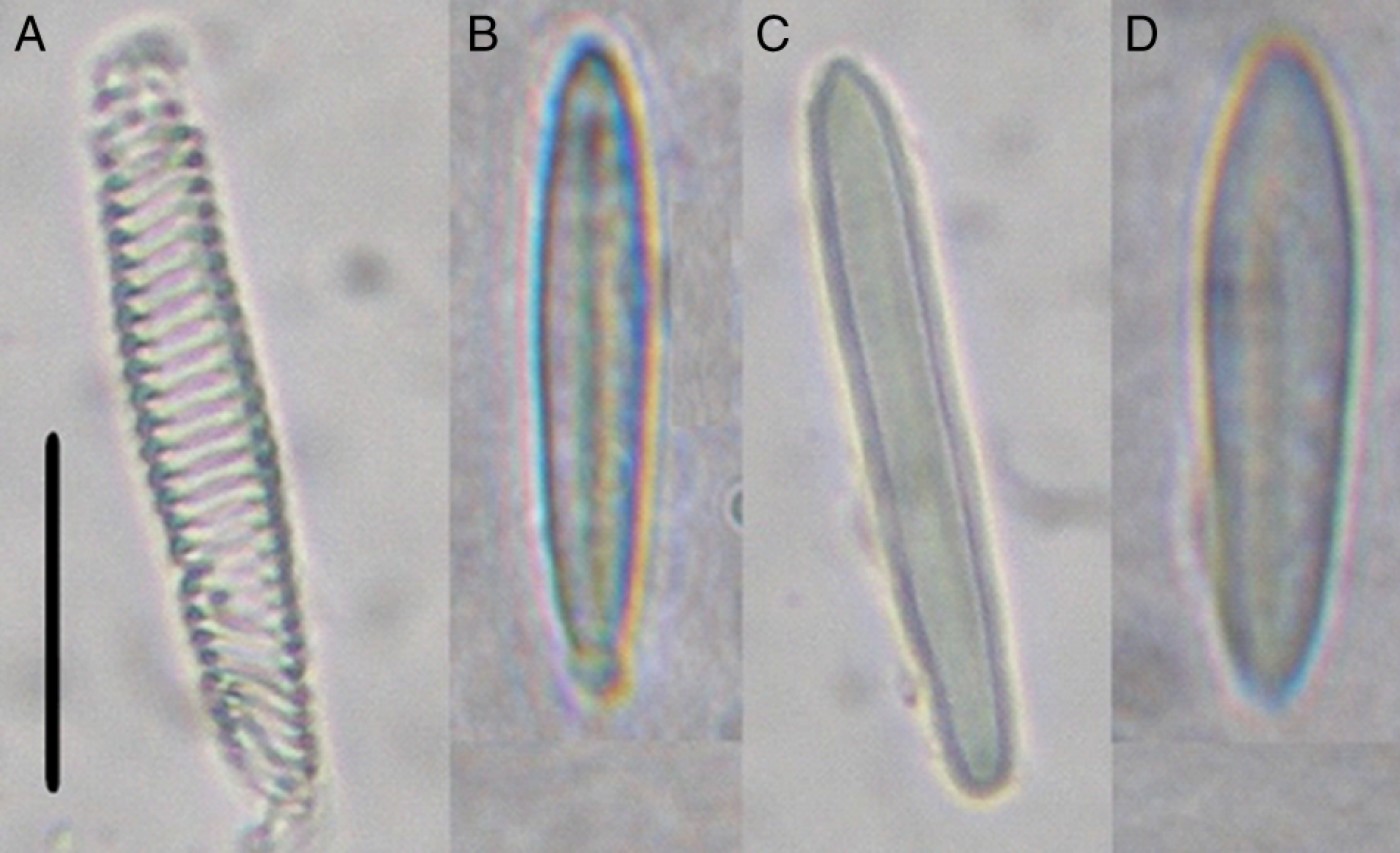
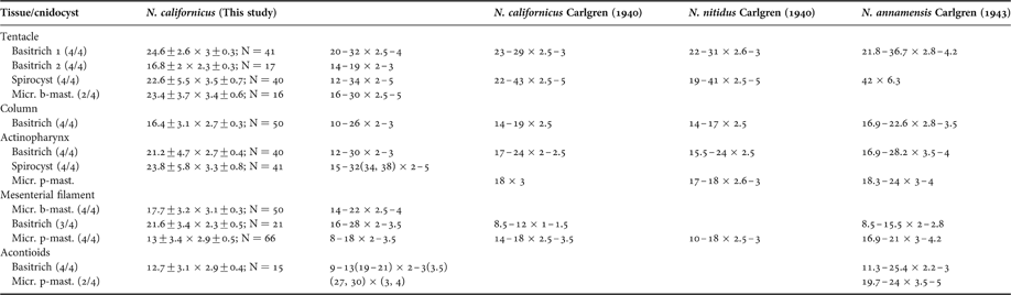
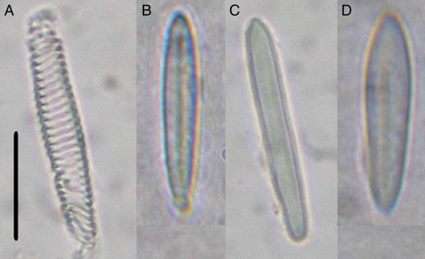
Cnidom: spirocysts, microbasic p-mastigophores, basitrichs and microbasic b-mastigophores (Figure 3). Table 1 shows the composition, size and distribution of cnidae of N. californicus. The data were compared with those of specimens of N. californicus, N. nitidus and N. annamensis studied by Carlgren (Reference Carlgren1940, Reference Carlgren1943).
Types of cnidocysts of Nemanthus californicus. (A) Spirocyst of tentacle; (B) microbasic p-mastigophore of mesenterial filament; (C) basitrich of mesenterial filament; (D) microbasic b-mastigophore of mesenterial filament. Scale bar: 10 µm.
Cnidae sizes (µm) of Nemanthus californicus, compared with data of Carlgren (Reference Carlgren1940, Reference Carlgren1943) for the three valid species of Nemanthus. Numbers in parentheses in the first column refer to the ratio of individuals possessing a particular cnidocyst type to the total number examined for that type. In the second column the mean, standard deviation and number of measured capsules are shown. In the third column, numbers in parentheses refer to individual values.

Micr. p-mast., microbasic p-mastigophore.
REPRODUCTION
This species is able to colonize the branches of the antipatharian by asexual reproduction. The individuals were so closely packed that the borders of the pedal disc touched each other intimately. We found small buds and tiny sea anemones at the basal disc of several specimens. Carlgren (Reference Carlgren1940) observed this type of reproduction in one individual of N. californicus.
DISCUSSION
The genus Nemanthus has three valid species (Fautin, 2008): N. nitidus (Sagami Bay and Bonin Islands, Japan; Korea Strait and Cheju Island area, Korea); N. californicus (Arena Bank, Sea of Cortéz, California; Isla San José of Archipiélago Islas Murciélago, Guanacaste, Costa Rica, this report); and N. annamensis (Bay of Nhatrang, S. Annam, Vietnam; Réam, Cambodia). Lavaleye & den Hartog (Reference Lavaleye and den Hartog1995) also mentioned the last species for the coast of Kenya, the Seychelles and the Maldives Islands.
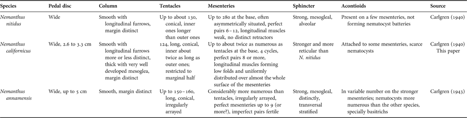
In Table 2 the basic morphological characteristics of valid species of genus Nemanthus are compared. According to external and internal morphology and geographical distribution, we identified our specimens as N. californicus. Carlgren (Reference Carlgren1940) observed about 124 tentacles while we counted about 94, but these differences could be due to age or size of individuals studied. Concerning the cnidae, we included more types than those provided by Carlgren (Reference Carlgren1940). For tentacles, we found two ranges for the basitrichs; basitrich 2 is scarce but present in all examined individuals. Also, we found microbasic b-mastigophores in two individuals. Curiously, we observed abundant spirocysts in the actinopharynx as well as basitrichs, but did not find microbasic p-mastigophores as recorded by Carlgren (Reference Carlgren1940). For mesenterial filaments, we differentiated between basitrichs and the very abundant microbasic b-mastigophores. The acontioids have basitrich and microbasic p-mastigophores but in low numbers. These nematocysts have been mentioned as numerous only for N. annamensis by Carlgren (Reference Carlgren1943). The ranges of most of cnidocysts mentioned in this study are in agreement with the data given by Carlgren (Reference Carlgren1940, Reference Carlgren1943) for the three species (Table 1). However, it is important to remark that high variability among nematocyst sizes from individuals of the same species was demonstrated by Acuña et al. (Reference Acuña, Excoffon, Zamponi and Ricci2003, Reference Acuña, Ricci, Excoffon and Zamponi2004); therefore they have only a relative value for species identification, being used only to complement other characteristics.
Comparison of the basic morphological characteristics of valid species of genus Nemanthus.

Members of genus Nemanthus normally live attached to gorgonians, their bases surrounding the branches; hence, they sometimes are referred to by the vernacular name ‘gorgonian wrappers’ (Kerstitch, Reference Kerstitch1989). Solitary or grouped sea anemones of this genus were also observed attached to branches of Anthipathes sp. colonies (Tazioli et al., Reference Tazioli, Bo, Boyer, Rotinsulu and Bavestrello2007). A particular case of N. annamensis which had settled on the carapace of the crab Lauridromia intermedia (Laurie, 1906) was reported by Lavaleye & den Hartog (Reference Lavaleye and den Hartog1995). A similar habitat was mentioned by Manuel (Reference Manuel1981) for the sea anemone Amphianthus dohrnii (Von Koch, 1878), which occurs on various gorgonians, hydroid stems, worm tubes, and similar rod-shaped organic structures. We observed N. californicus settled on the antipatharian Myriopathes panamensis with the pedal disc surrounding the branch in part or totally; both sides of the pedal disc may encounter each other at the limbus, but there is no tissue junction, as was observed for the ‘ring sea anemones’ by Ocaña et al. (Reference Ocaña, den Hartog and van Ofwegen2004). Notwithstanding, the relationship between N. californicus and the black coral could be similar, since important damage to colonies is caused in both cases, including the death of the black coral in our study. In fact, live black coral tissue was observed between the anemones on infested branches. Some colonies had entire sections that were alive and anemone-free, while other sections had anemones and the coral tissue was dead underneath (J. Cortés, personal observations). Ocaña et al. (Reference Ocaña, den Hartog and van Ofwegen2004) called this type of relationship parasitism.
We found traces of asexual reproduction (mostly budding), an expected adaptation to colonize the branches of M. panamensis; the bud arises as a protrusion from the parental pedal disc. Budding in Actiniaria was a restricted process to boloceroidid sea anemones, where it involves the autotomy of tentacles and the formation of new individuals (Fautin, Reference Fautin2002). Wassilieff (Reference Wassilieff1908) has described longitudinal fission in N. nitidus, but Carlgren (Reference Carlgren1940) considered ‘budding’ to be more appropriate, since he found a small bud at the base of one specimen of N. californicus, similar to the buds observed in our specimens. We use the term budding according to Hughes (Reference Hughes1989), who considers that budding is the formation of new individuals from primordial tissue by growth and cellular differentiation, without division of the parent.
Differences between N. californicus and N. nitidus consist mainly of colour and the development of muscles of the oral disc and the mesenteries and the sphincter. Both species differ from N. annamensis in the abundance of nematocysts (numerous basitrichs) in the acontioids of the latter species (Carlgren, Reference Carlgren1940, Reference Carlgren1943). Lavaleye & den Hartog (Reference Lavaleye and den Hartog1995) observed that the three species are unidentifiable on the basis of the available descriptions. Morphological and anatomical differences given by Carlgren (Reference Carlgren1940, Reference Carlgren1943) are in degree rather than substantial (Table 2), and therefore possibly represent only individual variation. Based on these comments and our results, we suggest that a complete revision of the genus is needed to clarify the differences between the three species, or to establish that it could be a monospecific genus with a wide Indo-Pacific distribution.
ACKNOWLEDGEMENTS
We are grateful to Oficina de Asuntos Internacionales (Programa Académicos Visitantes) of Universidad de Costa Rica for granting local expenses to F.H.A. and A.C.E. in San José, Costa Rica during 2007 and 2008, respectively. To Asociación Universitaria Iberoamericana de Posgrado (AUIP) for granting travel expenses to F.H.A. during 2007. To Dennis Opresko for the black coral identification and Odalisca Breedy for her kind help at Museo de Zoología (UCR). To Daniel Brown (Instituto Nacional de Investigación y Desarrollo Pesquero) and Laboratory of Histology (UNMdP) for microphotography and histology facilities, respectively. Finally we are grateful to Meg Daly and Marjorie Reaka for their constructive comments and English version improvement on this manuscript. This work was partially funded by a grant to F.H.A. (CONICET PIP 5504).