Introduction
Branchial cleft anomalies are the most frequent congenital lesions of the neck. Besides being an important source of morbidity, they can also be a diagnostic and therapeutic challenge.
They are localised most frequently anterior to the sternocleidomastoid muscle, often under the angle of the mandible, but can also be found anterior and posterior to the pinnae of the ear and more distally on the neck.
Three different clinical, and possibly also aetiological, malformations are recognised: cysts, sinuses and fistulae.Reference Glosser, Pires and Feinberg1 Most authors also consider cartilage remnants to be branchial cleft anomalies. Branchial cleft cysts are closed, epithelium-lined cavities without external or visceral openings. Sinuses are tracts with or without cysts communicating to either the skin or the pharynx. Fistulae are tube-like structures open at each end (i.e. at the skin and the pharyngeal mucosa).
The external opening of the typical branchial cleft fistula is located anterior to the sternocleidomastoid muscle in the lower part of the neck; from here, the fistula extends along the anterior border of the sternocleidomastoid muscle, then passes inward between the carotid bifurcation and ends in the posterolateral wall of the pharynx, just below the tonsillar fossa.Reference Agaton-Bonilla and Gay-Escoda2
These lesions can cause intermittent discomfort because of swelling, inflammation and, in the case of fistulae and sinuses, discharge from the skin opening.
In the adult population, branchial cleft cysts are a diagnostic challenge because of the possibility of cystic metastasis from occult carcinoma.Reference Gourin and Johnson3 Therefore, the accuracy of pre-operative diagnosis is of great concern. While quite a few earlier studies have reported the sensitivity of pre-operative diagnosis of branchial cleft anomalies, to our knowledge its positive predictive value has not previously been assessed.Reference Kenealy, Torsiglieri and Tom4–Reference Daoud6
The purpose of this study was twofold: first, to examine the accuracy of pre-operative diagnosis of branchial cleft anomalies, and second, to describe the occurrence, clinical presentation, management and outcome of these anomalies in paediatric and adult Danish subpopulations.
Materials and methods
A retrospective review was performed of the health records of all patients who had been diagnosed with a second branchial cleft anomaly (i.e. cyst, sinus, fistula or cartilage remnant) as a result of at least one visit to the department of otolaryngology – head and neck surgery, Roskilde County Hospital, Denmark, over a 10-year period between January 1997 and December 2006. The hospital is a regional referral centre, and all paediatric and adult patients in Roskilde County suspected of having a branchial cleft anomaly would have been referred to our department. In the reviewed time period, the mean population for which our clinic provided care was approximately 232 100.
A search of the electronic health records archive, using the International Classification of Disease 10 diagnosis Q18.0 and the Procedural Code ENB 40 (i.e. extirpation of branchial cyst or fistula) yielded 139 patients. No third or fourth branchial pouch anomalies were diagnosed in the study period.
Thirteen patients were excluded, for the following reasons: (1) the referral diagnosis of branchial cyst could not be confirmed at the hospital (n = 4); or (2) an initial clinical diagnosis of branchial cleft anomaly was changed after fine-needle aspiration biopsy (FNA) and/or imaging (n = 9).
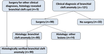
The remaining 126 patients comprised either individuals who had been diagnosed as having a branchial cleft anomaly on the basis of clinical and paraclinical examination (121 patients), or individuals who had undergone surgery on suspicion of another lesion but who had received a histological diagnosis of branchial cleft cyst (Figure 1).
Flow chart depicting patient recruitment and diagnosis.

Data collection included demographic data, age at symptom debut, referral diagnosis, treatment before referral, symptoms and signs, diagnostic investigation, treatment, peri-operative and histological findings, hospitalisation time, complications, and follow-up time.
Data analysis was performed using the Statistical Package for the Social Sciences software program. Where the sample distribution was approximately normal, the mean and standard deviation were used as descriptive statistics; otherwise, median and range were used. Confidence intervals (CIs) for proportions were calculated according to the efficient-score method (corrected for continuity) described by Newcombe.Reference Newcombe7 One sample binomial tests were used to test whether proportions of two-level categorical variables significantly differed from 0.5.
Results and analysis
Accuracy of pre-operative diagnosis
As illustrated in Figure 1, 99 patients underwent surgery based upon a pre-operative diagnosis of branchial cleft anomaly. A further 22 patients clinically diagnosed with branchial cleft anomaly did not undergo surgery, for the following reasons: the cyst disappeared after incision and antibiotic treatment (n = 1); the cyst disappeared spontaneously or after FNA (n = 8); the patient did not attend for follow up (n = 2); the surgery was postponed because of the patient's age (n = 6); or the patient did not accept surgery (n = 5).
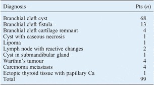
Of the 99 patients operated upon for a branchial cleft anomaly, the diagnosis was confirmed by histological examination in 85 (Table I); this corresponded to an overall positive predictive value for pre-operative diagnosis of 85/99 = 0.856 (95 per cent CI, 0.771–0.918). As all of the fistulae and sinuses (n = 13) and cartilage remnants (n = 4) were correctly diagnosed pre-operatively, the specific positive predictive value for branchial cleft cysts (n = 68) was 68/82 = 0.829 (95 per cent CI, 0.727–0.900).
Histological diagnoses after pre-operative BCA diagnosis

BCA = branchial cleft anomaly; Pts = patients; Ca = carcinoma
Five of the 14 patients misdiagnosed pre-operatively had a malignant disease (specifically, one case of papillary carcinoma in ectopic thyroid tissue and four cases of squamous cell carcinoma (SCC) (three primary tonsil carcinomas and one primary rhinopharyngeal carcinoma)). The age of the patients with SCC ranged between 45 and 66 years, while the patient with papillary carcinoma in ectopic thyroid tissue was 25 years old. Fine-needle aspiration had been performed in all these patients, but panendoscopy and positron emission tomography had not.
The total number of patients with histologically verified branchial cleft anomalies was 90, as five patients who had undergone surgery for other pre-operative diagnoses (i.e. abscess (n = 2), hypertrophic lymph node (n = 2) and malignancy (n = 1)) were shown to have branchial cysts on histological examination. Thus, pre-operative diagnosis had an overall sensitivity of 0.944 (i.e. 85/90; 95 per cent CI, 0.869–0.979) and a cyst-specific sensitivity of 0.932 (i.e. 68/73; 95 per cent CI, 0.841–0.975).
Patients with verified branchial cleft anomaly
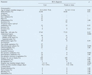
The demographic data, clinical presentation, diagnostic investigation, treatment and complications of patients with a histologically verified diagnosis of branchial cleft anomaly are summarised in Table II, and discussed in more detail below.
Histologically verified BCA cases: demographic and clinical data

BCA = branchial cleft anomaly; pts = patients; y = years
As there were only four cases of cartilage remnants, statistics were not computed for this subgroup.
Demographics
In the 61 patients with histologically verified branchial cysts and the 10 patients with verified branchial fistulae, the median age at the time of the first symptom was 32.7 years (range, 1.0–69.3 years) for the former and 0.0 years (range 0.0–29.0 years) for the latter. Two girls and two boys were diagnosed with cartilage remnants at birth. The median operation age for this group was 7.3 years (range, 3.4–14.4 years). There was a clear difference in the median operation age of the fistulae and sinus patients compared with the cyst patients, but the sex distribution for these anomalies was the same.
Clinical presentation
Eighty-one per cent of histologically verified branchial cleft anomalies presented as cysts, 14 per cent as fistulae or sinuses, and 5 per cent as cartilage remnants. Only one patient had a bilateral anomaly (fistulae). The main symptom of branchial cleft cysts was a swelling in the neck, while the cardinal symptom for fistulae and sinuses was secretion. Whereas virtually all cysts occurred cranially on the neck (in the ‘carotid triangle’), the majority of the fistulae and sinuses had their opening distally (‘supraclavicular’). Clinically, the greatest diameter of the cysts ranged from 1.2 to 8 cm, and 10 per cent of them appeared inflamed at first presentation. Although there was a slight tendency in favour of development on the right side, the observed proportion of left-sidedness to right-sidedness was not statistically significantly different from 0.5, either for cysts (p = 0.29) or for sinuses and fistulae (p = 0.092).
Investigation
Fine needle aspiration biopsy was performed in virtually all patients with cysts. This showed epithelial cells in 78 per cent of cases, polymorphonuclear cells in 69 per cent and macrophages in 36 per cent. Cholesterol crystals were seen in only two cases. Microbiological culture was performed for 34 per cent of the cysts, but only yielded growth of pathogens in one case (Klebsiella pneumoniae). Specimen collection and investigation for atypical mycobacteria (via culture and polymerase chain reaction) was performed for seven patients with cysts; all results were negative. Radiological studies were performed for 16 patients with cysts (i.e. 15 ultrasonographies and one magnetic resonance imaging scan). Fistulography was performed in about one-quarter of the patients with fistulae or sinuses.
Management
At the hospital, 23 per cent of the patients with cysts received antibiotics and 12 per cent of the cysts were drained by incision or aspiration. Tonsillectomy was performed in approximately one-half of the patients with a fistula or sinus; in these patients, two incisions were required to facilitate total excision of the tract. Seven of the patients with inflamed cysts were initially treated with incision and drainage, followed by excision surgery after the inflammation had resolved. In the patients with branchial cysts, re-operation was performed in two cases: in one case due to bleeding, and in the other case becuase of a spinal accessory nerve lesion (a nerve transplantation was done). The only re-operation in the fistula and sinus group was performed because of recurrence.
Complications
Wound infection occurred in 4 per cent of the patients operated upon for cysts and 8 per cent of patients operated upon for a fistula or sinus. There was one case of post-operative haemorrhage in the cyst group which required surgery. Four cases had recorded nerve lesions, involving variously the spinal accessory nerve, the great auricular nerve, the mandibular branch of the facial nerve, and a combination of the spinal accessory nerve and the mandibular branch of the facial nerve. The facial nerve mandibular branch lesions were temporary but the remaining nerve lesions were permanent. All of these neurological deficits occurred after surgery on inflamed cysts. No cases of aesthetic sequelae were recorded.
Discussion
Cystic neck lumps are a diagnostic challenge for the head and neck surgeon. Incorrect pre-operative diagnoses of histologically verified branchial cleft anomalies reported in the literature include thyroglossal duct cyst, cervical lymphadenitis, cervical abscess, toxoplasmosis, tuberculosis, dermoid, dermal inclusion cyst, hydatid cyst, lymphangioma and malignant neoplasm.Reference Kenealy, Torsiglieri and Tom4, Reference Daoud6
Quite a few studies have reported the sensitivity of pre-operative diagnosis of branchial cleft anomalies. Reported values for a mixed population of both children and adults range from 41 to 52 per cent.Reference Daoud6, Reference Titchener and Allison8 In the paediatric population, the sensitivity of pre-operative diagnosis was 76 per cent in one study and 85 per cent in another.Reference Kenealy, Torsiglieri and Tom4, Reference Schroeder, Mohyuddin and Maddalozzo5 In contrast to these results, our study found a rather high overall sensitivity of pre-operative diagnosis for a mixed population, i.e. 94 per cent.
Clinically, the positive predictive value of a pre-operative diagnosis of branchial cleft anomaly is more important than its sensitivity, at least in the adult population, because of the possibility of cystic degeneration in a metastatic lymph node, and the still debated possibility of brachiogenic malignancy. Both SCC (most frequently of the tonsil) and papillary carcinoma of the thyroid may mimic a branchial cleft cyst.Reference Hardee and Hutchison9–Reference Granstrom and Edstrom12
To our knowledge, the positive predictive value of a pre-operative diagnosis of branchial cleft anomaly has not previously been assessed. Gourin and Johnson reported an incidence of malignancy of 9.9 per cent among patients presenting with a solitary lateral cervical cyst; however, all patients with a non-SCC histological diagnosis had been excluded from their study.Reference Gourin and Johnson3 In our patients, we found a positive predictive value of 86 per cent.
Four of our patients initially considered to have a branchial cleft cyst were ultimately diagnosed with SCC, emphasising the importance of considering metastatic SCC in the differential diagnosis of cystic neck swellings, even in the absence of other clinical signs or symptoms. All our patients with metastatic SCC were over 40 years of age, in accordance with earlier findings.Reference Gourin and Johnson3, Reference Thompson and Heffner13
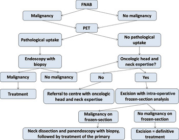
The approach to dealing with a solitary cystic neck mass in patients over 40 years of age in whom clinical examination (including nasoendoscopy) reveals no signs of malignancy will depend on the local conditions – most importantly, whether there is local oncological head and neck surgery expertise or not. If there is access to positron emission tomography, this investigation should be performed before any surgery. A possible clinical strategy is illustrated in Figure 2.
Suggested clinical strategy for dealing with a solitary cystic mass in the neck of a patient over 40 years of age in whom clinical examination (including nasoendoscopy) reveals no sign of malignancy. FNAB = fine needle aspiration biopsy; PET = positron emission tomography

The sex distribution of branchial cleft anomalies did not differ significantly from 0.5 in our study. Some previous series have shown a higher incidence in women, while others have found a higher incidence in men.Reference Agaton-Bonilla and Gay-Escoda2, Reference Kenealy, Torsiglieri and Tom4, Reference Schroeder, Mohyuddin and Maddalozzo5, Reference Titchener and Allison8, Reference Kadhim, Sheahan, Colreavy and Timon14, Reference Deane and Telander15 No significance tests were performed in these studies, so branchial cleft anomalies are probably equally frequent in men and women.
Our data indicate that branchial fistulae and sinuses are diseases of childhood, while cysts occur predominantly in adults. This is in accordance with the results of Deane and Telander.Reference Deane and Telander15 In contrast, Agaton-Bonilla and Gay-Escoda have found a similar age distribution for cysts (mean 23.6 years) and fistulae (mean 24.6 years).Reference Agaton-Bonilla and Gay-Escoda2
Earlier studies have reported that two-thirds of branchial cysts occur on the left side of the neck, but this side predilection could not be confirmed in a recent study, in which right-sided branchial cysts were actually more common than left-sided branchial cysts (54 vs 45 per cent; p = 0.399; 1 per cent were bilateral).Reference Agaton-Bonilla and Gay-Escoda2, Reference Maran and Buchanan16, Reference Doshi and Anari17 We too found that branchial anomalies were more frequently localised on the right side of the neck, but this difference in distribution was not statistically significant.
Ten per cent of cysts were found to be inflamed in hospital, but 33 per cent had received antibiotics before referral. Other authors have reported rates of inflammation ranging from 23 to 35 per cent.Reference Kadhim, Sheahan, Colreavy and Timon14, Reference Deane and Telander15, Reference Fleming18 Interestingly, microbiological culture of aspirates from inflamed cysts seldom yields growth. In our study, only one of the 25 microbiological cultures yielded growth of pathogens. In another study, microbiological culture of aspirates from 11 inflamed cysts failed to yield causative organisms.Reference Kadhim, Sheahan, Colreavy and Timon14 The same low frequency of growth was reported by Daoud, who found bacterial growth in three out of 16 cultures from inflamed cysts (all three Staphylococcus aureus).Reference Daoud6 We hypothesise that the majority of cysts that clinically appear inflamed are either sterile or the site of a viral infection, and that only seldom is there a bacterial infection in the cysts.
Cytological examination of FNA aspirate is an essential element in the investigation of neck lumps. However, as it has a reported sensitivity of 73 per cent for the diagnosis of any type of cystic cervical metastasis, and a sensitivity of 60 per cent for the diagnosis of cystic metastases masquerading as branchial cleft cysts, it cannot stand alone.Reference Sheahan, O'Leary, Lee and Fitzgibbon10 Indeed, all of our patients ultimately diagnosed with malignant disease underwent FNA before surgery, but no signs of malignancy were seen on cytological examination. The reported cytological features of branchial cysts include squamous cells, cellular debris, lymphocytes and macrophages.Reference Frierson19 Cholesterol crystals can also be found in aspirates from branchial cysts, but this finding was seen in only two FNA specimens (3 per cent) in our series. Kadhim et al. found cholesterol crystals in 12 per cent of their aspirates, whereas Daoud reported a frequency of 50 per cent.Reference Daoud6, Reference Kadhim, Sheahan, Colreavy and Timon14
Occasionally, a clinically diagnosed branchial cleft cyst will disappear after drainage (either aspiration or incision), and this was indeed the case with nine of our patients.Reference Deane and Telander15 However, the definitive treatment for branchial cysts, fistulae and sinuses is complete surgical excision. Other treatment modalities (such as repeated incision and drainage or the use of sclerosing agents) are considered non-curative and can increase the recurrence rate after surgical excision.Reference Glosser, Pires and Feinberg1, Reference Agaton-Bonilla and Gay-Escoda2 The timing of surgery depends on the age of the patient, but surgery should ideally be performed before inflammation occurs and distorts the surgical planes of dissection.Reference Glosser, Pires and Feinberg1, Reference Schroeder, Mohyuddin and Maddalozzo5 Complete excision of branchial cysts is important in order to avoid recurrence; in particular, the surgeon must look for, and remove if present, a fistulous tract from the cyst to the lateral pharyngeal wall. Such a tract was found in 6 per cent of our cyst patients. The same principle of complete excision applies to fistulae and sinuses. Two or even three incisions may be required to facilitate total excision of a long tract that has its origin low in the neck.Reference Deane and Telander15 None of our patients required three incisions, but a secondary incision was required in approximately half of our fistula and sinus operations.
There is generally agreement in the literature that surgery on an inflamed cyst is associated with a higher risk of incomplete excision (and thus of recurrence) and complication. Indeed, all the neurological complications in our series occurred after surgery on inflamed cysts. However, the appropriate management of inflamed cysts is not clear, and the issue has only sporadically been addressed in the literature.
Kadhim et al. have proposed that all patients with inflamed cysts should be hospitalised in order to receive intravenous antibiotics.Reference Kadhim, Sheahan, Colreavy and Timon14 If inflammation resolves, then surgery should be undertaken after an interval of six weeks. Otherwise, aspiration of the cyst is added, with continuation of intravenous antibiotics. If the inflammation does not resolve in spite of maximal antibiotic treatment and aspiration, incision and drainage is needed. In Kadhim and colleagues' series, excision after an interval of six weeks was performed in all cases.
• Branchial cleft anomalies are the most frequent congenital disease of the neck
• They are an important source of morbidity, and a diagnostic and therapeutic challenge
• Branchial fistulae and sinuses are commonest in childhood, branchial cysts in adulthood
• The positive predictive value of a pre-operative diagnosis of branchial cleft anomaly is <100 per cent thus, any cystic neck lesion should be presumed cancerous until proven otherwise
Schroeder et al. have also reported treating infected cysts aggressively with intravenous antibiotics, while incision and drainage were reserved for recalcitrant cases.Reference Schroeder, Mohyuddin and Maddalozzo5
While the wisdom of delaying surgery until the inflammation has resolved cannot be questioned, it is not clear why aggressive intravenous antibiotic therapy should be the first line of treatment for inflamed cysts. First, as mentioned above, microbiological cultures from cyst aspirates very rarely yield growth of pathogens. Second, the universally accepted therapy for abscesses or empyema is drainage in the first instance, with antibiotics as auxiliary treatment. Third, the existing studies are all retrospective, and one cannot exclude the possibility that the inflammation would have resolved spontaneously without aggressive intravenous therapy. Thus, prospective, randomised studies are necessary to elucidate the nature of the inflammation of branchial cysts, and whether intravenous antibiotics are necessary or not.
Conclusion
In our study, a pre-operative diagnosis of branchial cleft cyst had a positive predictive value of 86 per cent. This finding emphasises the importance of considering every cystic lesion on the neck in the adult population as a potential cystic metastasis until proven otherwise. Branchial fistulae and sinuses seem to be diseases of childhood, whereas branchial cysts occur mainly in adulthood. Branchial cleft anomalies are equally frequent in men and women, and equally distributed on the left and right side of the neck. Surgery should be postponed until inflammation has resolved. Prospective, randomised studies are needed to clarify the most appropriate management of inflamed cysts.