Introduction
A centrosome is the major microtubule organizing centre (MTOC) at which the minus ends of the cellular microtubules are concentrated. During mitosis, the centrosomes function as spindle poles to separate a complete set of the chromosomes into each daughter cell. A centrosome consists of a pair of centrioles embedded in a proteinous matrix called pericentriolar material. However, no such typical centrosome is present in the mouse oocytes and early embryos. The centrioles disappear from the oocyte centrosome at about the time when the mouse oocyte enters a resting dictyate stage of meiosis. The centrosome in the mouse oocyte consists of vesicle aggregates that reside next to the germinal vesicle (Calarco, Reference Calarco2000; Albertini & Barrett, Reference Albertini and Barrett2004). In many mammals including human, a centriole is provided by sperm during fertilization. However, this is not the case in the mouse and the centriole is synthesized de novo much later at the blastocyst stage. Therefore, the acentriolar centrosomes should function as spindle poles during meiosis as well as mitosis of the mouse oocytes and early embryos.
Nek2 was initially identified as a mammalian homolog of the mitotic kinase called NIMA in Aspergillus nidulans (Schultz et al., Reference Schultz, Fry, Sutterlin, Reid and Nigg1994). The best known function of Nek2 may be regulation of the centrosome separation (Fry et al., Reference Fry, Meraldi and Nigg1998a). C-Nap1 is the substrate of Nek2 responsible for this function (Fry et al., Reference Fry, Mayor, Meraldi, Stierhof, Tanaka and Nigg1998b). Involvement of Nek2 on other regulatory functions has been also known, suggesting that Nek2 is a mitotic kinase with multiple cellular functions (reviewed in Fry, Reference Fry2002). In fact, we identified centrobin/Nip2, a substrate of Nek2, which is required for the MTOC activity of the centrosome (Jeong et al., Reference Jeong, Lee, Kim, Yoo and Rhee2007; Lee et al., Reference Lee, Kim, Jeong and Rhee2009). Centrobin is a daughter centriole-specific and is also critical for centriole duplication (Zou et al., Reference Zou, Li, Bai, Gunning, Wazer, Band and Gao2005).
We have investigated importance of Nek2 and centrobin during the mouse early embryogenesis (Sonn et al., Reference Sonn, Khang, Kim and Rhee2004, Reference Sonn, Jeong and Rhee2009). Centrobin is associated with spindle poles throughout mitosis of the mouse early embryos. When the centrobin protein was depleted by double-stranded RNA (dsRNA) injection, development of the mouse early embryos was blocked mostly at the 4-cell stage. Many of the blastomeres in the centrobin-suppressed embryos showed defects in spindle assembly (Sonn et al., Reference Sonn, Jeong and Rhee2009). These phenotypes suggested that centrobin is critical for spindle assembly during the mitosis.
In order to investigate biological functions of Nek2 in meiosis, we examined expression and knockdown phenotypes of Nek2 and its substrates, centrobin and C-Nap1, in the mouse oocyte. Our results indicate that Nek2 and centrobin is critical for spindle assembly during meiosis.
Materials and Methods
Oocyte collection, microinjection and culture
Fully grown, germinal vesicle (GV)-intact oocytes were obtained from 21-day-old Fvb mouse ovaries and freed of attached cumulus cells. The collection medium was bicarbonate-free minimal essential medium (Earle's salt) supplemented with 25 mM HEPES and polyvinylpyrrolidone (3 mg/ml), pH 7.3. Germinal vesicle breakdown (GVBD) was inhibited by addition of 0.2 mM 3-isobutyl-1-methyl-xanthine (IBMX). Oocytes were cultured in Whitten's medium containing 0.01% polyvinyl alcohol, 5 mM NaHCO3, 15 mM HEPES and 0.2 mM IBMX. The microinjected oocytes were cultured in the CZB medium (Chatot et al., Reference Chatot, Ziomek, Bavister, Lewis and Torres1989) containing 0.2 mM IBMX for 16 h and then washed through 10 drops of IBMX-free CZB and matured in vitro in CZB for 18 h in an atmosphere of 5% CO2 in air at 37°C.
Double-stranded RNA preparation
Double-stranded RNA was prepared by annealing two complementary RNAs transcribed by T7 or SP6 RNA polymerase in vitro. The cDNA fragments for dsRNA were initially subcloned into the pGEM-T vector. The Nek2 dsRNA was a 501-bp fragment at the kinase domain common to both Nek2A and Nek2B starting from 5′-CGAACCAACACAACCCTGTA-3′ to 5′-GCCATCAGAGTAGCGGTAGG-3.′ The centrobin dsRNA was a 408-bp fragment starting from 5′-GAAAGGCAGGCCTGGATCAA-3′ to 5′-TTGGTGCTGTCCACTCAGTT-3.′ The C-Nap1 dsRNA was a 690-bp fragment starting from 5′-GAGGACCTAGAACAGATCAA-3′ to 5′-CTCAGCACACCGTTGTTGAA-3.′ The MmGFP dsRNA was a 443-bp fragment starting from 5′-CACATGAAGCAGCACGACTT-3′ to 5′-ACGAACTCCAGCAGGACCAT-3′.
After RNAs were synthesized using the T7 and SP6 RNA polymerase (Roche), DNA templates were removed with the DNase I treatment. The RNA products were extracted with phenol/chloroform and ethanol-precipitated. To anneal sense and antisense RNAs, equimolar quantities of both sense and antisense RNAs were mixed in the annealing buffer (10 mM Tris, pH 7.4, 0.1 mM EDTA) to a final concentration of 2 μM each, heated for 1.5 min at 94°C and incubated at room temperature for several hours. To remove leftover single-stranded RNA, the mixture was treated with 2 μg/ml RNase T1 (Calbiochem) and 1 μg/ml RNase A (Sigma) for 30 min at 37°C. The dsRNA was treated with 140 μg/ml proteinase K (Sigma), phenol/chloroform extracted, ethanol precipitated, washed in 75% ethanol and dissolved in water. The quality of dsRNA was confirmed by running an agarose gel. The dsRNA samples were diluted to a final concentration of 2–4 mg/ml and stored at –70°C before use.
Quantification of mRNA in the oocyte
Total RNAs were isolated by the acid guanidium thiocyanate-phenol-chloroform extraction method as reported previously (Khang et al., Reference Khang, Sonn, Park, Rhee, Park and Kim2005). Ten embryos were added into a tube containing 300 μl of solution D (4 M guanidium thiocyanate, 25 mM sodium citrate, pH 7.0, 0.5% N-lauryl sarcosyl and 0.1 M 2-mercaptoethanol) on ice and immediately plunged into liquid nitrogen for storage until use. After thawing, 0.1 volume of 2 M sodium acetate (pH 4.0), one volume of water-saturated phenol and 0.2 volume of chloroform-isoamyl alcohol (49:1) were added. After vortex-mixing, the mixture was incubated on ice for 10 min. Total RNA was then fractionated by centrifugation at 12,000 rpm for 15 min at 4°C and precipitated from supernatant in the presence of one volume of isopropanol. The pellet was washed with 75% ethanol and dissolved in water.
For reverse transcription, the RNA sample was heated in the presence of 100 pmol random hexanucleotides in a final volume of 8 μl at 65°C for 5 min. After brief centrifugation at 4°C, 12 μl of master mix [200U RNaseH-MMLV reverse transcriptase, 4 μl dNTP mix (2.5 mM each), 1 μl RNasin (26 U/μl), 2 μl 0.1 M DTT and 4 μl of 5× RT buffer (50 mM Tris–HCl, pH 8.3, 75 mM KCl, 3 mM MgCl2 and 10 mM dithiothreitol)] was added and the reaction mixture was incubated at 37°C for 1 h. The reaction was terminated by incubating the sample at 75°C for 15 min.
The amounts of specific mRNA were determined with reverse transcription-PCR based methods. To quantify the centrobin mRNA, we used a pair of the following primers that generate a 457-bp fragment; 5′-CTGATGATCACAGGGCCGAA-3′ to 5′-TGTTCCAGCCTCTTCAGGAT-3′. For quantification of C-Nap1 mRNA, we used a primer set that resulted in a 579-bp fragment: 5′-GAGCAGTGCAGGTCAGTCTT-3′ and 5′-CGTCCTCTGATCTTCCAGAA-3′.
The PCR amplification was carried out with 2 μl of RT reaction mixture in 20 μl of PCR reaction solution containing 2 μl of 10× PCR buffer, 1.6 μl dNTP mix (2.5 mM each), 10 pmol each of PCR primers and 1U Ex-Taq polymerase (Takara). The sample was subjected to a 35-cycle amplification on Gene Amp PCR System 2400 (Perkin Elmer). Six-μl samples of PCR products were analysed on 1% agarose gel electrophoresis.
Immunocytochemical staining of the oocytes
Oocytes were fixed in 3.7% formaldehyde in PBS for 10 min at room temperature, neutralized with 50 mM NH4Cl in PBS for 10 min and post-permeabilized with 0.25% Triton X-100 in PBS for 10 min. Immunocytochemical staining was performed by incubating the fixed samples with the anti-centrobin or anti-C-Nap1 polyclonal antibodies (Sonn et al., Reference Sonn, Jeong and Rhee2009) in PBST (PBS containing 0.1% Triton X-100 and 3% BSA) for 60 min, followed by an incubation in FITC-conjugated anti-rabbit IgG antibodies (KPL) diluted 1:200 in PBST for 40 min. Immunocytochemical staining was also performed by incubating the fixed samples with the anti-β-tubulin mouse monoclonal antibody or anti-γ-tubulin goat polyclonal antibody diluted 1:200 in PBST for 60 min, followed by an incubation in FITC-conjugated anti-mouse IgG antibodies (KPL) diluted 1:200 (in PBS/Triton X-100) for 40 min. The samples were observed under a fluorescence microscope (Zeiss).
Immunoblot analysis
Protein samples from the mouse oocytes were solubilized in the Laemmli sample buffer, resolved by 8% SDS-PAGE and blotted onto a nitrocellulose membrane. The membrane was blocked by soaking in Blotto (Tris-buffered saline with 0.3% Triton X-100 and 5% non-fat dried milk) for 1 h 30 min and incubated overnight with the primary antibody in the blocking solution. The membrane was then washed three times with TBST (Tris-buffered saline with 0.3% Triton X-100), incubated with a secondary antibody conjugated with horseradish peroxidase for 45 min and washed five times with TBST. The signal was detected with the ECL western blotting detection reagents (Amersham) following the manufacturer's recommendations.
Results
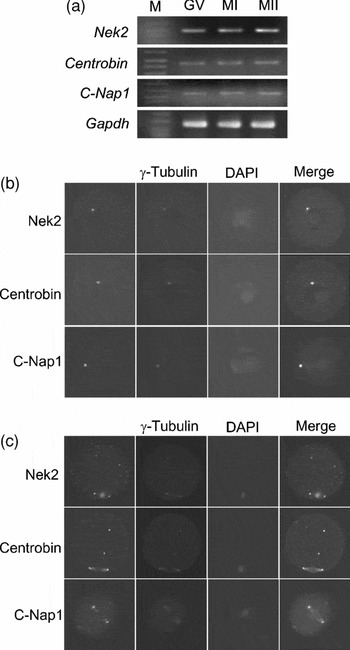
We began our study by determining expression of Nek2 and its substrates, centrobin and C-Nap1, in the mouse oocyte. The RT-PCR analysis showed that Nek2, centrobin and C-Nap1 were expressed in the mouse oocytes of all meiotic stages (Fig. 1a). Subcellular localizations of the proteins were determined by immunocytochemistry with the specific antibodies. In diplotene stage oocytes, Nek2, centrobin and C-Nap1 were localized at the acentriolar centrosomes which were co-immunostained with the γ-tubulin antibody (Fig. 1b). In the metaphase II-arrested oocytes, all three proteins were localized at the meiotic spindle poles (Fig. 1c). In addition, Nek2 and centrobin were also detected at the γ-tubulin particles present in the cytoplasm (Fig. 1c; Schuh & Ellenberg, Reference Schuh and Ellenberg2007). Nek2 association with the metaphase chromosomes was evident in the mouse oocytes, as reported previously in the spermatocytes and early embryos (Fig. 1c; Rhee & Wolgemuth, Reference Rhee and Wolgemuth1997; Sonn et al., Reference Sonn, Khang, Kim and Rhee2004).
Expression and subcellular localization of Nek2, centrobin and C-Nap1 in mouse oocytes. (a) RT-PCR analysis. Total RNA was isolated from the oocytes with intact germinal vesicle (GV) and those arrested at meiosis I (MI) and meiosis II (MII). The RNA was reverse-transcribed and PCR-amplified with primers specific to Nek2, centrobin, C-Nap1 and Gapdh. The oocytes at (b) GV or (c) meiosis II stages were immunostained with antibodies specific to Nek2, centrobin or C-Nap1. γ-Tubulin was co-immunostained and DNA was stained with DAPI.

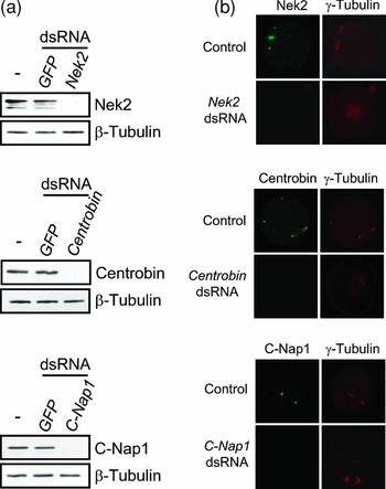
The microinjection of double-stranded RNA is known to be an efficient way to suppress the target gene expression in the mouse oocytes (Svoboda et al., Reference Svoboda, Stein, Hayashi and Schultz2000). The immunoblot analysis revealed that microinjection of the Nek2-, centrobin- and C-Nap1-specific dsRNAs reduced the corresponding proteins below the detection levels in the mouse oocytes (Fig. 2a). The immunostaining analysis showed that the Nek2-, centrobin- or C-Nap1-specific signals disappeared at the spindle poles of the dsRNA-injected oocytes (Fig. 2b). The Nek2 signals at the metaphase chromosomes also disappeared in the Nek2-suppressed oocytes (Fig. 2b). The γ-tubulin signals were evident in the spindle poles of the control and C-Nap-1-suppressed oocytes, but not in those of the Nek2- and centrobin-suppressed oocytes, indicating defects in spindle pole formation (Fig. 2b).
Suppression of Nek2, centrobin and C-Nap1 expression in the mouse oocytes. (a) The mouse oocytes were microinjected with dsRNAs specific to Nek2, centrobin or C-Nap1 and cultured in vitro. Sixteen hours later, the oocytes were subjected to immunoblot analyses for detection of the Nek2, centrobin, or C-Nap1 proteins. Un-injected oocytes were used as a control and β-tubulin was detected as a loading control. (b) The oocytes microinjected with dsRNA specific to Nek2, centrobin or C-Nap1 were immunostained with antibodies specific to Nek2, centrobin or C-Nap1, along with the γ-tubulin antibody. Un-injected oocytes were used as a control. DNA was stained with DAPI.

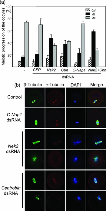
In order to determine involvement of Nek2, centrobin and C-Nap1 in the oocyte maturation, we cultured the dsRNA-injected oocytes in the IBMX-containing medium for 16 h. During this period, the target protein levels should be reduced sufficiently while meiosis of the oocytes was arrested at the diplotene stage. The oocytes were then transferred to a fresh medium without IBMX for resumption of oocyte maturation. In fact, over 90% of non-injected or GFP-injected oocytes resumed meiosis in the fresh medium (Fig. 3a). In these conditions, over 80% of the Nek2-, centrobin- or C-Nap1-suppressed oocytes also resumed meiosis, suggesting that the target proteins may not be required for resumption of the oocyte maturation (Fig. 3a). However, the number of oocytes reaching to meiosis II was reduced significantly in the Nek2- or centrobin-suppressed oocytes (Fig. 3a). Simultaneous suppression of both Nek2 and centrobin expression reduced the number of meiosis II oocytes a little more profoundly, but did not affect on the meiotic resumption rate (Fig. 3a). These results revealed that Nek2 and centrobin are not required for initiation of oocyte maturation, but they are critical for progression of meiosis. The C-Nap1-suppressed oocytes reached to metaphase II with a comparable rate with the control oocytes, suggesting that C-Nap1 is dispensable for oocyte maturation (Fig. 3a).
Effects of Nek2, centrobin and C-Nap1 suppression on oocyte maturation in vitro. (a) The mouse oocytes were microinjected with dsRNA specific to C-Nap1, Nek2 and/or centrobin (Cbn) and cultured for 24 h in the presence of 0.2mM IBMX. The oocytes were then transferred into a fresh medium and observed oocyte maturation in vitro. Un-injected and GFP dsRNA-injected groups were included as controls. The number of oocytes with germinal vesicle (GV) and those arrested at meiosis I (MI) or meiosis II (MII) was counted. The experiments were repeated four times and data were presented as mean and standard error. (b) Representative morphology of the oocytes in which Nek2, centrobin or C-Nap1 expression was suppressed by RNAi. The oocytes were co-immunostained with antibodies against β-tubulin and γ-tubulin. DNA was stained with DAPI.

Phenotypes of the Nek2-, centrobin- and C-Nap1-suppressed oocytes were analyzed. The un-injected and C-Nap1-injected oocytes matured normally to the metaphase II stage of meiosis. In contrast, Nek2- and centrobin-suppressed oocytes showed similar meiotic defects (Fig. 3b). First, meiotic spindle poles in the Nek2- and centrobin-suppressed oocytes were not focused but disorganized (Fig. 3b). The γ-tubulin signal was frequently absent at the spindle pole but are dispersed randomly near spindles, forming multiple poles and monopoles (Fig. 3b). As results, the meiotic chromosomes did not congress at the metaphase plate but scattered along the disorganized spindles (Fig. 3b). These results indicate that Nek2 and centrobin are required for prometaphase progression of the meiosis in the mouse oocytes.
Discussion
Here, we observed that Nek2, centrobin and C-Nap1 were localized at the acentriolar centrosomes in the GV-arrested oocytes and at the spindle poles in the metaphase II-arrested oocytes. Meiosis of the Nek2- and centrobin-suppressed oocytes resumed in vitro, but eventually were blocked at the prometaphase of meiosis with defects in spindle formation. These knockdown phenotypes are basically identical to those observed in the mouse early embryos in which embryogenesis was arrested at the 4-cell stage with defects in spindle assembly (Sonn et al., Reference Sonn, Khang, Kim and Rhee2004, Reference Sonn, Jeong and Rhee2009). No visible phenotype was observed in the C-Nap1-suppressed oocytes and embryos (Fig. 3, Sonn et al., Reference Sonn, Jeong and Rhee2009).
The knockdown phenotypes of Nek2 in the mouse oocytes are distinguished from those observed in the tissue culture cells. The most visible phenotype in the Nek2-suppressed culture cells may be premature centrosome separation (Fry et al., Reference Fry, Meraldi and Nigg1998a). C-Nap1 is a substrate of Nek2 responsible for this phenotype, as evidenced that premature splitting of centrosomes was induced by microinjection of the C-Nap1 antibody (Mayor et al., Reference Mayor, Stierhof, Tanaka, Fry and Nigg2000). In the current study, we did not observe premature centrosome separation in the Nek2- and C-Nap1-suppressed oocytes. Rather, knockdown phenotypes in the Nek2-suppressed mouse oocytes are comparable with those in the Xenopus embryos in which Nek2-depletion caused abortive cleavage with abnormal spindle formation (Uto & Sagata, Reference Uto and Sagata2000). Considering that Nek2 is involved in multiple functions during the cell cycle, one can expect diverse knockdown phenotypes of Nek2 (reviewed in Fry, Reference Fry2002). The Nek2 activity may be the most critical for the spindle assembly process in the oocytes and early embryos in which a strong MTOC activity is required to produce bipolar spindles in a centrosome-independent manner (Fig. 3, Sonn et al., Reference Sonn, Khang, Kim and Rhee2004).
Even if a defect in centriole duplication is the prominent phenotype of the centrobin-suppressed cells, this cannot be observed in the mouse oocytes which lack centrioles (Zou et al., Reference Zou, Li, Bai, Gunning, Wazer, Band and Gao2005). We previously observed that the centrobin-suppressed cells had a reduced MTOC activity, resulting in cell shrinkage in interphase cells and spindle assembly defects in mitotic cells (Jeong et al., Reference Jeong, Lee, Kim, Yoo and Rhee2007). Defects in spindle assembly are also observed in the centrobin-suppressed mouse oocytes and early embryos, indicating the importance of centrobin in both the meiotic and mitotic spindle formation (Sonn et al., Reference Sonn, Jeong and Rhee2009). Simultaneous knockdown of Nek2 and centrobin did not produce additive phenotypes, suggesting that Nek2 and centrobin may participate in an identical regulatory pathway for controlling meiotic spindle formation.
A microscopic imaging of live maturing oocytes proposed a comprehensive model for acentrosomal spindle assembly during meiosis (Schuh & Ellenberg, Reference Schuh and Ellenberg2007). Once meiosis resumes, acentriolar MTOCs start to form de novo from cytoplasmic microtubule networks. Over 80 MTOCs then move toward each other and form larger MTOC clusters. After germinal vesicle breakdown, the spindle number increases massively and spindle bipolarization is achieved by progressive clustering of multiple poles that is forced, in part, by microtubule motor proteins. Chromosome capturing and orientation occurs simultaneously along with the spindle bipolarization. Our knockdown phenotypes suggest that Nek2 and centrobin are involved in the acentriolar spindle assembly process. As knockdown of Nek2 and centrobin frequently result in monopolar and multipolar spindles, they may be required for clustering of the MTOCs. Another possibility may be that Nek2 and centrobin are required for stable spindle formation that is especially critical for oocytes and embryos. We currently determine biochemical properties of centrobin in association with microtubule stability.
Acknowledgements
This study was supported by a grant of the Korea Healthcare Technology R&D Project, Ministry for Health, Welfare & Family Affairs, Republic of Korea (A090558). S. Sonn was supported by the second stage of the Brain Korea 21 Project in 2007.