Introduction
The Spionidae family (Grube, Reference Grube1850) is represented by 39 checked genera, among which Spiophanes (Grube, Reference Grube1860) comprises 31 named species plus three more species whose nomenclature is still incomplete (Meißner, Reference Meißner2005; Meißner & Blank, Reference Meißner and Blank2009). Seven species of Spiophanes are currently reported for the Mediterranean Sea: Spiophanes afer Meißner, Reference Meißner2005; Spiophanes algidus Meißner, Reference Meißner2005; Spiophanes bombyx (Claparède, Reference Claparède1870); Spiophanes kroyeri Grube, Reference Grube1860; Spiophanes mediterraneus Meißner, Reference Meißner2005; Spiophanes reyssi Laubier, Reference Laubier1964; and Spiophanes wigleyi Pettibone, Reference Pettibone1962. Among these, four species are cosmopolitan (i.e. S. bombyx, S. kroyeri, S. reyssi and S. wigleyi). Spiophanes bombyx is common in shallow waters up to subtidal depths, usually on sandy bottoms (Lardicci, Reference Lardicci1989; Meißner, Reference Meißner2005); this species shows variation in important morphological characters in specimens from different localities of the world, so it might represent a species complex (Meißner & Blank, Reference Meißner and Blank2009). Spiophanes kroyeri occurs from subtidal to bathyal depths on sandy-muddy bottoms, probably preferring mud (Lardicci, Reference Lardicci1989; Meißner, Reference Meißner2005), however the distribution of this species should be revised due to misidentifications in the past. Spiophanes reyssi was firstly described from bathyal mud of the French Mediterranean coasts as a subspecies of S. kroyeri (Laubier, Reference Laubier1964) and then raised to species level, although its original description shows inconsistency of some characters usually observed in other Spiophanes species (Meißner, Reference Meißner2005); it occurs from subtidal to bathyal depths (Lardicci, Reference Lardicci1989; Çinar et al., Reference Çinar, Dağlı and Şahin2014). Spiophanes wigley occurs from subtidal to bathyal bottoms (Meißner & Hutchings, Reference Meißner and Hutchings2003) although in the Mediterranean it has been found only on subtidal sandy-muddy bottoms (Lardicci, Reference Lardicci1989). Spiophanes mediterraneus distribution is restricted to the Eastern Basin (Israel) (Meißner, Reference Meißner2005) and the Aegean coasts of Turkey (Çinar et al., Reference Çinar, Dağlı and Şahin2014) in bathyal soft bottoms while the other two species, S. afer and S. algidus, have a more recent history in the Mediterranean. Spiophanes afer was firstly described from the Mediterranean Spanish coasts and subsequently recorded in Israel, Turkey (Dağlı, Reference Dağlı2008; Dağlı et al., Reference Dağlı, Çinar and Ergen2011), Sea of Marmara (Dağlı, Reference Dağlı2013) and Adriatic Sea (Mikac, Reference Mikac2015), although the last author also suggests a possible misidentification with the widespread S. kroyeri. Spiophanes algidus, originally described from the Southern Ocean, has been recently reported only along the Aegean coasts of Turkey and added to the list of alien species of the Mediterranean Sea (Çinar et al., Reference Çinar, Bilecenoglu, Ozturk, Katagan, Yokes, Aysel and Erdogan2011; Dağlı et al., Reference Dağlı, Çinar and Ergen2011; Zenetos et al., Reference Zenetos, Çinar, Crocetta, Golani, Rosso, Servello and Verlaque2017).
Spiophanes species, like other Spionidae, are typical surface deposit feeders having a pair of long peristomial tentacles or palps that are used for selecting particles (Rouse & Pleijel, Reference Rouse and Pleijel2001). They live in burrows formed by fragile mucoid secretion (Day, Reference Day1967). This genus differs from the other genera by having large crook-like hooks in the first neuropodium, body subdivided into three different regions and absence of branchiae.
The classification of the Spiophanes species underwent revisions and integrations, with revaluation of diagnostic characters (Pettibone, Reference Pettibone1962; Laubier, Reference Laubier1964; Imajima, Reference Imajima1991; Blake et al., Reference Blake, Hilbig and Scott1996; Maciolek, Reference Maciolek2000). In a recent review of the genus, Meißner & Hutchings (Reference Meißner and Hutchings2003) described a new diagnostic character for the identification of species: the ‘chaetal spreader’. This character is related to glandular openings present in the middle body region (usually in chaetiger 4–8/9) of Spiophanes. Five different shapes of glandular organs were described: ‘2 + 3 type’ with an undulate glandular opening; the ‘1 + 2 type’ with an undulate glandular opening; the ‘0 + 1 type’ with semicircular or subtriangular glandular opening; the ‘0 + 1 type’ with an undulate glandular opening; and ‘0 + 1 type’ with a simple horizontal glandular opening. The numbers refer to the lobes arising from anterior plus posterior margins of the gland opening (Meißner & Hutchings, Reference Meißner and Hutchings2003). Spiophanes tcherniai is actually the only species of the genus where gland opening and chaetal spreader do not appear (Meißner & Hutchings, Reference Meißner and Hutchings2003).
Even if phenotypic analysis is the main approach for polychaete taxonomy (Musco et al., Reference Musco, Çinar and Giangrande2005; Tovar-Hernández et al., Reference Tovar-Hernández, Licciano and Giangrande2007; D'Alessandro et al., Reference D'Alessandro, Cosentino, Giacobbe, Andaloro and Romeo2014), it is currently accepted that, due to polymorphism diversity and high levels of homoplasy, species were often overlooked (Westheide & Schmidt, Reference Westheide and Schmidt2003). For this reason, in the last decades DNA barcode has been considered a well-accepted taxonomic method for discrimination of closely related species (Barroso et al., Reference Barroso, Klautau, Solé-Cava and Paiva2010; Nygren & Pleijel, Reference Nygren and Pleijel2010; Carr et al., Reference Carr, Hardy, Brown, Macdonald and Hebert2011). Based on the assumption that intraspecific genetic divergence is always lower than interspecific divergence (Herbert et al., Reference Hebert, Cywinska, Ball and de Waard2003), the widely used mitochondrial Cytochrome c Oxidase Subunit I (COI) facilitates species identification. In this article a combination of morphological and DNA barcoding analysis was carried out to describe the new species Spiophanes adriaticus sp. nov. from the Adriatic Sea.
Material and methods
Sampling and taxonomic analyses
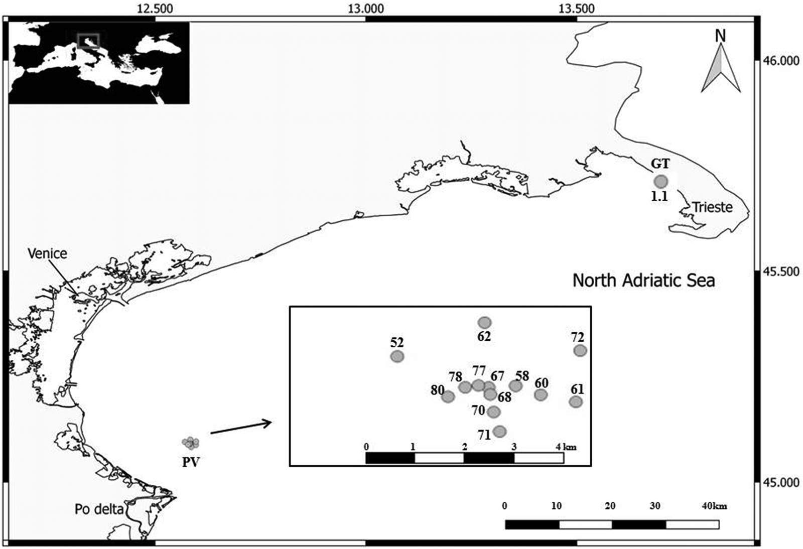
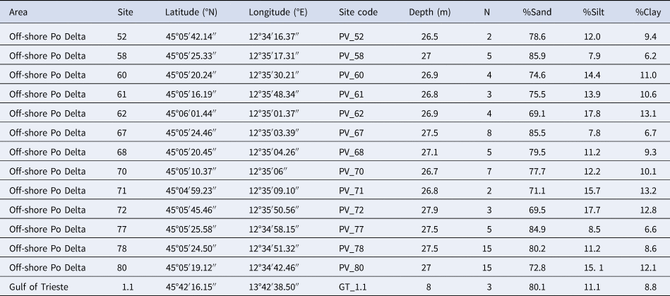
Samples of Spiophanes adriaticus sp. nov. were collected in November 2016 and July 2017, in two areas of the northern Adriatic Sea: Miramare Marine Reserve (Gulf of Trieste) and Po River Delta offshore (Figure 1). Coordinates, depth, sediment type and number of specimens belonging to Spiophanes adriaticus sp. nov. are reported in Table 1.
Maps with sampling points.

Locations, sampling details, sediment characteristics of sampling stations and occurrence of specimens (N)

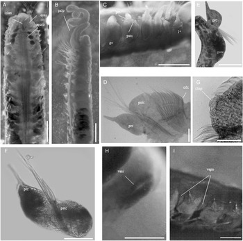
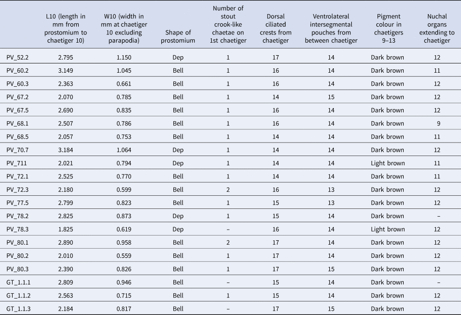
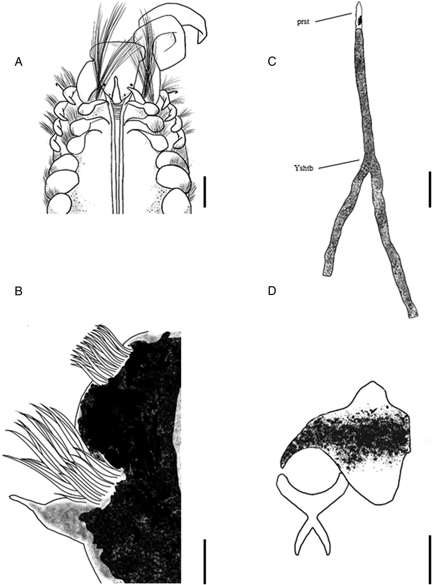
Spiophanes adriaticus sp. nov. was found in 14 out of 42 sediments collected in the two sampling areas. Sediments were taken by van Veen grab (0.1 m2 sampling surface). Samples were sieved on a 1.0 mm mesh and were immediately fixed in ethanol 95% and subsequently preserved in ethanol 90%. Specimens were observed by means of a Zeiss discovery 2.0 stereomicroscope (8 × 120 magnification) equipped with Zeiss, AxioCam ERc 5s software and a Leica DM 2500 optical microscope (Figures 2 & 3). Methyl green and Shirlastain A were used to observe some diagnostic characters as length of ciliated thin bands, dorsal crests, chaetal spreader and staining pattern. For 20 specimens, the length from the prostomial tip to chaetiger 10 (L10 in mm), the width at chaetiger 10 excluding parapodia (W10 in mm) as well as the main morphological characters were assessed and are reported in Table 2. All the measures were taken with AxioCam ERc 5s. Drawings were made by means of a drawing tablet and Krita software (Figure 4).
Spiophanes adriaticus sp. nov.: (A) anterior end, dorsal view; (B) anterior end, lateral view; (C) parapodia from chaetigers 1 to 6, lateral view; (D) parapodium 4th, frontal view; (E) parapodium 11th, frontal view; (F) parapodium 16th, frontal view; (G) chaetal spreader of 5th parapodium; (H) vertical slit from 11th parapodium; (I) ventrolateral intersegmental pouches of 16th parapodium, ventral view. Scale bar: (A); (B); (C) 500 µm; (D); (E); (F) 200 µm; (G) 50 µm; (H); (I) 200 µm. Legend: chsp, chaetal spreader; cyan, cirriform occipital antenna; crlc, crook-like chaeta; nuco, nuchal organs; prc, prechaetal lobe; pstc, postchaetal lobe; vepo, ventrolateral intersegmental pouches; vesi, vertical slit. Holotype (PV_68.3): (B); (C); (D); (E); (F); (G); (H); (I) Paratype (PV_80.1): (A).

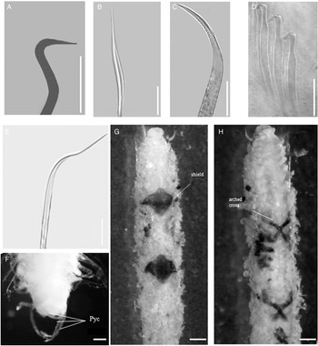
Spiophanes adriaticus sp. nov.: (A) stout, crook-like chaeta from 1st parapodium; (B) short bilimbate chaeta from parapodium 6th; (C) crook-like chaeta from parapodium 4th; (D) hooks without hood from parapodium 15th; (E) capillary chaeta from parapodium 20th; (F) pygidium; (G) tube with shield, ventral view; (H) tube with arched cross, dorsal view. Scale bar: (A, B, C, E): 200 µm; (D): 50 µm; (F): 100 µm; (G, H): 500 µm. Holotype (PV_68.3): (A, B, C, D, E, F). Paratype (PV_80.1): (G; H). Legend: pyc, pygidial cirri.

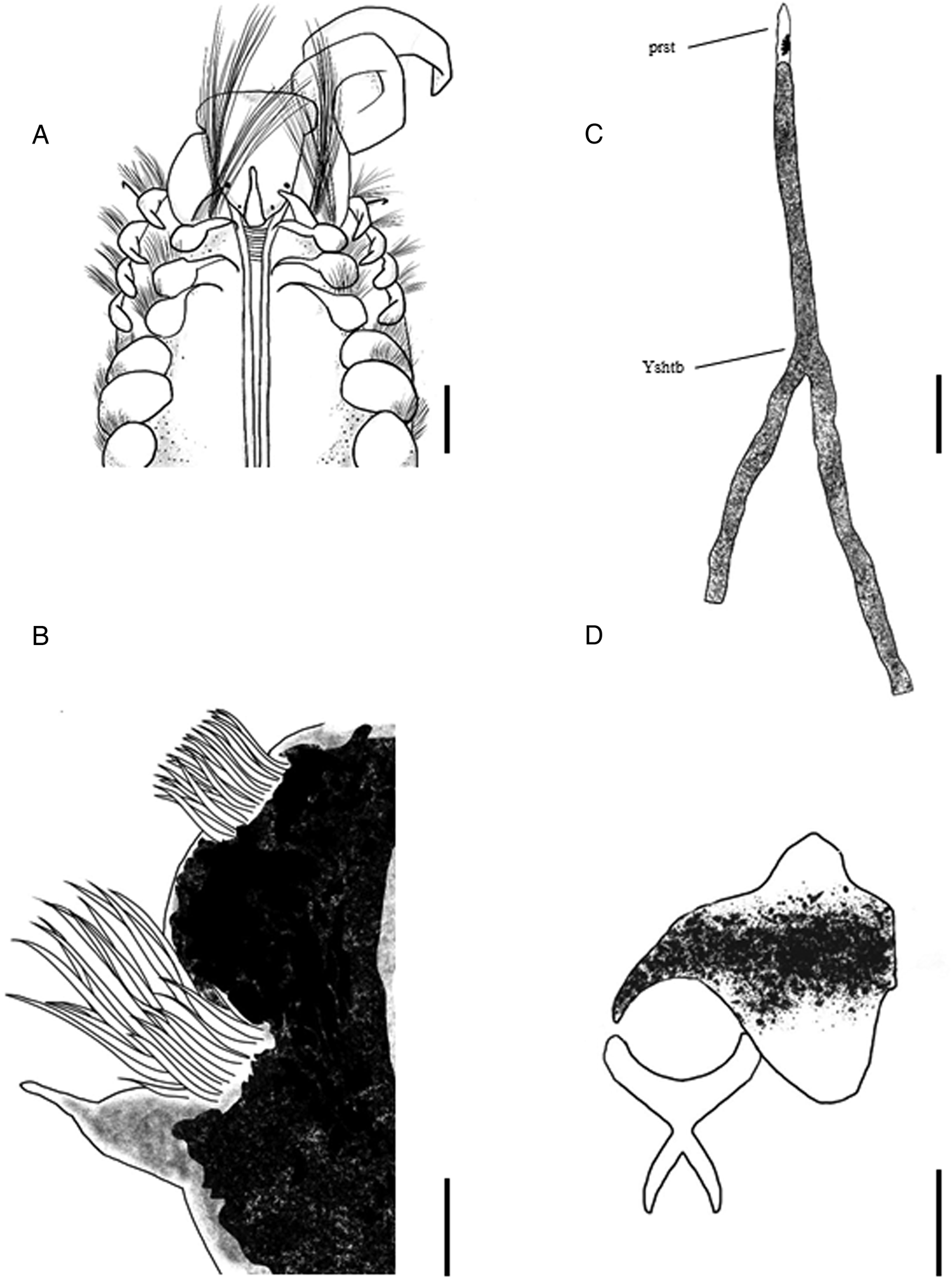
Spiophanes adriaticus sp. nov.: (A) anterior end, dorsal view; (B) parapodia 10th, frontal view; (C) tube, dorsal view; (C) shield, three-dimensional view. Scale bar (A), (B), (C) 200 µm; (C) 500 µm. Legend: prst, prostomium; Yshtb, Y shaped tubes.
Comparison of morphological and morphometric characters in 20 specimens of Spiophanes adriaticus sp. nov. collected in two areas (PV and GT) of the Adriatic Sea

Bell, bell-shaped with short antero-lateral projections; Dep, bell-shaped anteriorly depressed into two frontal lobes; –, not clear visible in the specimen.
Granulometric analysis
At each sampling location, aliquots of top layer sediment (10–15 g) were collected for the grain size analyses. Small pebbles and shell fragments were separated from other fractions by sieving at 2 mm. Each sample was disaggregated and the organic matter oxidized using 10% hydrogen peroxide (Carlo Erba, Rodano, Italy) at 60°C for 24 h. The analyses were performed using a Malvern Mastersizer 2000 equipped with Hydro 2000s (Malvern Instruments Ltd, Malvern, UK). Data are expressed as percentages of sand, silt and clay.
Molecular analyses
Three specimens of Spiophanes adriaticus sp. nov. were preserved in EtOH (90%) for DNA extraction. DNA extraction was conducted using PureLink™ Genomic DNA Mini Kit (InVitrogen). The mitochondrial cytochrome c oxidase I (COI) gene was amplified using LCO1490 and HCO2198 (Folmer et al., Reference Folmer, Black, Hoen, Lutz and Vrijenhoek1994). PCR reactions (25 µl) contained 2 µl DNA template, reaction buffer, 1.0 mM MgCl2, 250 µM of each dNTP, 10 pmol of each primer, and 0.5 U Red TAq Polymerase. The PCR temperature profile consisted of the following steps: initial denaturation at 94°C for 1 min; 35 cycles of 30 s at 94°C, 30 s at 50°C and 1 min at 72°C; and 5 min at 72°C. The amplified products were resolved on a 1% agarose gel purified using a QIAquick PCR purification Kit (QIAGEN) and sequenced on an ABI PRISM 310 automated sequencer (Applied Biosystems). DNA sequences were multiply aligned and compared with Spiophanes spp. present in Bold System and GenBank using CLUSTAL X alignment software (Thompson et al., Reference Thompson, Higgins and Gibson1994; Larkin et al., Reference Larkin, Blackshields, Brown, Chenna, McGettigan, McWilliam and Thompson2007) on a default setting and thereafter the sequence alignment was manually rechecked to allow an interpretation of the observed genetic distances. Multiple outgroup COI sequences from GenBank belonging to Spionidae family were analysed in order to select the most closely related taxon to Spiophanes genus to be used as outgroup and Prionospio steenstrupi was shown to be the most reliable to solve phylogenetic relationships (GenBank accession number GU672396). Mean genetic divergence values within and among different species were calculated using Kimura 2-parameter model as implemented in MEGA vers 6 (Tamura et al., Reference Tamura, Stecher, Peterson, Filipski and Kumar2013). For phylogenetic tree reconstruction we used Neighbour-joining (NJ) and Maximum parsimony (MP) method. The robustness of the phylogenies obtained by NJ and MP were tested by bootstrap analysis (Felsenstein, Reference Felsenstein1985) with 1000 replications. All specimens are deposited in the Polychaeta collection of National Institute of Oceanography and Experimental Geophysics (OGS).
Results
Systematics Class
POLYCHAETA Grube, Reference Grube1850
Order SPIONIDA sensu Rouse & Fauchald, 1997
Family SPIONIDAE Grube, Reference Grube1850
Genus Spiophanes Grube, Reference Grube1860
Spiophanes adriaticus sp. nov. (Figures 2–4)
Type material
Holotype entire with 70 chaetigers LT: 17.15 mm; L10: 2.25 mm; W10: 0.84 mm, code PV_68.3.
Paratypes code: Gulf of Trieste, 3 specimens (GT_1.1); Po River Delta, 2 specimens (PV_52); Po River Delta, 5 specimens (PV_58); Po River Delta, 4 specimens (PV_60); Po River Delta, 3 specimens (PV_61); Po River Delta, 4 specimens (PV_62); Po River Delta, 8 specimens (PV_67); Po River Delta, 5 specimens (PV_68); Po River Delta, 7 specimens (PV_70); Po River Delta, 2 specimens (PV_71); Po River Delta, 3 specimens (PV_72); Po River Delta, 5 specimens (PV_77); Po River Delta, 15 specimens (PV_78); Po River Delta, 15 specimens (PV_80).
Description
Prostomium subtriangular, bell-shaped, with short blunt anterolateral projections, two plump palps up to about 10th chaetiger and a cirriform occipital antenna (Figures 2A, B, 4A). Two pairs of black eyes present (Figures 2A, 4A). Peristomium moderately developed as lateral bulges (Figures 2A, 4A). Nuchal organs as two thin bands, divergent in the first parapodia and running parallel along the dorsum, to about chaetigers 11–12 (Figures 2A, 4A). Dorsal ciliated crests from chaetiger 16. Parapodia of chaetigers 1 to 4 oriented dorsolaterally (Figures 2C, 4A). Prechaetal lamellae of chaetiger 1 cirriform oriented dorsally; postchaetal lamellae cirriform with a wide base, prechaetal and postchaetal lamellae equal in length (Figure 2C). Prechaetal lobe of chaetiger 2 triangular with slender tip, postchaetal lamellae sublunate with small cirriform tip (Figure 2C). Prechaetal lobe of chaetigers 3 and 4 triangular with a slender tip, postchaetal lamellae sublunate with a rounded tip (Figure 2C, D). Chaetigers from 5 to 8 with reduced notopodial lamellae triangular with rounded tip, neuropodial lamellae rounded reduced in size (Figure 2C). From chaetiger 9 up to 13, notopodial lamellae with small triangular base and slender tip; neuropodial lamellae rounded (Figures 2E, 3B). In chaetiger 14 notopodial lamellae triangular with slender tip, longer than those of the previous chaetiger, neuropodial lamellae rounded. From chaetiger 15 to 18 notopodial lamellae ovoid with slender tip, longer than those of the previous chaetiger, neuropodia lamellae rounded (Figure 2F). From chaetiger 19 notopodial lamellae smaller than previous ones, ovoid with long slender tip; neuropodial lamellae smaller than previous ones rounded.
Chaetal spreader of the ‘2 + 3 type’ from chaetiger 5 to 7, with undulate glandular opening well developed on chaetigers 5–7 (Figure 2G); glandular organs in chaetigers 9–14 open as lateral vertical slits (Figure 2H). Ventrolateral intersegmental pouches present from between chaetigers 14–15 (Figure 2I). Bacillary chaetae were not observed. Notochaetae of first chaetiger capillary, neurochaetae with 1 stout, crook-like chaeta (Figure 3A) in addition to capillary and long limbate chaetae (Figure 3B). In parapodia 2 and 3, notochaetae capillary and neurochaetae bilimbate. Notochaetae of chaetiger 4 capillary; neurochaetae bilimbate with 1 crook-like chaeta (Figure 2D). Notochaetae of first four parapodia longer than neurochaetae and longer than the following ones (Figure 2B, C). Notochaetae 5–14 of two types: long capillary limbate and short capillary limbate (Figure 3B). Neurochaetae in neuropodia 4–14 bilimbate with one stout curved (90°) crook-like chaeta (Figures 2D, 3C). From chaetiger 14, capillaries with narrow sheath in notopodia, arranged in tufts (Figure 2F). Notopodia of chaetiger 4–18 with long and short notopodial bilimbate chaetae. In neuropodia, from chaetiger 15 neurochaeta presented 7 hooks without hoods, with a broadening in subapical region (Figure 3D), and 1 stout pointed neurochaeta, slightly curved. Hooks progressively decreasing in number along body, becoming 3 in the second last neurochaeta and 2 in the last one, still accompanied by 1 stout pointed chaeta. From chaetiger 18, capillary chaetae in the notopodia. In the posterior region (from 60° chaetiger), curved notopodial chaetal with slender tip (Figure 2E). Pygidium with 3 pairs of cirri in terminal part (Figure 3F).
Tube formed by an agglomeration of grain sand, Y shaped (Figures 3G, H, 4C). The tube has an internal chitinous structure composed of two pieces: a brownish shield with pointed arched wings and a smaller arched cross (Figures 3G, H, 4D). This particular structure is present along the tube and could have a support function.
Methyl green staining pattern
Inconspicuous.
Distribution and habitat
Currently known only from the North Adriatic Sea, specifically within the Gulf of Trieste and offshore the Po delta, in sandy, silty-sandy sediments of the sublittoral zone (between 8 and 27.5 m of depth).
Pigmentation
Conspicuous dark brownish pigmentation on parapodia in chaetigers 9–13 encompasses notopodium and neuropodium, a slightly dark region observable along ventral interramal region in chaetiger 1 to 7 (sometimes to 6).
Molecular analyses
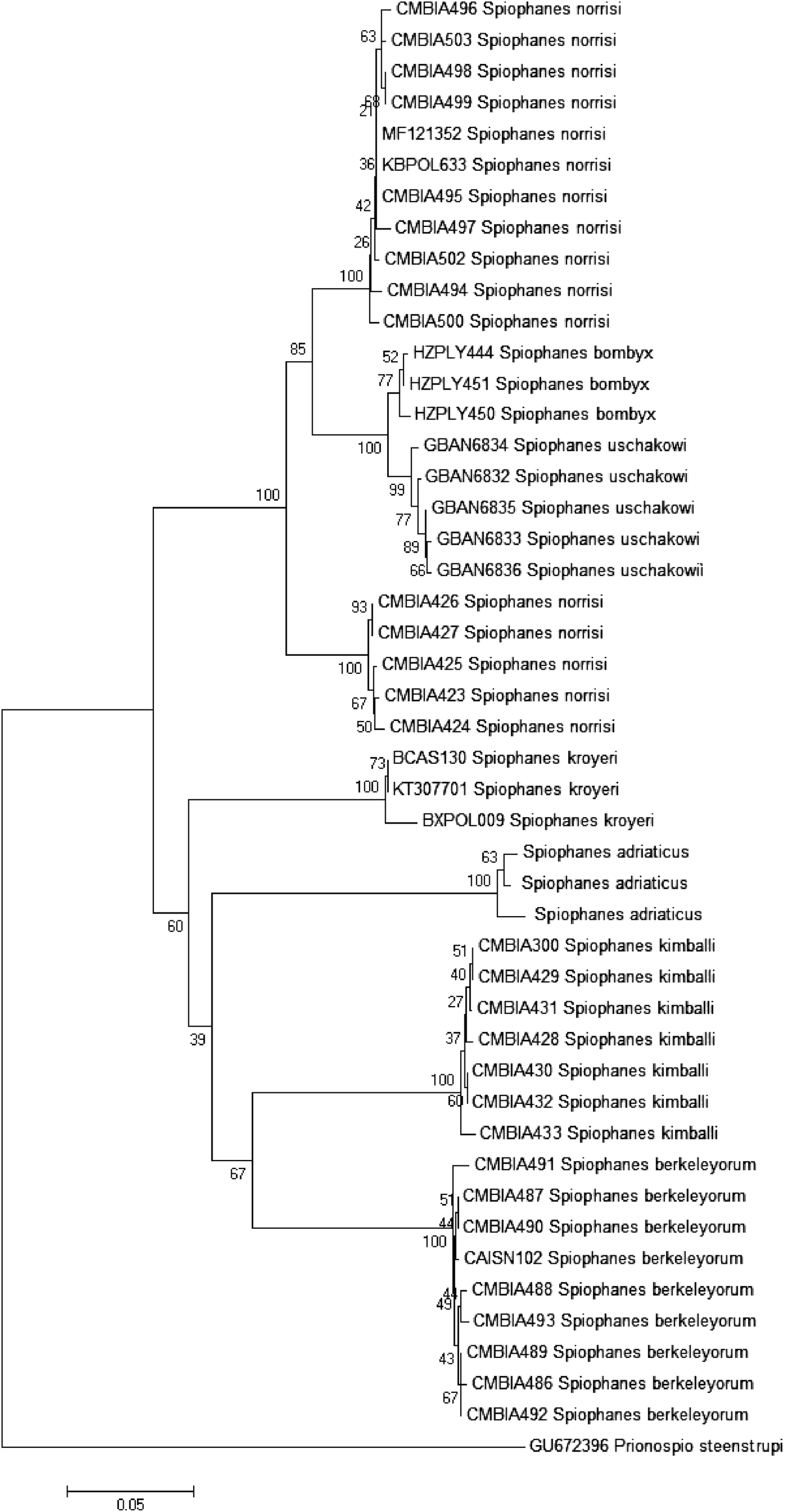
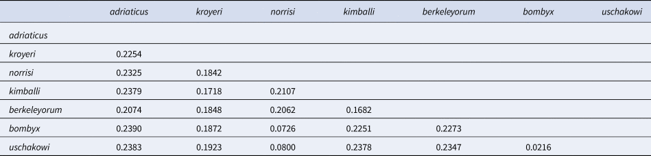
The sequence size has a variation of 550–600 base pairs (bp). Alignment was conducted with S. berkeleyorum, S. bombyx, S. kimballi, S. kroyeri, S. norrisi and S. uschakowi for a total of 46 sequences. Some sequences ascribed to S. bombyx specimens were excluded from our analyses due to a species misidentification (Meißner & Blank, Reference Meißner and Blank2009). Of 536 aligned base pairs, 222 (41%) resulted as variable, of which 178 (33%) were informative and 44 singleton (8%). The mean genetic divergence within species ranged from 0.0–0.56%, while the pairwise genetic divergence between species ranges from 0.021 between S. bombyx and S. uschakowi and 0.239 between S. bombyx and our specimens (Table 3). Phylogenetic tree constructed under NJ and MP showed a similar topology (Figure 5). The NJ tree showed well-supported distinct clades corresponding with discrete species; Spiophanes specimens from the Adriatic Sea (present study) grouped together forming a specific distinct unit, close to S. kimballi and S. berkeleyorum and S. kroyeri. From this group, S. norrisi, S. bombyx and S. uschakowi branched off.
Neighbour-joining tree based on Kimura 2-parameter distance. The percentage of replicate trees in which the associated taxa clustered together in the bootstrap test (1000 replicates) are shown next to the branches.

Estimates of molecular divergence among species calculated with Kimura 2-parameter model

Remarks
The paratypes showed some intraspecific morphological variability. In particular, the prostomium is bell-shaped with short anterolateral projections or anteriorly depressed into two frontal lobes, chaetiger 1 bears 1 or 2 stout crook-like chaetae, the first appearance of dorsal ciliated crests ranged from the 14th to the 17th chaetiger, ventrolateral intersegmental pouches present from between chaetiger 13–14 to between chaetiger 15–16, pigmentation in chaetigers 9–13 varies from light brown to dark brown, nuchal organs extend to chaetigers 9–12. All the specimens showed 4 eyes and chaetal spreader developing from chaetiger 5. No specimen was observed with gametes. As for morphometric characters, L10 ranged from 1.825–3.184 mm with a mean value of 2.480 mm, while W10 ranged from 0.559–1.150 mm with a mean value of 0.820 mm.
Etymology
Spiophanes adriaticus sp. nov. refers to the occurrence of the species along Italian coasts and offshore of North Adriatic Sea.
Discussion
Spiophanes adriaticus sp. nov. differs from the other species of Spiophanes by having a combination of the following characters: two pairs of black eyes, a cirriform occipital antenna, nuchal organs usually extending to chaetigers 11–12 (rarely to chaetiger 9), dorsal ciliated crests from chaetigers 14–17, chaetal spreader ‘2 + 3 type’, undulate glandular opening, ventrolateral intersegmental pouches from between chaetigers 14–15 (rarely 13–14 or 15–16), hooks without hood from 15th chaetiger and Y shaped tubes.
There are only five species of Spiophanes with chaetal spreader ‘2 + 3 type’: S. afer, S. duplex, S. kimballi, S. modestus and S. viriosus. Spiophanes afer differs from Spiophanes adriaticus sp. nov. by the absence of eyes in adult specimens, length of dorsal ciliated organs extending to about chaetigers 13–15 and the shape of parapodia. In addition, in S. afer dorsal ciliated crests appear moderately developed from chaetigers 18–19. Spiophanes afer also shows fair pigment (pinkish to light brown) developed on chaetigers 14–15 and glandular areas dorsally on parapodia of chaetigers 9–14, more intensively visible after staining with methyl green. Spiophanes duplex differs from Spiophanes adriaticus sp. nov. in lacking occipital antenna and ventrolateral intersegmental pouches, in having dorsal ciliated organs extending to about chaetigers 13–14 or, alternatively, diverging and converging on chaetigers 15–17 usually diverging at the end, in having dorsal ciliated crests distinct from chaetigers 17–18, pygidium with two lateral anal cirri, and in showing pigmentation on chaetigers 9–14. Spiophanes modestus differs from Spiophanes adriaticus sp. nov. for the presence of distinct prostomial horns, short occipital antenna, dorsal ciliated organs becoming divergent and undulate between chaetigers 15–17, dorsal ciliated crests apparent from chaetiger 15, neuropodial hooks without hood from 14th parapodia, for the absence of ventrolateral intersegmental pouches, pygidium with 2 dorsolateral anal cirri and for absence of conspicuous pigmentation. Spiophanes viriosus differs from Spiophanes adriaticus sp. nov. for the presence of distinct prostomial horns, for nuchal organs as two parallel lines running until parapodia 17–18 and slightly diverging at the end, dorsal ciliated crests apparent from chaetiger 18, and for colouration on vertical slit of parapodia 9–12. Spiophanes kimballi differs from Spiophanes adriaticus sp. nov. for the presence of only one pair of eyes, for the absence of ventrolateral intersegmental pouches (except in gravid specimens), dorsal ciliated crests visible from chaetiger 17, for pigmentation more distinct on chaetigers 11–13 and different methyl green staining pattern.
From an ecological point of view, Spiophanes adriaticus sp. nov. inhabits subtidal sandy and silty-sandy bottoms, but we cannot exclude its occurrence deeper and in other substrates. Among the Spiophanes species found in the Mediterranean, Spiophanes adriaticus sp. nov. shares its depth range with S. afer, S. bombyx, S. wigleyi, as well as S. kroyeri and S. reyssi, although the last two species can also be found down to bathyal bottoms.
Species belonging to Spiophanes genus are difficult to identify and many possible mistakes were recently reported by several studies, underlining that some intraspecific variations, linked to diagnostic features, could lead to erroneous classification (Meißner & Blank, Reference Meißner and Blank2009; Mikac, Reference Mikac2015). Our data indicate that the values of sequence divergence within and between species agree with the assumption of the barcode gap, strengthening the effectiveness of COI for species identification. Further in all the pairwise comparisons of S. adriaticus sp. nov. the sequence divergence values are the highest ones obtained; this result, together with the monophyletic clade shown in the phylogenetic tree and the morphological features, support the validity of the new species Spiophanes adriaticus sp. nov.
With this new finding the number of Spiophanes species recorded for the Mediterranean Sea increases to eight, emphasizing that the species richness within polychaetes is ever-increasing, thanks both to the improvement of observation techniques and the use of molecular analyses (Dağlı et al., Reference Dağlı, Çinar and Ergen2011; Mikac, Reference Mikac2015; D'Alessandro et al., Reference D'Alessandro, Romeo, Castriota, Cosentino, Perzia and Martins2016).
An integrated taxonomic approach based on traditional morphological characters together with molecular marker species identification is needed to fully delineate species boundaries, quantifying diversity, and clarifying geographic distributions in the polychaetes considered (Di Camillo et al., Reference Di Camillo, Gravili, De Vito, Pica, Piraino, Puce and Cerrano2018). DNA barcoding has proved to be a useful method for species identification and consequently for rapid global biodiversity assessment (Hebert et al., Reference Hebert, Cywinska, Ball and de Waard2003). Regarding polychaetes, Meyer & Paulay (Reference Meyer and Paulay2005) and Carr et al. (Reference Carr, Hardy, Brown, Macdonald and Hebert2011) supported the strength of COI for species identification as well as for the detection of overlooked species, underlining the importance of molecular analyses as a support to morphological taxonomic research. A broader application of the integrated approach in polychaetes taxonomy with the subsequent implementation of a DNA barcode biodiversity library could rapidly increase taxonomic and biodiversity knowledge on the group, identifying cryptic species as well as solving species complexes.
Key to Mediterranean species of Spiophanes
1. Occipital antenna absent, neuropodial hooks from chaetiger 15 with reduced hood, ventrolateral intersegmental pouches absent, dorsal ciliated organs short at most to chaetiger 4 ……… 2
– Occipital antenna present, neuropodial hooks from chaetiger 15 without hood, ventrolateral intersegmental pouches present or absent, ciliated organs long ……… 3
2. Prostomium with long anterolateral horns. Usually 2 pairs of eyes present. Dorsal ciliated organs as two wide short dorsal bands to the end of chaetiger 2. Dorsal ciliated crests from chaetiger 3. Reddish pigment in neuropodia of the middle body region sometimes in chaetigers 15–18………Spiophanes bombyx (Claparède, Reference Claparède1870)
– Prostomium without horns. Usually 2 pairs of eyes present. Dorsal ciliated organs as a pair of dorsal loops extending to chaetiger 4. Dorsal ciliated crests from chaetigers 15 or 16. Notopodial postchaetal lamellae of chaetigers 9–15 with reddish-brown pigment in proximal region, darkest on chaetigers 12–15……… Spiophanes wigleyi Pettibone, Reference Pettibone1962
3. Chaetal spreader of the ‘0 + 1 type’ ……… 4
– Chaetal spreader of the ‘2 + 3 type’ ……… 7
4. Presence of glandular organ opening (vertical slit) in chaetiger 8. Up to 4 red eyes present in juvenile specimens. Dorsal ciliated crests distinct from chaetigers 15–19……… Spiophanes reyssi Laubier, Reference Laubier1964
– Absence of glandular organ opening (vertical slit) on chaetiger 8 opening ………. 5
5. Eyes absent. Dorsal ciliated organs as two thin bands to about chaetiger 13. Ventrolateral intersegmental pouches present from between chaetigers 14–15. Dorsal ciliated crests from about chaetiger 18. Dark brown pigment around glandular opening on parapodia of chaetigers 9–14……… Spiophanes mediterraneus Meißner, Reference Meißner2005
– Ventrolateral intersegmental pouches present from between chaetigers 15–16 ……… 6
6. Eyes absent. Dorsal ciliated organs to chaetigers 14–16. First four parapodia oriented dorsally, anterior postchaetal lamellae foliaceous. Dorsal ciliated crests from chaetigers 16–18. Light brown pigment in 1–7 postchaetal lamellae; brown pigment in neuropodia of chaetigers 7–14, most conspicuous in chaetigers 8–11 ……… Spiophanes kroyeri Grube, Reference Grube1860
– Eyes absent. Dorsal ciliated organs to about chaetigers 14–15. Only first parapodium dorsally oriented, anterior neuropodial postchaetal lamellae subulate to subtriangular, notopodial postchaetal lamellae long cirriform. Dorsal ciliated crests distinct from chaetigers 17–18. Grey to brownish pigment laterally on parapodia of chaetigers 9–15, best seen on chaetigers 12–14……… Spiophanes algidus Meißner, Reference Meißner2005
7. Eyes usually absent. Dorsal ciliated organs to chaetigers 13–15. First and second parapodium in dorsal position, 3rd and 4th in dorsolateral to lateral position. Fully developed ventrolateral intersegmental pouches present from between chaetigers 15–16. Dorsal ciliated crests distinct from chaetigers 18–19. Dark brown pigment on parapodia of chaetigers 9–13, faint brownish or pink pigment on chaetigers 14–15………Spiophanes afer Meißner, Reference Meißner2005
– Two pairs of black eyes present. Dorsal ciliated organs to chaetigers 9–12 divergent in first two parapodia. Parapodia 1 to 4 oriented dorsolaterally. Ventrolateral intersegmental pouches present from between chaetigers 14–15, rarely 13–14 or 15–16. Dorsal ciliated crests from chaetiger 14–17. Dark brown pigment on neuropodia of chaetigers 9–13, dark pigment along ventral interramal region in chaetigers 1–7. ……… Spiophanes adriaticus sp. nov. D'Alessandro et al., 2018
Acknowledgements
The authors wish to thank Marco Segarich and Carlo Franzosini for the logistical support and Dr L. Ferrante and M.D. Porporato PhD for laboratory analysis and map drawing. A special thanks to Matteo Bazzarro for grain-size analysis.
Financial support
This work was funded by Adriatic LNG S.r.l. (limited liability company established by Edison, Qatar Petroleum and ExxonMobil) as a part of the prescribed monitoring programme for the potential impact of the offshore gravity based structure for unloading, storing and re-gasifying Liquefied Natural Gas (LNG).