INTRODUCTION
Peripheral neuropathic pain, manifested as spontaneous pain, hyperalgesia and allodynia, arises as a result of many forms of nerve damage; for example, diabetic neuropathy, HIV neuropathy, post-herpetic neuralgia, drug-induced neuropathy and traumatic nerve injury (Jensen et al., Reference Jensen, Gottrup, Sindrup and Bach2001; Costigan et al., Reference Costigan, Scholz and Woolf2009). The dorsal root ganglia (DRG) containing the primary sensory neurons play a key role in neuropathic hypersensitivity (Woolf and Mannion, Reference Woolf and Mannion1999; Woolf and Salter, Reference Woolf and Salter2000; Zimmermann, Reference Zimmermann2001). The primary sensory neurons send off the afferent axons; their peripheral branches course through the peripheral nerves to the target tissue; and central branches of the afferent axons run dominantly through the dorsal roots into the spinal cord. The bodies of the primary sensory neurons are enveloped by flattened satellite glial cells (SGC), and the neuron-SGC units are limited by the basal laminae from surrounding interstitial spaces (Pannese, Reference Pannese1981; Hanani, Reference Hanani2005). Like glial cells of the central nervous system (CNS), SGC also react to a neuron injury.
Chronic constriction injury (CCI) of nerve by chromic gut (Bennett and Xie, Reference Bennett and Xie1988) is a widely used experimental model with characteristic signs of neuropathic pain found in human. There is compelling evidence that neuropathic pain following experimental nerve injury is associated with changes of neuronal excitability in DRG (Kirita et al., Reference Kirita, Takebayashi, Mizuno, Takeuchi, Kobayashi, Fukao, Yamashita and Tohse2007). The changes in the DRG are related to proliferation and activation of SGC (Humbertson et al., Reference Humbertson, Zimmerman and Leedy1969; for a review see Hanani, Reference Hanani2005) and up- and downregulation of genes (Wang et al., Reference Wang, Sun, Della Penna, Benz, Xu, Gerhold, Holder and Koblan2002; Xiao et al., Reference Xiao, Huang, Zhang, Bao, Lu, Guo, Yang, Huang, Fu, Xu, Cheng, Yan, Zhu, Zhang, Chen and Han2002) as well as proteins (Zhou et al., Reference Zhou, Rush and McLachlan1996, Reference Zhou, Chie, Deng, Zhong, Xue, Rush and Xian1999; Honore et al., Reference Honore, Rogers, Schwei, Salak-Johnson, Luger, Sabino, Clohisy and Mantyh2000). In response to nerve injury, activated SGC are able to synthesize numerous neuroactive agents and their receptors for reciprocal interrelationships with neighbouring neurons (for a review see Hanani, Reference Hanani2005). Although it is generally accepted that CNS glial cells play an important role during creation and maintenance of neuropathic pain following peripheral injury (Milligan et al., Reference Milligan, Twining, Chacur, Biedenkapp, O'Connor, Poole, Tracey, Martin, Maier and Watkins2003; Watkins et al., Reference Watkins, Hutchinson, Johnston and Maier2005, Reference Watkins, Hutchinson, Milligan and Maier2007), little is known about the responses and role of SGC in sensory ganglia for development of neuropathic pain.
Recently, there has been considerable interest in contribution of pro- and anti-inflammatory cytokines in the induction and maintenance of neuropathic pain derived from their changes predominantly in the DRG neurons (Sommer and Schafers, Reference Sommer and Schafers1998; Sommer et al., Reference Sommer, Schmidt and George1998; Schafers et al., Reference Schafers, Geis, Svensson, Luo and Sommer2003; for a review see Sommer and Kress, Reference Sommer and Kress2004). Interleukin-6 (IL-6) belongs to a family of neuropoietic cytokines which also includes ciliary neurotrophic factor, leukemia inhibitory factor, oncostatin M, cardiotrophin-1, cardiotrophin-like cytokine and IL-11 (Kishimoto et al., Reference Kishimoto, Akira, Narazaki and Taga1995; Hibi et al., Reference Hibi, Nakajima and Hirano1996). It is well known that IL-6 is a key component of the nervous system's injury response. For example, IL-6 plays a role in the neuroprotection (Hama et al., Reference Hama, Kushima, Miyamoto, Kubota, Takei and Hatanaka1991; Ikeda et al., Reference Ikeda, Iwasaki, Shiojima and Kinoshita1996) and modulation of pain (Arruda et al., Reference Arruda, Sweitzer, Rutkowski and DeLeo2000; DeLeo and Yezierski, Reference DeLeo and Yezierski2001; McMahon et al., Reference McMahon, Cafferty and Marchand2005; Obreja et al., Reference Obreja, Biasio, Andratsch, Lips, Rathee, Ludwig, Rose-John and Kress2005; Schoeniger-Skinner et al., Reference Schoeniger-Skinner, Ledeboer, Frank, Milligan, Poole, Martin, Maier and Watkins2007).
IL-6 first binds to its specific membrane-bound IL-6 receptor (IL-6R), which is then associated with gp130 (Rose-John et al., Reference Rose-John, Scheller, Elson and Jones2006). These complexes bring together the intracellular regions of gp130 to initiate a signal transduction cascade through certain transcription factors, Janus kinases and signal transducer and activator of transcription 3 (STAT3). Phosphorylated STAT dimers then translocate to the nucleus and initiate transcription of target genes (Taga, Reference Taga1996; Heinrich et al., Reference Heinrich, Behrmann, Muller-Newen, Schaper and Graeve1998).
OBJECTIVE
Injury to the peripheral nerve may stimulate an increase of various cytokines and their receptors in the DRG, including IL-6 and its receptors (Murphy et al., Reference Murphy, Grondin, Altares and Richardson1995; Gardiner et al., Reference Gardiner, Cafferty, Slack and Thompson2002). Our knowledge regarding the cellular distribution of IL-6 protein and its receptors is still scarce, however, and questions as to their expression in SGC, in particular, are much debated.
The aim of the present study was to provide evidence that activated SGC display enhanced expression of IL-6 and of IL-6 receptors as well as activation of STAT3 in response to nerve injury leading to neuropathic pain.
METHODS
Animals and surgical procedures
Fifty-two pathogen-free Wistar rats (male, 250–300 g) used for the experiments were housed on a 12 h light/dark cycle at 22–24°C under specific pathogen-free conditions in the animal housing area of the Masaryk University. Sterilized standard rodent food and water were available ad libitum. All experimental procedures were carried out aseptically and according to protocols approved by the Animal Investigation Committee of the Faculty of Medicine, Brno, Czech Republic and followed ethical guidance (Zimmermann, Reference Zimmermann1983).
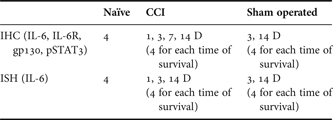
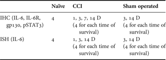
Surgical procedures were performed under deep anaesthesia with a mixture of equal volumes of ketamine (40 mg/ml) and xylazine (4 mg/ml) administered intraperitoneally (0.2 ml/100 g). For the CCI, the right sciatic nerve was exposed at the level of the mid-thigh by blunt dissection just proximal to its trifurcation, and three ligatures (3–0 Ethicon) were tied around the nerve with 1 mm spacing to reduce nerve diameter by approximately one-third. Animals operated on to create CCI were left to survive for 1, 3, 7 and 14 days. DRG removed from intact rats were used as naïve controls and the right sciatic nerves of sham-operated rats were only exposed without any lesion. The sham-operated rats were allowed to survive for 3 and 14 days. An overview of rats used for immunohistochemical staining and in situ hybridization is presented in Table 1.
An overview of naïve, CCI and sham-operated rats used for immunohistochemical (IHC) and in situ hybridization (ISH) staining. Number of animals surviving for 1, 3, 7 and 14 days (1D, 3D, 7D and 14D) is indicated in parentheses.
Behavioural tests
To ensure rats developed clear signs of neuropathic pain, mechanical allodynia and thermal hyperalgesia were measured in hind paws before and after operation. The withdrawal thresholds of mechanical allodynia were measured by Dynamic Plantar Aesthesiometer (Ugo Basile) and thermal hyperalgesia by Plantar Test (Ugo Basile) in both ipsi- and contralateral hind paws of operated rats. Baseline measurements were obtained for all animals one day prior to surgery.
Rats were first acclimated in clear Plexiglas® boxes for 30 min prior to testing. The paws were tested alternately with 5 min intervals between tests one day before operation and at each period of survival. Five latency measurements were taken for each paw during each test session. In the case of thermal hyperalgesia, withdrawal time was measured and the intensity of radiance was set at the value of 50. Data for mechanical allodynia and thermal hyperalgesia were expressed as mean ± SE of withdrawal thresholds in grams and withdrawal latency in seconds, respectively. Behavioural data were evaluated using Kruskal–Wallis one-way analysis of variance (STATISTICA, StatSoft, Inc., USA), and P values less than 0.05 were considered to be significant.
Sections and immunohistochemical staining
Immunohistochemical staining for IL-6, its receptors and pSTAT3 was performed on L4-L5 DRG from four naïve rats, CCI rats left to survive for 1 (n = 4), 3 (n = 4), 7 (n = 4) and 14 (n = 4) days, and sham-operated rats surviving for 3 (n = 4) and 14 (n = 4) days.
For tissue harvesting, the rats were deeply anaesthetized with a lethal dose of sodium pentobarbital (70 mg/kg body weight, i.p.) and then perfused transcardially with 500 ml of phosphate-buffered saline (PBS, 10 mM sodium phosphate buffer, pH 7.4, containing 0.15 M NaCl) followed by 500 ml of Zamboni's fixative (Zamboni and DeMartino, Reference Zamboni and DeMartino1967).
The L4–L5 DRG from both sides were detected within their intervertebral foramina following total laminectomy and foraminotomy. The DRG were removed, collected to samples of ipsilateral and contralateral L4–L5 DRG for each group of rats (naïve, CCI-operated and sham-operated), and fixed in Zamboni's fixative at 4°C overnight. The samples were then washed in 20% phosphate-buffered sucrose for 12 h. Pairs of ipsilateral and contralateral DRG were blocked in Tissue-Tek® OCT compound (Miles, Elkhart, IN) and cut together. Serial longitudinal cryostat sections (12 µm) through DRG were mounted to chrome-alum covered slides and processed for indirect immunohistochemical staining.
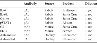
Briefly, the sections were washed with PBS containing 0.05% Tween 20 (PBS-TW20) and 1% BSA for 10 min, treated with 5% normal donkey serum in PBS-TW20 for 30 min and incubated with 25 µl of primary antibody (Table 2) in a humid chamber at room temperature (21–23°C) for 18 h. Then, after washing, the immunoreaction was visualized by treatment with FITC- or TRITC-conjugated and affinity purified donkey anti-rabbit or anti-mouse secondary antibodies (Table 2) for 90 min at room temperature. Sections of naïve DRG and those removed from CCI rats for all periods of survival as well as from sham-operated rats were incubated simultaneously under the same conditions. The control sections were incubated with omission of the primary antibody and displayed no immunostaining.
Primary and secondary antibodies used for immunofluorescence detection in rat DRG.

To identify precisely SGC and distinguish them from satellite macrophages (Dubovy et al., Reference Dubovy, Tuckova, Jancalek, Svizenska and Klusakova2007), a part of the DRG sections was incubated with a mixture (1:1) of rabbit polyclonal antibody against the detected molecule and mouse monoclonal anti-glutamine synthetase (GS) or anti-ED-1 with final dilution for single immunostaining. A mixture (1:1) of affinity purified TRITC-conjugated donkey anti-rabbit and FITC-conjugated donkey anti-mouse secondary antibodies (Table 1) was applied with a final dilution of 1:100 at room temperature for 90 min. The control sections were incubated while omitting the primary antibodies.
Immunostained sections were rinsed, stained with Hoechst 33342 to detect positions of the cell nuclei, and mounted in a Vectashield aqueous mounting medium (Vector Laboratories Inc., Burlingame, CA). The immunostained sections were observed and analysed using a Leica DMLB epifluorescence microscope equipped with appropriate filter combinations, a Leica DFC-480 camera (Leica Microsystems, Wetzlar GmbH, Germany), and a stabilized power supply for the lamp housing. The sizes of DRG neurons were categorized as small (<25 µm), medium (25–40 µm) and large (>40 µm) according to diameters calculated from areas measured using a LUCIA-G image analysis system. Only the neuronal profiles containing nuclei were counted.
Inasmuch as the same immunostaining patterns were observed in both L4 and L5 DRG, the results are described together for both segments in ipsi- and contralateral sides for each group of animals (naïve, CCI-operated and sham-operated rats).
In situ hybridization
For DRG tissue harvesting, four naïve rats and next four CCI rats for each period of survival (1, 3 and 14 days), and sham-operated rats surviving for 3 (n = 4) and 14 (n = 4) days were deeply anaesthetized with a lethal dose of sodium pentobarbital (70 mg/kg body weight, i.p.) and perfused transcardially with 500 ml PBS (10 mM sodium phosphate buffer, pH 7.4, containing 0.15 M NaCl and 0.1% diethylpyrocarbonate [DEPC]) followed by 500 ml of 4% paraformaldehyde containing 0.1% DEPC. The DRG samples were then washed in 20% phosphate buffered sucrose for 12 h and blocked in Tissue-Tek® OCT compound (Miles, Elkhart, IN). Serial longitudinal cryostat sections (12 µm) through the DRG were mounted on chrome-alum covered slides.
To localize the IL-6 gene transcript, in situ hybridization was performed according to published protocol by Harnicarova et al. (Reference Harnicarova, Kozubek, Pachernik, Krejci and Bartova2006). We used two 50-mer oligoprobes synthesized for the target IL-6 gene transcript. Digoxigenin (DIG)-dT was used for probe labelling, the sites of which are indicated by asterisks. Sequences of oligoprobes were as follows: (1) 5′DIG-CGC TGT TCA T AC AAT* CAG AAT TGC CAT* TGC ACA ACT CT*T T TCT CAT TTC C-3′DIG; (2) 5′DIG-TCA AGT GCT TTC AAG AT*G AGT TGG ATG GTC TTG GT*C CTT AGC CAC TCC TTC-3′DIG (VBC-Biotech). All solutions used in this procedure were prepared in double-distilled water treated with DEPC. DIG was detected by DIG Colorimetric Nucleic Acid Detection Kit (Roche). The sections were mounted in a Vectashield aqueous mounting medium (Vector Laboratories Inc.) and analysed using a Leica DMLB microscope equipped with a Leica DFC-480 camera (Leica Microsystems, Germany). The control sections incubated with DIG-labelled sense probe or omission of the DIG-oligonucleotide (anti-sense) probes displayed no colour staining.
Image analysis of immunofluorescence brightness and density of mRNA staining
To detect changes of immunofluorescence brightness or density of mRNA staining in SGC of DRG from naïve and CCI rats, sections of the same thickness (12 µm) incubated under the same conditions (see above) were used for image analysis measurement. Immunofluorescence brightness for IL-6, IL-6R, gp130 and pSTAT3 in SGC was measured in at least 30 sections of naïve L4–L5 DRG and ipsilateral or contralateral L4–L5 DRG after CCI for 3 and 14 days, representing early and later periods of survival. Density of IL-6 mRNA staining was measured in SGC of L4–L5 DRG from rats sham operated for 3 and 14 days to prove increased reaction of SGC to tissue injury.
The measurement of immunofluorescence brightness and density of IL-6 mRNA staining was performed using a LUCIA-G v4.21 image analysis system (Laboratory Imaging Ltd., Prague, Czech Republic) according to our protocol (Dubovy et al., Reference Dubovy, Klusakova and Svizenska2002). Briefly, immunostained or mRNA stained SGC were identified for measurement by thresholding technique after subtracting background by manual editing. The immunofluorescence brightness for investigated molecules or density of mRNA staining were expressed as mean ± SD. Data were tested for normal distribution, and differences between brightness (density) in SGC of DRG from naïve and experimental rats were tested by Mann–Whitney U test using STATISTICA 5.5 software (StatSoft, Tulsa, OK, USA) with P < 0.05 as the level of significant differences.
RESULTS
Behavioural tests
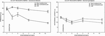
All rats operated upon to create CCI of the sciatic nerve and used for immunohistochemical assay or in situ hybridization displayed a decreased withdrawal threshold of mechanical allodynia and thermal hyperalgesia in hind paws ipsilateral to nerve ligatures, thus indicating neuropathic pain. Contralateral hind paws did not exhibit statistically significant changes of withdrawal thresholds for mechanical allodynia when compared with 1 day before operation. In contrast, withdrawal thresholds of thermal hyperalgesia decreased bilaterally only at post-operative day 3 (Fig. 1). Sham-operated rats did not exhibit mechanical allodynia or thermal hyperalgesia either ipsilaterally or contralaterally to CCI (data not shown).
Results of behavioural tests in CCI rats. A progressive development of evoked mechanical allodynia and thermal hyperalgesia was found in the ipsilateral hind paws. A transient thermal hyperalgesia was measured in contralateral hind paws 3 days from CCI operation. Data were expressed as mean ± SE of withdrawal thresholds in grams and withdrawal latency in seconds for mechanical allodynia and thermal hyperalgesia, respectively. *Indicates statistically significant difference (P < 0.05) when compared with measurement one day before operation.

IL-6 protein and mRNA in DRG of naïve and CCI-operated rats
IL-6 immunohistochemical staining
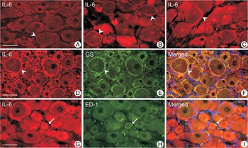
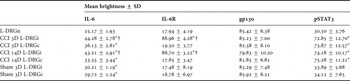
Sections of naïve L4-L5 DRG revealed very weak immunofluorescence staining for IL-6 protein in small neurons and SGC enveloping large neuronal bodies (Fig. 2A). In comparison to sections of the naïve DRG, an elevation of immunofluorescence intensity for IL-6 was found not only in small- and medium-sized neuronal bodies but also in SGC of L4–L5 DRG removed from both sides of CCI rats for all periods of survival (Fig. 2B,D,G). The SGC immunolabelled for IL-6 were observed predominantly around large neuronal bodies, which displayed weak immunostaining in comparison with small- and medium-sized ones. Immunofluorescence brightness for IL-6 in SGC was significantly higher in L4–L5 DRG from both sides of CCI rats when compared with those from naïve rats. In addition, SGC of ipsilateral L4–L5 DRG displayed IL-6 immunostaining of higher intensity than did SGC of contralateral counterparts (Table 3). Immunostaining pattern and brightness were similar in sections of L4–L5 DRG removed from sham-operated rats and those of contralateral DRG from CCI-operated rats (Fig. 2B,C; Table 3). Double immunostaining with IL-6 polyclonal and GS monoclonal antibodies proved that SGC displayed IL-6 protein (Fig. 2D–F). In contrast, the ED-1+ macrophages invading the neuronal area did not display immunoreaction for IL-6 (Fig. 2G–I). No immunostaining for IL-6 was observed in neuronal somata and SGC of control sections from L4 to L5 DRG removed from CCI rats (data not shown).
Representative sections immunostained for IL-6 in L4–L5 DRG removed from naïve rat (A) and from ipsilateral (D,G) and contralateral (B) sides of CCI rats for 3 (D–F) or 14 (B,G–I) days. Immunofluorescence staining for IL-6 in section of ipsilateral L4 DRG of sham-operated rat for 7 days is illustrated in C. A faint IL-6 immunofluorescence was present in neurons and SGC of naïve DRG (A), but this was enhanced in small- and medium-sized neuronal bodies and SGC (arrowheads) following CCI (B,D,G) and sham operation (C). Immunostaining for IL-6 was co-localized with GS immunofluorescence (D–F) but was absent from ED-1+ macrophages (arrows) invading DRG (G–I). Scale bars = 50 µm.

A comparison of immunofluorescence mean brightness for IL-6, IL-6R, gp130 and pSTAT3 in SGC from L4–L5 DRG ipsilateral (DRGi) or contralateral (DRGc) to CCI for three (3D) and fourteen (14D) days.

*Indicates statistically significant difference (P < 0.05) when compared with naïve DRG.
†Indicates statistically significant difference (P < 0.05) when DRGi was compared with DRGc.
In situ hybridization for IL-6 mRNA
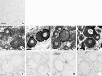
No neuronal bodies and their SGC displayed detectable signal for IL-6 mRNA in sections of L4–L5 DRG removed from the naïve rats. A weak staining for IL-6 mRNA was detected only in the blood vessels of the DRG (Fig. 3A). Unilateral CCI of the sciatic nerve-induced bilateral elevation of staining for IL-6 mRNA in L4–L5 DRG 1, 3 and 14 days after operation. In comparison to naïve DRG, a distinct intensity of IL6 mRNA staining was observed, particularly in neuronal somata of all sizes and in their SGC. Intense staining for IL-6 mRNA in neuronal bodies did not usually allow to distinguish staining in the SGC; this was possible mainly in SGC surrounding the large neuronal bodies (Fig. 3B–E).
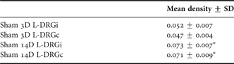
Moderate staining for IL-6 mRNA was found in SGC and small neuronal bodies of both ipsi- and contralateral L4–L5 DRG removed after 3 days from sham-operated rats. The SGC and blood vessels, but no neuronal bodies, displayed an increased density of the staining for IL-6 mRNA in sections of L4–L5 DRG from rats 14 days after sham operation (Fig. 3F–I; Table 4). The results provide unequivocal evidence that SGC are able to synthesize IL-6 protein.
(A–H) Detection of IL-6 mRNA byin situhybridization in sections of L4–L5 DRG from naïve (A), CCI (B–E) and sham-operated (F–I) rats. Bilateral elevation of staining for IL-6 mRNA was induced in neuronal bodies of all sizes and SGC (arrows) by unilateral CCI of the sciatic nerve for 3 (B,C) and 14 days (D,E). In contrast, sections of L4–L5 DRG removed after 3 (F,G) or 14 (H,I) days from sham-operated rats displayed only moderate IL-6 mRNA staining in SGC (arrows) predominantly surrounding the large neurons. A higher intensity of IL-6 mRNA staining was observed in sections of DRG removed 14 (H,I) than 3 (F,G) days from sham operation. Scale bars = 20 µm.

A comparison of mean density of in situ hybridization staining for IL-6 mRNA in SGC from L4 to L5 DRG ipsilateral (DRGi) or contralateral (DRGc) to sham operation for three (3D) and fourteen (14D) days.
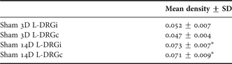
*Indicates statistically significant difference (P < 0.05) when compared with sham operation for 3 days.
Immunohistochemical staining for IL-6R and gp130 protein in DRG of naïve and CCI-operated rats
Like many membrane receptors, both IL-6R and gp130 have their soluble forms, sIL-6R and sgp130, that can be detected in the blood and extracellular fluids (Scheller and Rose-John, Reference Scheller and Rose-John2006). Processing of tissue sections for immunostaining allows detecting only membrane-bound forms of IL-6R and gp130.
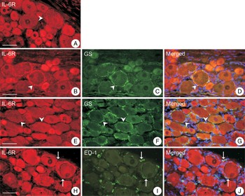
Sections of naïve lumbar DRG showed immunofluorescence staining for IL-6R in small- or medium-sized neuronal bodies while SGC did not display a distinct IL-6R immunoreaction (Fig. 4A). A similar pattern of IL-6R immunostaining was observed in L4–L5 DRG removed from CCI rats after 1 day. The unilateral CCI of the sciatic nerve for 3, 7 and 14 days induced an enhanced IL-6R immunofluorescence in SGC surrounding neuronal somata of ipsilateral L4–L5 DRG (Fig. 4B–D), but contralateral counterparts did not display increased immunostaining in SGC (Fig. 4E–G, Table 3). Results of double immunostaining for IL-6R and ED-1 revealed that ED-1+ macrophages infiltrating L4–L5 DRG did not exhibit immunohistochemical detection of IL-6R protein (Fig. 4H–J).
Representative sections immunolabelled for IL-6R in L4–L5 DRG of naïve rat (A) and L4–L5 DRG of ipsilateral (B,H) and contralateral (E) sides harvested from rats 3 (B) or 14 (H) days from unilateral CCI of the sciatic nerve. Double immunostaining for IL-6R and GS revealed that membrane-bound IL-6R protein was induced in SGC of DRG ipsilateral (B–D), but not in those contralateral, to CCI (E–G). Immunostaining for IL-6R was absent from ED-1+ macrophages (arrows) infiltrating ipsilateral DRG (H–J). Scale bars = 50 µm.

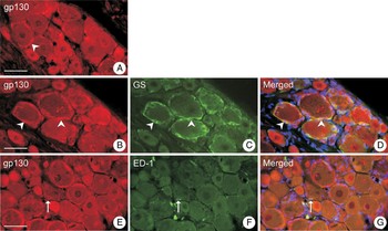
Moderate immunofluorescence staining for gp130 was localized in the small- and medium-sized neuronal bodies of DRG from naïve rats. In the same sections, a distinct immunostaining for gp130 was observed in the SGC enveloping weakly stained large neuronal bodies (Fig. 5A). A similar pattern and intensity (Table 3) of gp130 immunofluorescence was found in the sections of both ipsi- and contralateral L4–L5 DRG removed from CCI rats for all periods of survival. Double immunostaining for gp130 and GS or ED-1 proved that gp130 protein was detected in SGC (Fig. 5B–D) but not in ED-1+ macrophages (Fig. 5E–G). Sections of L4–L5 DRG removed from sham-operated rats displayed similar patterns and brightness of immunostaining for IL-6R and gp130 in the SGC when compared with those in naïve DRG (data not shown).
Representative sections illustrating immunostaining for gp130 protein in sections of L4–L5 DRG removed from naïve (A) and CCI-operated (B–G) rats. Small- and medium-sized neuronal bodies and SGC surrounding large neurons (arrowheads) displayed immunofluorescence staining of similar brightness when sections of DRG from naïve and CCI-operated rats were compared. Immunolabelling for gp130 and GS was co-localized (B–D) to verify expression of gp130 protein in SGC (arrowheads), but gp130 immunostaining was missing in ED-1+ macrophages (arrows) (E–G). Scale bars = 50 µm.

Immunohistochemical staining for pSTAT3 in DRG of naïve and CCI-operated rats
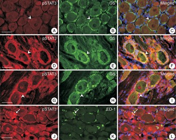
To detect STAT3 phosphorylation, we used an antibody that specifically recognizes the phosphorylated residue tyrosine 705. A faint pSTAT3 immunostaining was observed in small neuronal bodies and in some SGC surrounding neuronal somata of large diameter in sections of naïve DRG (Fig. 6A). A distinct elevation of pSTAT3 immunofluorescence was detected only in SGC of L4–L5 DRG removed from both sides of CCI rats for all periods of survival when sections were incubated under the same conditions as those of naïve DRG (Fig. 6D,G,J, Table 3). The presence of pSTAT3 protein in SGC was verified by double immunostaining for pSTAT3, and GS was used as a marker of SGC (Fig. 6D–I). Approximately 40% of ED1+ cells observed in the neuronal area of ipsilateral L4–L5 DRG were also immunolabelled with antibody against pSTAT3 (Fig. 6J–L). No increased pSTAT3 immunofluorescence was found in sections of DRG from sham-operated rats when compared with those from naïve rats.
Representative sections illustrating double immunostaining for pSTAT3 and GS in L4–L5 DRG of naïve rat (A–C) and L4–L5 DRG from ipsilateral (D–F,J–L) and contralateral (G–I) sides harvested from rat 7 days from unilateral CCI of the sciatic nerve. A faint immunofluorescence was found in SGC (arrowheads) of naïve DRG, but strong immunostaining was induced in SGC (arrowheads) surrounding large neuronal bodies of both ipsi- and contralateral DRG. A part of ED-1+ macrophages (arrows) displayed co-localization of pSTAT3 and ED-1 immunostaining (J–L). Scale bars = 50 µm.

CONCLUSIONS
• Unilateral CCI of the sciatic nerve induced mechanical allodynia and thermal hyperalgesia in hind paws ipsilateral to nerve ligatures. The L4–L5 DRG of CCI rats displayed elevation of IL-6 mRNA as well as immunostaining for IL-6, IL-6R and pSTAT3 proteins in SGC while the level of gp130 protein remained similar when compared with SGC of naïve DRG. Elevation of IL-6 mRNA and protein as well as pSTAT3 protein were observed bilaterally in SGC of L4–L5 DRG, but increased immunofluorescence for IL-6R protein was found only in SGC of ipsilateral counterparts from CCI rats.
• The results suggested that IL-6 signalling by membrane-bound IL6R in SGC of ipsilateral DRG might be associated with neuropathic pain induction. On the other hand, bilateral elevation of IL-6 and pSTAT3 indicated bilateral neuroinflammatory reaction of SGC to unilateral CCI.
DISCUSSION
All CCI rats used for immunohistochemical assay or in situ hybridization displayed signs of neuropathic pain in hind paws ipsilateral to nerve ligatures for all periods of survival. Contralateral hind paws exhibited only temporal significant changes of withdrawal thresholds for mechanical allodynia and thermal hyperalgesia. No neuropathic pain signs were found in hind paws of sham-operated rats.
Unilateral CCI of the sciatic nerve-induced bilateral elevation of IL-6 and pSTAT3 in SGC, but increased specific membrane-bound IL-6R was found in SGC only in ipsilateral L4–L5 DRG. In contrast, gp130 levels in SGC remained similar when compared with those of naïve DRG. Bilateral elevation of IL-6 mRNA staining was observed in both neurons and SGC of L4–L5 DRG while DRG from the sham-operated rats displayed increased IL-6 mRNA staining only in SGC.
Expression of IL-6 in SGC of DRG
IL-6 is a pleiotropic cytokine involved as a key signal molecule in the neuronal and immune responses to nerve injury. Moreover, there is considerable evidence for a role of IL-6 in nociception and hyperalgesia. For example, a direct injection of IL-6 causes hypersensitivity to thermal and mechanical stimuli (Oka et al., Reference Oka, Oka, Hosoi and Hori1995; DeLeo et al., Reference DeLeo, Colburn, Nichols and Malhotra1996; Brenn et al., Reference Brenn, Richter and Schaible2007). In addition, IL-6 −/− mice have shown reduced heat sensitivity in a hot plate behavioural test (Ramer et al., Reference Ramer, Murphy, Richardson and Bisby1998; Zhong et al., Reference Zhong, Dietzel, Wahle, Kopf and Heumann1999).
In response to sciatic nerve injury, IL-6 mRNA and protein have been found in bodies of sensory neurons in the corresponding ipsilateral DRG (Murphy et al., Reference Murphy, Grondin, Altares and Richardson1995, Reference Murphy, Borthwick, Johnston, Kuchel and Richardson1999). In our experiments, in addition to detecting IL-6 mRNA and protein in neurons, we observed enhanced staining for IL-6 mRNA and protein in SGC of L4–L5 DRG from both sides after unilateral CCI of the sciatic nerve as well as after sham operation. These results provide unequivocal evidence that unilateral CCI of the sciatic nerve induces synthesis of IL-6 protein bilaterally in the SGC of homonymous DRG. The bilateral elevation of IL-6 protein in DRG following unilateral CCI of the sciatic nerve has been confirmed by Western blot analysis (Brazda et al., Reference Brazda, Klusakova, Svizenska, Veselkova and Dubovy2009).
Increased staining for IL-6 mRNA and protein in SGC of L4–L5 DRG removed from sham-operated rats suggests that SGC may react to injury of any tissue. These results of activated SGC in DRG of sham-operated rats correspond with findings of SGC proliferation and activation in response to scarification or incision of the skin (Elson et al., Reference Elson, Simmons and Speck2004; Li et al., Reference Li, Xu, Liu, Zhang and Dai2008).
Expression of IL-6 receptors (IL-6R and gp130) in SGC of DRG
The biological activity of IL-6 is mediated by its binding to a specific membrane-bound IL-6R that associates with gp130, a transmembrane subunit necessary to activate IL-6 intracellular signalling. Alternatively, IL-6 may act via a soluble form of the IL-6R that is generated by limited proteolysis of the membrane-bound receptor or by alternative splicing of IL-6R mRNA (Jones et al., Reference Jones, Horiuchi, Topley, Yamamoto and Fuller2001). The complex of IL-6/sIL-6R acts as an agonist for cell types that do not express the cognate IL-6R but only the gp130 (Rose-John and Heinrich, Reference Rose-John and Heinrich1994).
Experiments with exogenous IL-6 in the presence of sIL-6R have suggested that neurons do not express cognate IL-6R, and the effects of IL-6 on neurons are believed to depend upon the presence of sIL-6R (Marz et al., Reference Marz, Otten and Rose-John1999; Obreja et al., Reference Obreja, Schmelz, Poole and Kress2002). In contrast to published assumptions, we found immunostaining for IL-6R in small- and medium-sized neuronal bodies of DRG removed from both naïve rats and those operated on CCI. In addition, immunostaining for IL-6R was induced in SGC of L4–L5 DRG ipsilateral to CCI of the sciatic nerve. Therefore, our results provide evidence that cognate IL-6R is constitutively expressed in small- and medium-sized DRG neurons and is induced by nerve injury in SGC.
Recently published findings describe expression of gp130 mRNA and protein in neuronal bodies rather than SGC of the rat DRG (Gardiner et al., Reference Gardiner, Cafferty, Slack and Thompson2002; Obreja et al., Reference Obreja, Biasio, Andratsch, Lips, Rathee, Ludwig, Rose-John and Kress2005). However, we found gp130 immunostaining in both neuronal bodies and SGC in naïve DRG and its unaltered levels in DRG after CCI, which is in accordance with our previously published results (Brazda et al., Reference Brazda, Klusakova, Svizenska, Veselkova and Dubovy2009). Our findings show that activated SGC in DRG ipsilateral to CCI of the nerve are endowed with IL-6 and its receptors to mediate IL-6 signalling in an autocrine manner in reaction to nerve injury. Moreover, the results suggest that IL-6 signalling by membrane-bound IL6R in SGC of ipsilateral DRG may have a potential role in neuropathic pain induction.
Expression of pSTAT3 in SGC of DRG
After IL-6R and gp130 assemblage, specific Janus kinases are activated which phosphorylate the STAT3 on specific residues (Ser727 and Tyr705). Phosphorylated STAT3 forms homo- or heterodimers with other STATs, which translocate into the nucleus where they exhibit transcriptional activities via binding to STAT binding elements in the promoter of specific genes involved in the cytokine response (Heinrich et al., Reference Heinrich, Behrmann, Muller-Newen, Schaper and Graeve1998).
Phosphorylation of STAT3 has been detected in neurons following nerve injuries (Schwaiger et al., Reference Schwaiger, Hager, Schmitt, Horvat, Streif, Spitzer, Gamal, Breuer, Brook, Nacimiento and Kreutzberg2000; Sheu et al., Reference Sheu, Kulhanek and Eckenstein2000; Qiu et al., Reference Qiu, Cafferty, McMahon and Thompson2005), but it is not yet known whether activated STAT3 occurs in SGC. We demonstrated that SGC display IL-6, gp130 and pSTAT3 immunostaining in DRG of both sides, while IL-6R immunoreaction was increased only in SGC of DRG ipsilateral to injured nerve. Since gp130 is expressed ubiquitously in DRG neurons and their SGC while STAT3 was activated bilaterally only in SGC by unilateral CCI, it might be expected that other molecules of the gp130-related family are involved in bilateral reaction of SGC to unilateral nerve injury. Alternatively, the bilateral activation of STAT3 in SGC might be mediated by IL-6 and its soluble IL-6R. A soluble complex of IL-6/sIL-6R circulating in blood can easily reach SGC of contralateral DRG because they do not contain a blood-nerve barrier (Arvidson, Reference Arvidson1979). We cannot exclude the trans-signalling pathway for activation of STAT3 in SGC also in DRG associated with damaged nerve. However, this alternative IL-6 signalling process needs to be confirmed by assessing the soluble complex of IL-6/sIL-6R in blood following CCI of the sciatic nerve.
Neuroinflammatory reaction of SGC to nerve injury and neuropathic pain induction
Generally, a nervous tissue injury alters the activities of somatic sensory pathways, resulting in behavioural hypersensitivity caused by both noxious stimuli (hyperalgesia) and normally innocuous stimuli (allodynia) (Scholz and Woolf, Reference Scholz and Woolf2002). Neuroinflammation is a proinflammatory cytokine-mediated process frequently associated with direct injury to the nervous system. An injury of peripheral nerves related with neuropathic pain is accompanied by neuroinflammation occurring also in DRG, where it involves activation of neurons as well as immune and glial cells (Hu and McLachlan, Reference Hu and McLachlan2002; Song et al., Reference Song, Zhang, Hu and LaMotte2003; Hanani, Reference Hanani2005).
The SGC are closely associated with the bodies of primary sensory neurons by transport and metabolism of various molecules, such as glutamate and glucose (Hanani, Reference Hanani2005), as well as by maintaining of ionic homeostasis (Vit et al., Reference Vit, Ohara, Bhargava, Kelley and Jasmin2008). Therefore, it is reasonable that SGC react immediately to the primary sensory neurons the afferent axons of which were lesioned following various types of peripheral nerve injuries. The changes include expression of molecules related with cytokine-mediated neuroinflammation. It is well known that SGC significantly influence activity of the primary sensory neurons in the sensory ganglia and modulate sensory function, including nociception (Cherkas et al., Reference Cherkas, Huang, Pannicke, Tal, Reichenbach and Hanani2004; Hanani, Reference Hanani2005; Takeda et al., Reference Takeda, Tanimoto, Kadoi, Nasu, Takahashi, Kitagawa and Matsumoto2007, Reference Takeda, Kitagawa, Takahashi and Matsumoto2008a,Reference Takeda, Takahashi and Matsumotob; Takeda et al., Reference Takeda, Takahashi and Matsumoto2009).
Neuroinflammatory response of SGC to nerve injury mediated by IL-6 may simultaneously contribute to induction of neuropathic pain and protection of neurons from further injury. Unilateral CCI of the sciatic nerve under aseptic conditions results in activation of SGC enveloping predominantly large DRG neurons to express IL-6, IL-6R and pSTAT3 while gp130 remains unaltered. Since IL-6R was induced only in SGC of DRG ipsilateral to CCI of the sciatic nerve, it is suggested that IL-6 signalling in SGC may contribute to hypersensitization of rat hind paw. On the other hand, enhanced IL-6 and pSTAT3 in SGC of DRG contralateral to injured nerve indicates their neuroinflammatory activation in relation to promoting, for example, contralateral DRG neurons to regenerate their axons following a conditioning lesion in vivo (Yamaguchi et al., Reference Yamaguchi, Ochi, Mori, Ryoke, Yamamoto, Iwata and Uchio1999; Ryoke et al., Reference Ryoke, Ochi, Iwata, Uchio, Yamamoto and Yamaguchi2000).
Acknowledgements
We thank Ms. Dana Kutějová, Ms. Marta Lněníčková, Ms. Stana Bártová and Mr. Lumír Trenčanský for their skilful technical assistance. This work was supported by grants 309/07/0121 and MSM0021622404.
Statement of interest
None.