Sudden cardiac death is a well-known complication in adult patients with aortic stenosis;Reference Campbell 1 however, the incidence and mechanism of sudden cardiac death in congenital aortic stenosis during childhood are less well defined, and thus controversy still exists about the beneficial effect of exercise restriction in preventing sudden cardiac death in this population.Reference Vahanian, Baumgartner and Bax 2 – Reference Brown, Dipilato and Chong 5 The controversy may be deepened with consideration of the possible beneficial effects of exercise in preventing cardiovascular events by putative mechanisms of ischaemic preconditioning.Reference Crisafulli, Mancardi, Marongiu, Rastaldo, Penna and Pagliaro 6
In this study, we report the case of a 15-year-old boy with aortic stenosis, in whom endurance exercise – football – may have contributed to the progression of severe left ventricular hypertrophy, which resulted in sudden cardiac arrest while he was warming up for football training. This case provides potentially important information when considering an indication of exercise restriction in children with aortic stenosis.
Case report
The patient was referred to another hospital because of heart murmur at the age of 1 year. He had a diagnosis of mild aortic stenosis with an estimated aortic peak instantaneous pressure gradient of 20 mmHg on echocardiography, and his annual follow-up examination showed a gradual increase in the peak instantaneous pressure gradient up to 55–60 mmHg at the age of 9 years. He started football club activity at the age of 10 years; however, there was neither worsening of the trans-aortic peak instantaneous pressure gradient nor development of left ventricular hypertrophy until he was 11 years old. Subsequently, he discontinued his annual consultations at the previous hospital while he continued his extracurricular activity of football. At the age of 13 years, when he entered junior high school, he showed normal electrocardiogram findings at the school health examination (Fig 1a). His school life was uneventful and without exercise restriction. The intensity and frequency, from weekly to daily, of his football training markedly increased after he started junior high school. At the age of 15 years, he experienced cardiopulmonary arrest during a warming-up exercise before football practice. He was successfully resuscitated with an automated external defibrillator and was transported to our hospital.
( a ) Electrocardiogram at the school health examination of the patient at 13 years of age. No significant sign of left ventricular hypertrophy is indicated. ( b ) Electrocardiogram on admission. A strain pattern is observed at V6, II, III, and aVf.

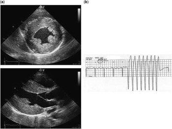
Echocardiography revealed severe aortic stenosis of the bicuspid aortic valve with a peak instantaneous pressure gradient of 126 mmHg and a mean estimated pressure gradient of 68 mmHg. The valve comprised a left coronary leaflet and a fused right and non-coronary leaflet. The left ventricular motion was excellent, whereas a marked thickening of the left ventricular wall was observed up to 18 mm in diastole (Fig 2a). Contrast-enhanced CT demonstrated a normal origin and course of the coronary arteries without apparent stenosis. The electrocardiogram showed a sharp contrast to that taken at the age of 13 years (Fig 1a), demonstrating a typical ST-T strain pattern in the left precordial leads (Fig 1b). Although he was waiting for aortic valve replacement, the electrocardiogram monitor showed non-sustained ventricular tachycardia without clinical symptoms (Fig 2b). At this point, we suspected the most possible cause for his cardiopulmonary arrest to be a ventricular tachycardia, because he could play football with full effort without showing any symptom of heart failure. Aortic valve replacement was successfully performed with an SJM17AP valve (St. Jude Medical Japan Co. Ltd., Tokyo, Japan). After the aortic valve replacement, there has been no episode of ventricular tachycardia for 3 years, and the left ventricular hypertrophy has gradually regressed.
( a ) Echocardiogram after the successful resuscitation of cardiopulmonary arrest. Marked ventricular hypertrophy is observed in the parasternal long-axis and short-axis views at the level of the papillary muscle. ( b ) Ventricular tachycardia observed at 2 weeks after resuscitation.

Discussion
The present report provides a potentially important evidence for considering exercise restriction in children with aortic stenosis. Several expert panels have recommended varying degrees of exercise restriction for patients with congenital aortic stenosis, including strenuous exercise and competitive sports, to prevent sudden cardiac death, restricting asymptomatic patients with moderate aortic stenosis, which is defined as a maximal instantaneous Doppler gradient of 40–70 mmHg, from some types of competitive sports, and restricting those with severe aortic stenosis – maximal instantaneous Doppler gradient of 70 mmHg – from all competitive athletics.Reference Vahanian, Baumgartner and Bax 2 – Reference Maron, Thompson and Ackerman 4 Nevertheless, these recommendations have been challenged by a recent study by Brown et al,Reference Brown, Dipilato and Chong 5 who showed an extremely low rate of sudden cardiac death in children with aortic stenosis, including severe aortic stenosis, who were not restricted from any exercise. In their study of 422 children with a wide range of aortic stenosis, including severe aortic stenosis with pressure gradient >70 mmHg, the authors reported that only 1 of 183 children (2541 patient-years) who were restricted from performing exercise died unexpectedly – during sleep but not during exercise.Reference Brown, Dipilato and Chong 5 The incidence was not different from that in patients with no exercise restriction.Reference Brown, Dipilato and Chong 5 On the basis of these results, they concluded that exercise restriction confers no advantage.
Even if our patient had not discontinued his annual checkup, exercise restriction would not have been necessarily recommended at the age of 13 years when his aortic stenosis was of moderate degree; however, the present case may suggest a potential benefit of exercise restriction in preventing progressive left ventricular hypertrophy in patients with moderate aortic stenosis who have a relatively low risk of sudden cardiac death. It is particularly notable that in our patient ventricular hypertrophy appeared to progress drastically during the last 2 years before cardiac arrest, from age 13 to 15 years, as clearly shown by the changes in electrocardiogram findings (Fig 1a and b) and aortic stenosis pressure gradient. During endurance and high-intensity exercise such as football, cardiac output increases markedly because of decreased peripheral vascular resistance and enhanced ventricular contractility. This results in an exaggerated valvular gradient and a marked increase in left ventricular systolic pressure during exercise. This can accelerate the progression of left ventricular hypertrophy,Reference Leischik, Spelsberg, Niggemann, Dworrak and Tiroch 7 which should increase left ventricular contractilityReference Senzaki, Iwamoto and Ishido 8 and the aortic stenosis pressure gradient, thus creating a vicious cycle exacerbating ventricular hypertrophy. Thus, from a viewpoint of delaying or preventing progressive left ventricular hypertrophy, exercise restriction may play a role even in patients with moderate aortic stenosis.Reference Baumgartner, Hung and Bermejo 9 This concept may be taken into account as a new indication criterion for exercise restriction in children with aortic stenosis, and deserves to be tested in further prospective studies.
In conclusion, although some recent research doubted the advantage of exercise restriction in children with aortic stenosis, we believe that exercise restriction prevents unexpected cardiac events not only by preventing possible ventricular ischaemic events and associated arrhythmia but also by potentially preventing progressive ventricular hypertrophy. Physical activity in these patients, if not restricted, should at least only be allowed with strict monitoring for hypertrophy progression.
Acknowledgements
The authors thank Dr Seiko Kuwata, Dr Clara Kurishima, Dr Yoich Iwamoto, Dr Hirotaka Ishido, and Dr Satoshi Masutani for helpful discussions, assistance with treatment, and critically reviewing the manuscript.
Financial Support
This research received no specific grant from any funding agency, commercial, or not-for-profit sectors.
Conflicts of Interest
None.
Ethical Standards
The family of the patient described provided their informed consent for the treatment of the child.