Introduction
Nodular fasciitis lesions are benign, reactive, proliferative masses localised in the subcutaneous tissue, and are usually of fibrous origin.Reference Enzinger and Weiss1 The condition was initially termed pseudosarcomatous fasciitis.Reference Badia, Rossi, Socri and Riminucci2 It is thought to occur as a reaction to trauma. Nodular fasciitis and related lesions are rapidly growing, painless, small, solitary masses which are usually diagnosed during the third to fifth decades of life.Reference Han, Hu, Yang, Wang and Huang3 The radiological findings of nodular fasciitis are generally non-specific. Nodular fasciitis lesions are commonly misdiagnosed as sarcoma, due to their rapid growth, fixation to underlying structures, increased mitotic activity and similar histopathological features.Reference Gelfand, Mirza, Kantor, Yu, Reale and Bondi4 They are most frequently located in the extremities and trunk, and maxillofacial lesions are relatively rare.Reference Leung, Shu, Chan, Chan and Chan5 Lesions are usually self-limiting, and may occasionally show spontaneous regression.Reference Gelfand, Mirza, Kantor, Yu, Reale and Bondi4
This paper presents the clinical, radiological and histopathological features of nodular fasciitis occurring in the parotid region, along with a literature review.
Case report
A 24-year-old man was referred to our hospital with a painful mass which had grown rapidly over the previous month, located in the right preauricular region.
Examination revealed a firm, immobile mass of nearly 2 cm diameter, located on the zygomatic bone near the upper border of the parotid gland. Its lateral surface was relatively well defined.
Ultrasonography showed a solid, 17 × 10 mm mass containing small, cystic areas. Ultrasonography failed to distinguish this mass from parotid tissue. No unusual blood flow was observed in the mass.
Fine needle aspiration cytological analysis showed the presence of proliferating fibroblasts, macrophages and adipocytes among the blood cells.
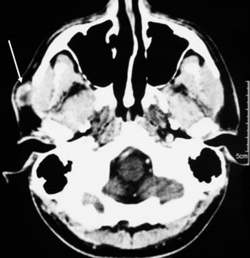
Axial computed tomography (CT) scanning of the maxillofacial region showed a contrast-enhanced, heterogeneous mass of approximately 2 cm diameter in anterosuperior contiguity with the parotid gland (Figure 1).
Contrast-enhanced, axial computed tomography scan showing nodular fasciitis in the right maxillofacial region in a 24-year-old man; scan shows a well defined soft tissue mass in the subcutaneous tissue, in anterosuperior contiguity with the parotid gland (arrow).

A superficial parotidectomy under general anaesthesia was planned. A modified Blair incision was performed, and the skin and the superficial musculo-aponeurotic system were elevated over the mass. It was noted that the mass lay at the upper border of the parotid gland, positioned over the zygomatic bone and frontal branches of the facial nerve. A plane of cleavage was noted between the lesion and the parotid tissue; therefore, a decision was made to proceed with excision rather than parotidectomy. The mass was gently dissected from the surrounding parotid tissue and underlying facial nerve branches, and then removed. Subcutaneous tissues and skin were sutured accordingly.
The post-operative period was uneventful.
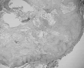
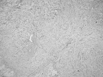
Histopathological analysis indicated nodular fasciitis (Figures 2 and 3).
Low power photomicrograph showing a rather circumscribed nodular fasciitis lesion with microcystic changes. (H&E; ×40)

Higher power photomicrograph showing that the lesion is composed of loosely arranged myofibroblast bundles, within an oedematous background with cheloid-type hyalinised fibrosis. (H&E; ×200)

Over a 21-month follow-up period, during which magnetic resonance imaging (MRI) findings were evaluated, no recurrence was detected.
Discussion
Nodular fasciitis is a benign, self-limiting proliferation of myofibroblasts and fibroblasts. It is often seen in the subcutaneous tissues, and rarely observed in the deep tissues.Reference Bernstein and Lattes6 The disease is thought to occur as a reaction to trauma or inflammation.Reference Han, Hu, Yang, Wang and Huang3 Lesions are usually located in the upper extremities (48 per cent), trunk (20 per cent), head and neck (20 per cent), and lower extremities (15 per cent).Reference Kim, Kim, Park, Baek, Byun and Kim7 In the head and neck region, lesions may be located in the facial skin, parotid gland, mandible, zygoma, tongue, cheek mucosa, throat and floor of the mouth.Reference Martínez-Blanco, Bagán, Alba and Basterra8 Nodular fasciitis may occur in all age groups, regardless of race or sex, but is most commonly observed during the third to fifth decades of life.
Nodular fasciitis is categorised into three subtypes, according to its anatomical localisation: subcutaneous, intramuscular and fascial. The subcutaneous form is most commonly encountered, with well circumscribed lesions. The intramuscular type is characterised by large-diameter lesions located within deep tissue, and often mimics soft tissue tumours. The fascial type involves lesions with irregular borders, located in the fascial plane.Reference Kim, Kim, Park, Baek, Byun and Kim7
The most important clinical symptom of nodular fasciitis is a fast-growing, solitary, firm mass. In general, it is not associated with pain or tenderness.Reference Han, Hu, Yang, Wang and Huang3 Pain, seizures and paraesthesia may occasionally be observed if peripheral nerve compression is present. Masses may lead to skin distortion and mimic malignant tumours. Lesions are usually smaller than 4 cm, and range in size between 0.5 and 10 cm.Reference Wang, De Schepper, Vanhoenacker, De Raeve, Gielen and Aparisi9 Our patient had a mass located in the subcutaneous tissue, of which he had been aware for one month prior to presentation.
Histologically, nodular fasciitis is characterised by fibroblasts with an immature appearance, arranged in short, irregular fascicles and bands, and accompanied by a dense reticulin network and varying amount of mature collagen.Reference Kransdorf and Murphey10 The lesion is usually attached to fascia, from which it extends into subcutaneous fat. The borders of the lesion are generally regular, although not encapsulated. While intense mitotic activity is present, cellular atypia is not observed. The lesion is highly vascularised, and inflammation is common.Reference Montgomery and Meis11 Nodular fasciitis can be classified as myxoid, cellular or fibrous, according to the type and amount of extracellular matrix. In young lesions, the myxoid component is generally diffuse, whereas in mature lesions the fibrotic component is more abundant.Reference Leung, Shu, Chan, Chan and Chan5 Lesions are often confused with sarcomas and other malignant tumours, due to their fast growth, hypercellularity and intense mitotic activity. However, several features of nodular fasciitis lesions are useful in the differential diagnosis, including their regular borders, relatively small size, myxoid content and absence of cellular atypia.Reference Han, Hu, Yang, Wang and Huang3
Nodular fasciitis lacks characteristic radiological features, and imaging findings are usually non-specific. However, imaging may be useful to exclude other elements of the differential diagnosis, including neurogenic tumours, minor salivary gland tumours, sarcoidosis, haemangioma, aggressive fibromatosis, dermatofibroma, and malignant fibrous histiocytoma. On CT and MRI scanning, subcutaneous nodular fasciitis usually appears as well defined lesions. Deeply located intramuscular lesions may not be so well defined, suggesting malignancy.Reference Shin, Lee, Cho, Han, Na and Choi12 In the case of solid lesions, hypercellularity and compact capillary networks may cause MRI and CT enhancement.Reference Wang, De Schepper, Vanhoenacker, De Raeve, Gielen and Aparisi9 In cystic lesions, MRI may show fluid-filled, mucoid spaces with related peripheral enhancement. Magnetic resonance imaging findings may vary depending on the type of nodular fasciitis. On T2-weighted MRI, hypercellular and myxoid lesions appears as hyperintense masses, while fibrous lesions are hypointense. This variation is due to differences in cellular, cytoplasmic and collagen content, and in extracellular fluid volume.Reference Wang, De Schepper, Vanhoenacker, De Raeve, Gielen and Aparisi9 In our case, only CT imaging was performed pre-operatively; this showed an enhancing lesion.
• Nodular fasciitis is a benign, self-limiting proliferation of myofibroblasts and fibroblasts, of possible traumatic aetiology
• Lesions can grow rapidly, and be misdiagnosed as sarcoma
• Radiological signs are non-specific
• Fibroblasts in the cytology specimen may indicate the diagnosis
• Surgical excision is the best treatment
• A rare parotid region lesion is reported
Treatment of nodular fasciitis includes total excision of the mass. Rarely, spontaneous regression may occur. After excision, recurrence is rarely reported.Reference Gelfand, Mirza, Kantor, Yu, Reale and Bondi4
Pre-operative suspicion of nodular fasciitis may arise based on a benign clinical course, history of trauma and cytological findings, and on the above-mentioned imaging signs. However, definitive diagnosis of nodular fasciitis depends on histopathological analysis.
We treated our patient with total excision rather than parotidectomy, based on the fact that the lesion was sited at the periphery of the parotid tissue. If the lesion had been in the middle of the parotid tissue, a superficial parotidectomy (at least) would have been warranted. Our patient experienced no recurrence over a 21-month follow-up period.