Introduction
South American camelids consists of at least three species, llamas (Lama glama), alpacas (Lama pacos), guanacos (Lama guanicoe) and vicunãs (Vicugna vicugna); their generic distinction is controversial. Traditionally, they were distributed at high altitudes (3600–5400 m) in South America where they are source of meat, hide, fibre and transport, and their feces are used for fuel and fertilizer (Díaz et al. Reference Díaz2016). In many Western countries, they are reared for recreation and the live animal is the commercial product.
Neonatal diarrhoea is a major concern in camelids. Coccidiosis is considered an important cause of neonatal diarrhoea in camelids, but there are also many reports of fatal coccidiosis in adult alpacas and llamas (Rosadio and Ameghino, Reference Rosadio and Ameghino1994; Cebra et al. Reference Cebra2007). More importantly, enteritis due to Eimeria species, especially E. macusaniensis, exposes camelids to secondary infections, including toxaemia due to Clostridium perfringens (Rosadio et al. Reference Rosadio2010).
Six species of Eimeria are named from camelids: E. macusaniensis, Eimeria lamae, Eimeria alpacae, Eimeria punoensis, Eimeria pervuiana and Eimeria ivitaensis (Yakimoff, Reference Yakimoff1934; Guerrero, Reference Guerrero1967; Guerrero et al. Reference Guerrero1971; Leguia and Casas, Reference Leguía and Casas1998; Cafrune et al. Reference Cafrune2009; Díaz et al. Reference Díaz2016). Of these, E. macusaniensis is the most prevalent and considered most pathogenic; mixed infections are common (Palacios et al. Reference Palacios2006; Cebra et al. Reference Cebra2007). Biologically, E. macusaniensis is unusual among all coccidian parasites in animals: its oocysts are the largest (>100 µm long) of any Eimeria, the prepatent period is very long (>30 days), and oocysts has been found in coprolites from prehistoric times in Peru and Chile (Levine and Ivens, Reference Levine and Ivens1970; Levine, Reference Levine1973; Foreyt and Lagerquist, Reference Foreyt and Lagerquist1992; Jarvinen, Reference Jarvinen2008; Fugassa et al. Reference Fugassa2008; de Souza et al. Reference de Souza2018).
Diagnosis of clinical coccidiosis in camelids is sometimes difficult because clinical signs can develop long before oocysts are detected in feces (Cebra et al. Reference Cebra2007). Eimeria macusaniensis is considered the most important coccidian of alpacas, and fatal infections have been reported in crias as young as 10 days old (Rosadio et al. Reference Rosadio2010). The pathogenesis remains unclear because schizonts preceding gamonts have not been clearly demonstrated. It is likely, that immature microgamonts have been misidentified as schizonts. Therefore, details of gametogony of E. macusaniensis in a naturally infected llama are described herein to facilitate the diagnosis of E. macusaniensis.
Materials and methods
A 5-year-old male llama (L. glama) from Illinois, USA, had a history of chronic weight loss. It eventually became so weak that it was euthanized in 1999 and necropsied by the attending veterinarian. The animal was well cared for, it had been dewormed and was housed with several other llamas by a private owner. Coccidiosis was suspected and formalin-fixed tissues were sent by the veterinarian to Dr D. M. Webb, Illinois Department of Agriculture, Animal Disease Laboratory (IDOA-ADL), Galesburg, Illinois, USA. All sections were stained with haematoxylin and eosin (HE). The initial diagnosis was E. macusaniensis-associated coccidiosis. No other cause was found, including toxaemia.
In December 1999, eight histological sections each containing two loops of the ileum of the llama were forwarded to me by post by Dr Webb, for diagnosis.
In 2017, review of the literature on camelid coccidia, I found that the description of endogenous stages of E. macusaniensis was not complete. Therefore, I requested more information on the llama from Illinois, but discovered that the IDOA-ADL is now permanently closed, files have been discarded and Dr Webb has retired. Therefore, no additional details are available about this case other than those presented here.
Retrospectively, three HE-stained slides were de-stained and re-stained with periodic Schiff reaction (PAS), counter stained with haematoxylin (PASH). All 16 sections of intestine were examined at 1000 × magnification, photographed and endogenous stages were measured using a digital camera (DP 73) fitted on an Olympus AX 70 microscope.
Results
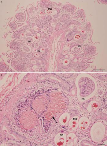
All histological sections were heavily infected with Eimeria. There was hyperplasia of epithelium with hypercellularity in the lamina propria and occasional plugging of the lumen of the gland of Lieberkühn in crypts (Figs 1A, B). Tissue structures were well preserved. There was no evidence of bacterial infection or necrosis. The parasitized host cell nucleus (probably an enterocyte) was indented but not hypertrophied. The heaviest infection was in crypts reaching to tunica muscularis mucosae.
Eimeria macusaniensis-associated lesions in the ileum of the naturally infected llama from Illinois, USA. Haematoxylin and eosin stain. Note microgamonts (mi) and macrogamonts (ma). (A) Hyperplasia with gametogonic stages in the lamina propria. Bar = 200 µm. (B) Plugging of lumen of a gland of Lieberkühn (arrow) with degenerating host cells. Bar = 50 µm.

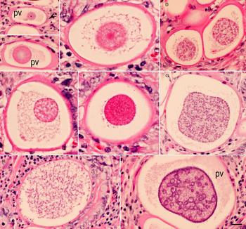
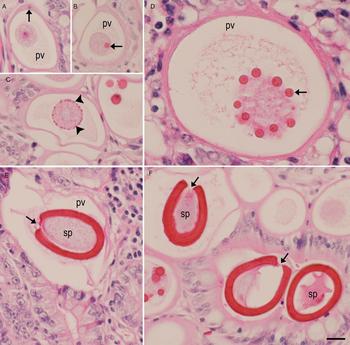
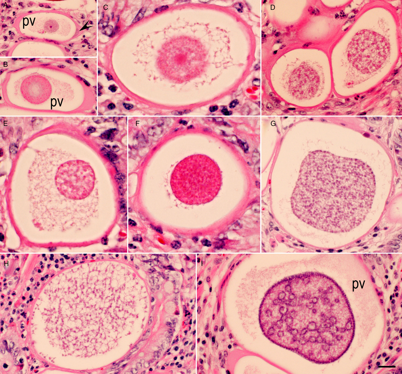
All endogenous stages of the coccidium were located in large parasitophorous vacuoles (pv) with empty spaces (Figs 1–5). Merozoites or merozoite-shaped early gamonts were not seen. The earliest gamont contained a central mass with a nucleus (Fig. 2A). At this stage, it was not possible to differentiate between micro- and macrogamonts (Fig. 2A). Some parasitized pv were up to 300 µm in long and the parasitophorous vacuolar membrane was 1–2 µm thick (Figs 2 and 3).
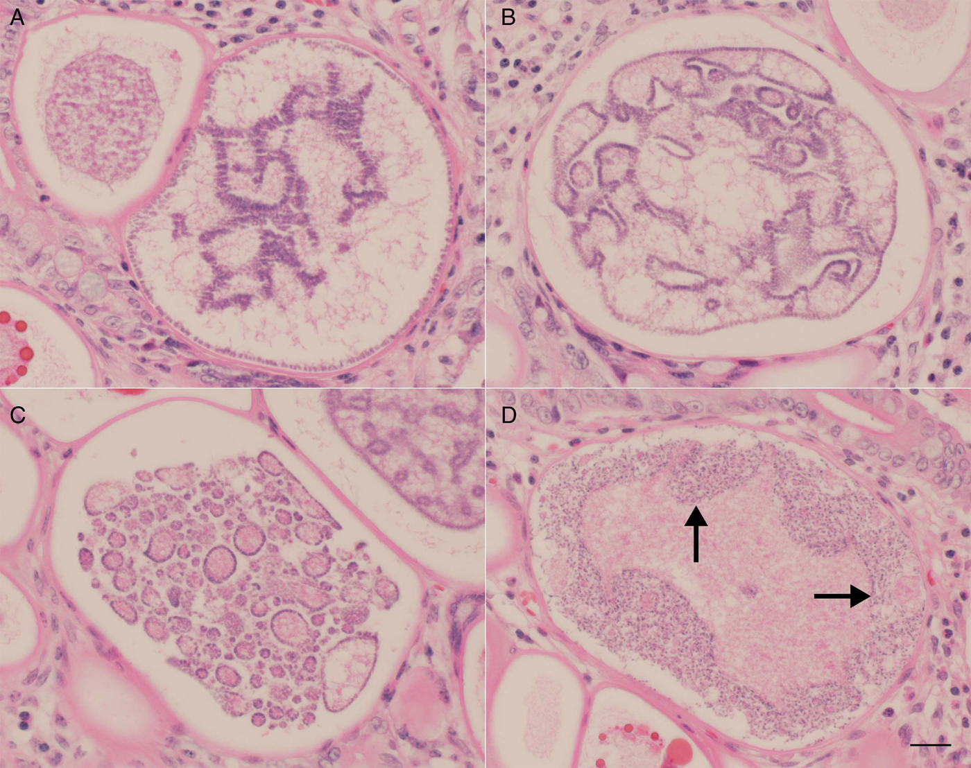
Immature stages of Eimeria macusaniensis microgamonts in the ileum of the llama. Haematoxylin and eosin stain. Bar = 20 µm and applies to all parts. The parasites are located intracellularly in large parasitophorous vacuoles (pv). The parasitized host cell nucleus (arrowhead) is indented but not hypertrophied. (A) Earliest macrogamont with a central nucleus and a nucleolus. (B) Microgamont with a nucleus with a lightly stained central area. (C) Strands of debris is present around the central nucleus. (D–F) Varying appearance of nuclei in microgamonts. (G,H) Numerous nuclei, some are at the periphery, whereas most are in the central mass. (I) Peripheral arrangement of nuclei. Some nuclei are in circles scattered throughout the macrogamont.
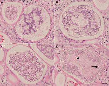
Maturing stages of Eimeria macusaniensis microgamonts in the ileum of the llama. Haematoxylin and eosin stain. Bar = 20 µm and applies to all parts. (A–C) Note different arrangements of nuclei in microgamonts. (D) Developing microgametes arranged peripherally (arrows). The central area contains amorphous material.
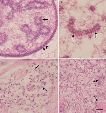
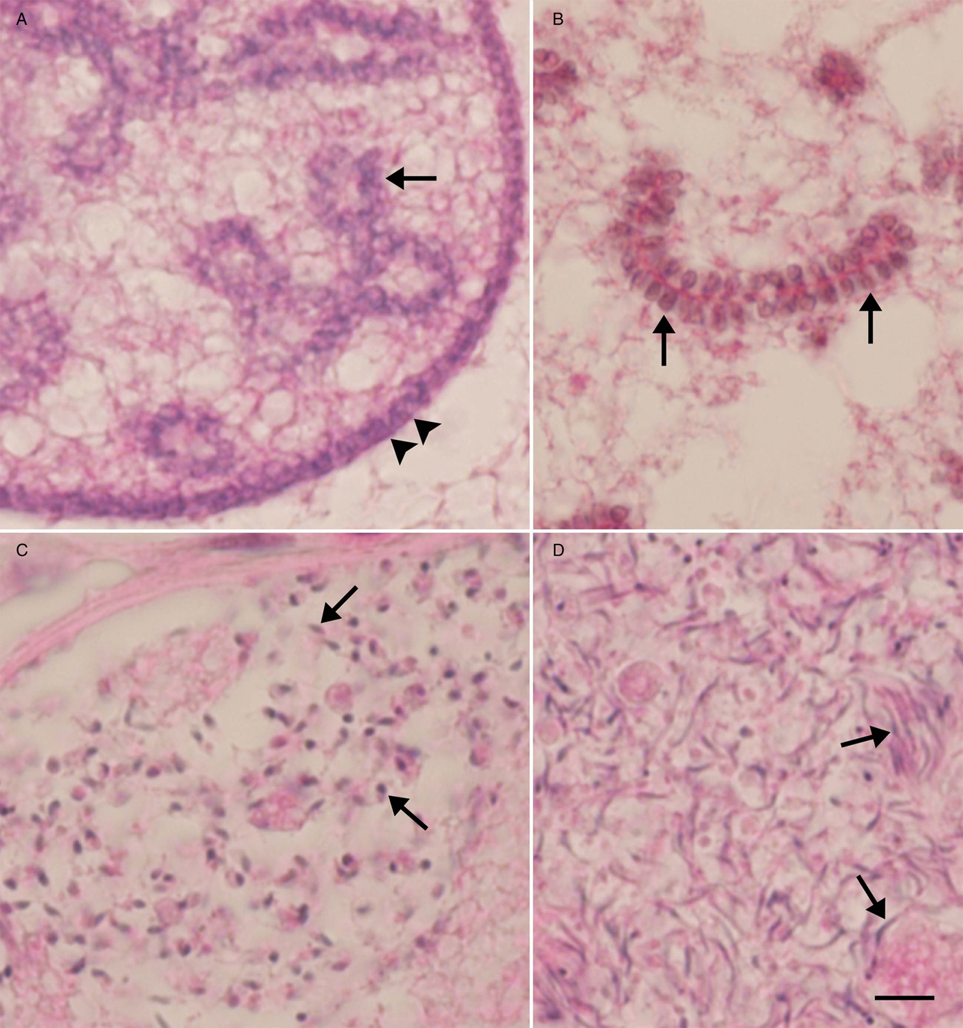
Higher magnification of Eimeria macusaniensis microgametes in the ileum of the llama. Haematoxylin and eosin stain. Bar = 20 µm and applies to all parts. (A) Peripherally arranged nuclei (arrowheads). Some nuclei are arranged in blastophores (arrow). (B) Linear arrangement of duplicating nuclei along a nuclear strand (arrows). (C) Developing microgametes (arrows). (D) Slender microgametes (arrows).
Developing stages of macrogamonts of Eimeria macusaniensis in the ileum of the llama. The parasites are located intracellularly in large parasitophorous vacuoles (pv). Haematoxylin and eosin stain. Bar = 20 µm and applies to all parts. (A) Early stage with a nucleus and a prominent nucleolus. The parasitized host cell nucleus (arrow) is indented but not hypertrophied. (B) Central mass with one eosinophelic body (EB, arrow). (C) Collapsed pv containing a central mass with peripherally arranged small EB (arrowheads). (D) Macrogamont with varying-sized EB. Also, note debris around the central mass. (E) Longitudinally cut oocyst with collapsed pv. Note micropyle and sporont (sp) occupying the whole interior of the oocyst. (F) Three oocysts, two of them have shrunken sporont (sp). Arrows in E and F point to micropyle.

The earliest microgamont had a 66 × 31 µm pv with a 17 µm diameter central mass (Fig. 2A). Subsequently, nuclei were differentiated as the parasite increased in size but nuclear staining varied in intensity. In some microgamonts, the nucleus/central mass were surrounded by debris (Fig. 2C, E). There were few to several hundred nuclei, some were arranged in whorls, others were in groups, in blatophores and still others were scattered at the periphery (Fig. 2D–I). Fissures were seen in nuclear arrangement, and some nuclei were arranged linearly (Figs 3A, B and 4A, B). In later stages of microgamonts, an amorphous mass was seen in between developing microgametes (Fig. 3D). Large microgamonts (excluding pv) measured 181 × 149 µm (164–199 × 114–166, n = 17). Mature microgametes were approximately 5 µm long; flagella were not identified (Fig. 4D). A residual body was not seen in any microgamont.
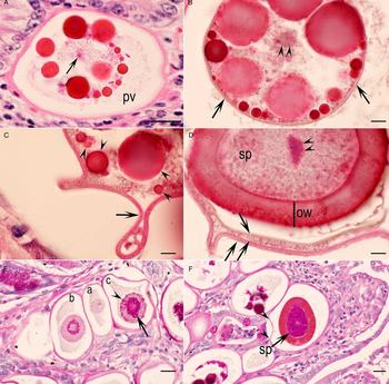
The earliest macrogamont-like structure had a 68 × 45 µm pv with a 25 µm diameter central nucleus with a prominent nucleolus (Figs 5 and 6). The earliest macrogamont could be recognized by PAS-positive amylopectin granules (Fig. 6E), and subsequently by eosinophilic wall-forming bodies (WFB) (Fig. 5B). Initially, the PAS-positive granules were located centrally around the nucleus (Fig. 6E), later spread to the entire gamont. The location, number and size of WFB varied (Figs 5C, D and 6A, B). The WFB varied in the intensity of staining and number. The WFB bodies were <1 to 19 µm in diameter (Figs 5C, D and 6A). Debris was present in some macrogamonts (Fig. 5D).
Higher magnification of macrogamonts and oocysts of Eimeria macusaniensis in the ileum of the llama. A–D = haematoxylin and eosin stain, bar in A, E and F = 20 µm, B–D = 5 µm. (A) Large pv containing 13 varying-sized eosinophilic bodies (EB) of varying staining intensity. Note a central nucleolus (arrow). Also, note the absence of an oocyst wall. (B) Part of a macrogamont with varying-sized EB of varying staining intensity. The EB are surrounded by a well-defined membrane (wall, arrows). Arrowheads point to nucleus. (C) Collapsed oocyst wall/membrane (arrow) enclosing varying-sized EB (arrowheads). (D) Part of a longitudinally cut oocyst. Note the sporont (sp), a central nucleus (double arrowheads) and thick oocyst wall (ow). Arrows point to parasitophorous vacuolar membranes. (E) Three young macrogamonts: (a) without WFB and PAS-positive granules, (b) with PAS-positive granules surrounding the nucleus and (c) with PAS-positive granules (arrow) and WFB (arrowhead). (F) An oocyst in the lamina propria. Note the entire sporont is PAS-positive. Also, note collapsed parasitophorous vacuolar membrane (arrowheads).

Formation of an oocyst wall was observed in a few specimens. Figure 6A shows a macrogamont with central nucleus and varying-sized WFB, but without oocyst wall. Figure 6B shows an early oocyst with a diameter of 51 µm surrounded by a membrane (wall) of uniform thickness; the largest light pink staining WFB are 19 µm in diameter and the smallest deep red staining are 2 µm in diameter; a central nucleolus is present. Figure 6C shows an oocyst with a crumpled wall but still containing the varying-sized WFB. Figure 6D shows a mature oocyst without WFB; the inner most oocyst layer surrounding the sporont is 9 µm thick. There are two other membranes (layers) that surrounded the thick-walled oocysts. The entire sporont was PAS-positive (Fig. 6F)
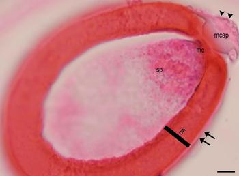
Oocysts were seen intracellularly in the lamina propria, and extracellularly in the lumen of glands (Fig. 5F). Some oocysts were degenerating and surrounded by inflammatory cells. Fully developed longitudinally cut oocysts were 72 × 50 µm (65–80 × 43–61, n = 13). The oocyst wall was up to 11 µm thick, homogenous and stained brilliantly with eosin. Oocysts had a micropyle and a micropylar cap was sometimes visible (Fig. 7).
Longitudinal section of a mature oocyst with a micropyle (mc), and micropylar cap (mcap) and thick oocyst wall (ow). There appears to be a thin parasitophorous vacuolar membrane (arrows) surrounding the oocyst.

Specimens deposited
Two sections of the ileum of the llama stained with HE (USNM no. 1473111) and PAS (USNM no. 1473112) were deposited in the National Museum of Natural History, Department of Invertebrate Zoology, Museum Support Center, MRC 534, Silver Hill Road, Suitland, Maryland, USA.
Discussion
There are discrepancies in the literature concerning the development of E. macusaniensis. It was initially found in a 5-month-old alpaca in Peru in 1967, but the species was not named until 1971 (Guerrero et al. Reference Guerrero, Hernandez and Alva1967, Reference Guerrero1971). The llama had a mixed infection with Eimeria species (Guerrero et al. Reference Guerrero, Hernandez and Alva1967). The endogenous stages could not be assigned to specific species of Eimeria. Hodgin et al. (Reference Hodgin1984) described gamonts and oocysts in the histological sections of a captive guanacos (L. guanicoe) that had died of leptospirosis and coccidiosis in Detroit Zoological Park, Detroit, Michigan, USA, and Schrey et al. (Reference Schrey1991) described endogenous stages in a 3-year-old llama from Wyoming, USA that had died of coccidiosis perhaps with concurrent viral infection. In both of these reports from the USA, the gamonts and oocysts found in the histological sections were attributed to E. macusaniensis. Hodgin et al. (Reference Hodgin1984) noted the absence of schizonts, whereas Schrey et al. (Reference Schrey1991) found meronts (schizonts). The macrogamonts, microgamonts and oocysts reported in both papers are essentially similar to the findings in the present study. Minor differences in the dimensions of parasites maybe related to several factors. The endogenous stages of E. macusaniensis are located in large pv that are difficult to compare because of shrinkage during fixation and autolysis. In the present study, the llama was euthanized and necropsied soon after death and there was no evidence of autolysis.
The main difference between the results reported by Schrey et al. (Reference Schrey1991) and the present study concerns their report of meronts. The pv containing meronts were 133–180 × 105–169 µm, and the meronts were 82–122 × 60–90 µm. The meronts contained blastophores, but the presence of merozoites was not mentioned. The illustration provided (Fig. 1 of Schrey et al. Reference Schrey1991) is identical to immature microgamonts illustrated in Figs 3 and 4 in the present study. It appears that the microgamonts were misidentified as meronts by Schrey et al. (Reference Schrey1991).
Several other scientists who reported fatal coccidiosis in alpaca and llamas also mentioned meronts and gamonts in the histological sections of small intestine (Rosadio and Ameghino, Reference Rosadio and Ameghino1994; Lenghaus et al. Reference Lenghaus, O'Callaghan and Rogers2004; Palacios et al. Reference Palacios2004, Reference Palacios2005, Reference Palacios2006; Rawdon et al. Reference Rawdon2006; Whitehead and Anderson, 2006; Cebra et al. Reference Cebra2007; Chigerwe et al. Reference Chigerwe2007; Schock et al. Reference Schock2007; Johnson et al. Reference Johnson, Stewart and Perkins2009; Rosadio et al. Reference Rosadio2010; Rojas et al. Reference Rojas2016), but did not describe the endogenous stages. It is likely that immature microgamonts were misinterpreted as schizonts.
To my knowledge, the schizogonic stages of E. macusaniensis are unknown and the pathogenesis of E. macusaniensis is partly unexplained. Cebra et al. (Reference Cebra2007) reported severe E. macusaniensis-associated coccidiosis in 49 camelids from Oregon, USA. The youngest affected alpaca was 21 days old. Histologic tissue sections of intestines were available from 13 camelids for which oocysts were not found in feces on first examination. Immature and mature gamonts were found in the tissues of 12 animals and oocysts in one of the 13 camelids on re-examination. In an outbreak of E. macusaniensis-associated coccidiosis on an Oregon farm, the earliest death occurred 20 days after camelids were moved to new farm; severe prepatent E. macusaniensis meront infection was confirmed histologically (Cebra et al. Reference Cebra2007). Thus, camelids can develop clinical coccidiosis before E. macusaniensis oocysts are found in feces.
As mentioned previously, oocysts of E. macusaniensis are morphologically distinctive, referred to as watermelon seeds or cut avacado. Oocysts in feces were 81–107 × 61–80 µm with 8.3–11.4 µm thick wall (Guerrero et al. Reference Guerrero1971). The wall consisted of three layers, an outer colourless 1.0 µm thick layer, middle 6.3–9.1 µm thick layer and 0.9 µm thin layer. In the present study, oocysts from feces were not available for comparison. However, in the histological sections stained with HE, two layers were recognized in mature oocysts. The outer most layer, interrupted at the micropyle, was lightly pink and <1.0 µm thick (Fig. 7). The main layer stained intensely with eosin was 7.0–11.0 µm thick and was interrupted at the micropylar end. The micropylar cap is stained light pink (Fig. 7).
In oocyst size, E. macusaniensis resembles another camelid Eimeria, E. ivitaensis. However, unlike E. macusaniensis oocysts, E. ivitaensis oocysts lack a micropyle. Differentiation of E. macusaniensis from E. ivitaensis in the histological sections may be problematic if oocysts are absent in feces or feces are not available for examination. Palacios et al. (Reference Palacios2006) reported dual fatal infections of E. macusaniensis and E. ivitaensis in histological sections of seven young (4–5 months old) alpacas from Peru. Schizonts (not illustrated) were detected in the jejunum and ileum; E. ivitaensis schizonts were said to be more pathogenic than those of E. macusaniensis. Location of endogenous stages of these coccidians differed; E. macusaniensis stages were in the jejunum, ileum, cecum and the ascending colon, whereas E. ivitaensis was confined to the jejunum and ileum (Palacios et al. Reference Palacios2006). The microgamonts and immature macrogamonts of both species of Eimeria were similar. However, their mature macrogamonts were different with respect to WFB. The WFB in E. macusaniensis were larger (13–18 µm) than those of E. ivitaensis (2–3.5 µm) (Palacios et al. Reference Palacios2006). The endogenous described in the present study were considered to be solely of E. macusaniensis. In the present study with E. macusaniensis, there was great variability in size and staining characteristics of WFB in E. macusaniensis.
Eimeria ivitaensis oocysts in feces are 83.5–98.6 × 49.3–59.1 µm (Leguía and Casas, Reference Leguía and Casas1998). The oocyst wall is 4.0–4.5 µm thick with three layers. Although E. ivitaensis oocysts are reported to lack a micropylar cap (Leguía and Casas, Reference Leguía and Casas1998), the micropylar cap may not be visible in the histological sections. In the present study, of all 16 sections examined, micropylar cap was found in only one E. macusaniensis oocyst. To my knowledge, morphology of developmental stages and oocysts has not been clearly described in the cases of E. ivitaensis-associated enteritis in camelids.
The development E. macusaniensis oocyst wall is unknown. In Eimeria species, the oocyst wall is initiated by the secretions of WFB. Most Eimeria species have two types of WFB, type 1 and type 2. The WFB1 are electron dense and larger in size than WFB2, and their characteristics are species-specific (Ferguson et al. Reference Ferguson2003). The WFB arise during the maturation of the macrogamont and form the oocyst wall. In some coccidians, there are additional veil-forming bodies (VFB); they form a veil-like outer covering on the oocyst wall that disappears when the oocysts are excreted in feces (Ferguson et al. Reference Ferguson2003). Transmission electron microscopy and staining with special stains are needed for the identification of WFB and VFB.
These WFB have not been defined for camelid coccidia. In the present study, the WFB varied in size and intensity of staining with HE; other stains were not used. Initially, the WFB were small (1–2 µm), few and were located at the periphery of the central mass. Subsequently, larger sized (19 µm) and deeply eosinophilic WFB were seen. The earliest indication of oocyst wall formation was a membrane (wall) surrounding the gamont; both small and large WFB were present at this stage but some lager WFB stained lighter than others. A crumpled wall (membrane) was seen around the macrogamont that had intact WFB. In fully formed oocysts, the WFB were absent. Transmission electron microscopic studies will be needed to study the oocyst wall formation in camelid coccidia.
The endogenous stages of E. macusaniensis described here resemble the development of Eimeria leuckarti of horses meticulously described by Barker and Remmler (Reference Barker and Remmler1972) in experimentally infected ponies euthanized 8, 10, 14, 23 and 28 to 52 days post inoculation (p.i.). Gamonts were seen 14 days p.i. Schizonts were not identified. The gametogony of E. leuckarti resembled gamonts of E. macusaniensis. However, the remarkable hypertrophy of host cell nucleus described by Barker and Remmler (Reference Barker and Remmler1972) was not found in E. macusaniensis. Results of Barker and Remmler (Reference Barker and Remmler1972) were confirmed by Hirayama et al. (Reference Hirayama2002) in the histological study of five horses naturally infected with E. leuckarti. Hirayama et al. (Reference Hirayama2002), using immunohistochemical methods, demonstrated that the hypertrophied host cell and the nucleus were enterocytes.
In conclusion, the morphological findings described in the present study should be helpful in the differential diagnosis of coccidiosis in camelids and removing the confusion between microgamonts and developing schizonts. Further studies are needed to describe asexual stages of camelid coccidia.
Acknowledgements
I would like to thank Dr D. M. Webb for sharing the case and permission to publish the results, and for his past collaborations. I am grateful to Camila Cezar, Fernando Antunes and Shiv Kumar Verma for help with illustrations and to Dr Ronald Fayer for advice.
Financial support
This research received no specific grant from any funding agency, commercial or not-for-profit sectors.