Introduction
Intrauterine growth restriction (IUGR) is a common perinatal outcome affecting approximately 5–10% of human infants worldwide. IUGR offspring not only exhibit increased neonatal mortality but are predisposed to postnatal metabolic complications and age-related disorders such as obesity or cardiovascular diseases.Reference Wang, Huo, Shi, Xu and Hutz 1 – Reference Visentin, Grumolato and Nardelli 3 Further, IUGR infants suffer from impaired development and function of organs involved in the digestion, absorption and metabolism of dietary nutrients.Reference Wang, Huo, Shi, Xu and Hutz 1
Epidemiological and experimental studies suggest that the poor postnatal growth and metabolic dysfunction of liver in IUGR individuals precede the later onset of pathologies.Reference Cianfarani, Agostoni and Bedogni 4 Several studies have shown impaired glucose regulation and insulin signaling as well as energy production in liver of IUGR newborns.Reference Lane, Crawford, Flozak and Simmons 5 , Reference Lane, Flozak, Ogata, Bell and Simmons 6 These abnormalities have in part, been attributed to redox imbalance and poor antioxidant defenses.Reference Wang, Chen and Li 7
The antioxidant defense systems are not fully developed in neonates and particularly deficient in IUGR infants increasing their vulnerability to oxidative stress.Reference Biri, Bozkurt and Turp 8 , Reference Hracsko, Orvos, Novak, Pal and Varga 9 The differences in oxidative stress response of digestive organs between IUGR and normal birth weight (NBW) infants have been previously reported.Reference Wang, Degroote and Van Ginneken 10 , Reference Mistry and Williams 11 Livers of IUGR animals also exhibit marked oxidative stress early in life as characterized by excessive reactive oxygen species (ROS) production, increased lipid peroxidation products and carbonyl protein levels, alterations in messenger RNA (mRNA) expression and activity of enzymatic antioxidants such as superoxide dismutase (SOD), catalase (CAT) and glutathione peroxidase (GPx).Reference Liu, Yao and Yu 12 , Reference Che, Xuan and Hu 13
The intensive ROS production in liver has mainly been attributed to the aerobic metabolism of dietary nutrients and xenobiotics exposure.Reference Circu and Aw 14 SOD, GPx and CAT which are the key components of enzymatic antioxidant detoxification machinery, develop during postnatally to protect vital functions of the liver against deleterious attack of ROS.Reference Xu, Wang, Guo, Liu and Feng 15 We therefore hypothesized that IUGR newborns are susceptible to pro-oxidant chemicals such as the dietary advanced glycation endproducts (AGEs). AGEs are present in heated infant formulas which are commonly consumed by IUGR newborns in order to match their postnatal growth to that of NBW infants. AGEs are products of Maillard reaction products (MRPs) which occurs as a result of condensation of reduced sugars and lysine-rich proteins during heat treatment of milk. Dietary MRPs induce free radical production through specific interaction with receptors for advanced glycation endproducts (RAGE) family, leading to pro-oxidative state and inflammatory response of target cells.Reference Kovacic and Somanathan 16 Elevated biomarkers of inflammation, oxidative stress and insulin resistance have been observed in human and animal models fed MRPs-rich diet.Reference Jaquet, Gaboriau, Czernichow and Levy-Marchal 17 , Reference Liu, Zheng and Zhang 18
MRPs-rich formula consumption plays a significant role in programming oxidative stress, especially by interfering with redox regulating physiological processes in the developing liver of IUGR neonates.Reference Elmhiri, Mahmood and Niquet-Leridon 19 These effects are mediated through the activation of RAGEReference Xu, Wang, Guo, Liu and Feng 15 and the nuclear transcription factor NFkB.Reference Elmhiri, Mahmood and Niquet-Leridon 19 , Reference Nass, Bayreuther and Simm 20 Our findings argue that among dietary MRPs, some are susceptible to considerably threat the long-term equilibrium of the redox system and their supply should be monitored in IUGR infants.Reference Abdennebi-Najar and Elmhiri 21 Interestingly, heated formulae contain also biologically active compounds among MRPs that may confer protection of the digestive system against dietary MRPs-induced oxidative stress.Reference Oh, Young Lee and Lee 22 The implication of microbiota on such protective effects are largely suggested.Reference Elmhiri, Hamoudi and Dou 23
Recently, the role of epigenetic processes in MRPs-induced programming effects has emerged with the discovery of microRNAs, an important class of RNA regulatory genes. MicroRNAs regulation is a novel mechanism by which changes in gene expression are brought at posttranscriptional levels by non-coding RNAs fragments of 19–24 nucleotides. MicroRNAs target-specific genes regulating cellular events such as cell cycle, differentiation, inflammation, energy metabolic pathways by promoting mRNAs degradation or inhibition of translation to proteins. MicroRNAs have also been incriminated to the MRPs-induced apoptosis and cellular effects in various cell typesReference Wu, Liu and Zeng 24 , Reference Li, Hou and Guo 25 and more recently to the control of the cellular oxidative stress.Reference Li, Zhou and Pei 26 , Reference Chang, Zhang and Tian 27 Although alterations in microRNAs expression play an essential role to the pathogenesis of digestive organs of IUGR offspring,Reference Lycoudi, Mavreli, Mavrou, Papantoniou and Kolialexi 28 , Reference Zhu, Wang and Yuan 29 a direct link among redox-sensitive microRNAs, dietary exposition to MRPs and oxidative stress response of IUGR consuming heated infant formula is still lacking.
In the present paper, we examined the impact of a high load of MRPs in milk formula on liver oxidative status and expression of microRNAs regulating oxidative stress pathways in adult IUGR piglets. Our findings link dietary MRPs intake to persistent hepatic oxidative stress as a result of altered antioxidant enzymes expression and activity with in IUGR juvenile pigs. We further show that these alterations are not mediated via microRNAs.
Materials and methods
Animals
At birth, 16 female piglets were selected from five sows accordingly to our previous experimentsReference Elmhiri, Mahmood and Niquet-Leridon 19 and kept with their mother to receive colostrum and mother milk during the first 2 weeks of postnatal life. At day 15, the piglets were randomly allocated to two groups of eight piglets and kept under observation for 72 h. Animals were fed with two different heated formula HHF (heated at 120°C for 20 min) or LHF (heated at 37°C for 5 min) from postnatal day 18 (PND18) till PND36. At PND36, weaned piglets were maintained under standard housing conditions and fed a regular diet ad libitum until euthanasia at PND54. Livers samples were collected, frozen in liquid nitrogen, and stored at −80°C for further analysis.
Ethical statement
The experimental protocol was designed in compliance with the guidelines for animal research of the French Ministry of Agriculture and all other applicable national and European guidelines and regulations for experimentation with animals (http://www2.vet-lyon.fr/ens/expa/acc_regl.html). The current experiment is a subset of a larger study investigating the effects of dietary glycation endproduct intake on long-term inflammation and oxidative stress in IUGR piglets. It was conducted in preclinical facilities of LaSalle institute that received the agreement from the French veterinary services to conduct animal research. The protocol was conducted in compliance with the relevant laws and institutional guidelines of French veterinary school of Nantes (Dr Thibaut Larcher, UMR703-PAnTher) and animal euthanasia and tissue collection were undertaken with the assistance of the scientific and technical staff of Unilasalle.
Measurement of AGEs in milk formula
The concentrations of the major AGEs, carboxymethyllysine (CML) in low and high temperature heated formula were determined by liquid chromatography tandem mass spectrometry method as described previously.Reference Niquet-Léridon and Tessier 30 CML was significantly higher in the HHF (147.32±1.53 µg/g protein) as compared to the LHF (12.56±0.35 µg/g protein) formula.
Determination of the total protein carbonylation levels
The analysis of liver protein carbonylation was performed by Oxiproteomic SAS. Total proteins were quantified by Bradford method and separated on sodium dodecyl sulfate polyacrylamide gel electrophoresis electrophoresis. Proteins were labeled with a fluorescent reagent SyproRubyTM (Life Technologies, USA) and detected at a specific wavelength of λex390/λem595 for total proteins and λex480/λem530 for carbonylated proteins. Fluorescence band intensities were quantified using Image J software (NIH, USA).
Antioxidant enzymes activity and total glutathione (GSH) levels analysis
Frozen liver tissue samples were weighed and divided into three samples. One sample (250 mg) was used to determine the total protein content, concentration of the total GSH and enzymatic activity of GPx, SOD, CAT. Liver tissue samples were ground and homogenized in a buffer solution, centrifuged and supernatants analyzed using a UV-light spectrophotometer microplate reader according to the manufacturer recommendations (Cayman Chemical Company). Activity values were normalized to proteins concentration (BCA protein assay kit; Sigma Aldrich).
Mn-SOD and GPx-1 gene expression analysis
Total RNA was isolated from 30 mg of liver samples using RNeasy PLUS mini kit (cat.74134; Qiagen, France) according to the manufacturer’s manual. The purity and concentration of total RNA were measured by a spectrophotometer at 260 and 280 nm. RNA isolated from each sample was reverse transcribed using QuantiTect Rev Transcription Kit (cat. 205311; Qiagen). The complementary DNA (cDNA) samples were diluted in nuclease free water (1:10). Real-time quantitative polymerase chain reaction (RT-qPCR) were conducted using SYBR Green dyes (Qiagen) and published primers for GPx-1, Mn-SOD and β-actin housekeeping gene used for gene expression normalization.Reference Elmhiri, Hamoudi and Dou 23 Each sample was analyzed in triplicate, and the expression was calculated according to the 2-ΔΔCt method.
MicroRNAs expression analysis
In total, 16 fresh livers were collected during surgery and were immediately frozen in liquid nitrogen. MicroRNAs were extracted from 20 mg of liver tissues using mirVana miRNA isolation Kit (AM1560; Thermo Fisher Scientific, France), according to the manufacturer’s protocol. The expression of microRNAs was measured by quantitative reverse transcription PCR using TaqMan MicroRNA Assays. The PCR primer pairs for miRs-21 and miRs-155 were obtained commercially from Thermo Fisher Scientific. In brief, cDNA was synthesized from total RNA by using the gene-specific primers according to the protocol of TaqMan MicroRNA Reverse Transcription Kit (Thermo Fisher Scientific). RT-PCR was performed by using a PCR primer (TaqMan MicroRNA Assays; Thermo Fisher Scientific) and reagents of TaqMan Universal PCR Master Mix (Thermo Fisher Scientific) in a StepOne apparatus (Software Version 2.3; Thermo Fisher Scientific). The 10 µl PCR reaction mixture included 2 µ RT product, 5 µl buffer TaqMan Universal Master Mix (Thermo Fisher Scientific) and 0.5 µl PCR primer (TaqMan MicroRNA Assays; Thermo Fisher Scientific). These reaction mixtures were incubated at 95°C for 20 s, followed by 40 cycles of 95°C for 1 s, 60°C for 20 s. The expression levels of miRs-21 and miRs-155 in each sample were measured in terms of threshold cycle (Ct) value and normalized to U6, which were used as an internal control. The relative quantitative was determined using the 2(−ΔΔCt) method.
Statistics
Effects of the dietary MRPs on proteins carbonylation, enzymes activities, GPx-1, Mn-SOD mRNA levels in liver tissues were analyzed by the Mann–Whitney tests using SPSS version 19 software (SPSS Inc., Chicago, IL, USA). Statistical analyses were performed using eight samples per treatment group except for protein carbonylation (n=5). Significant differences were noted at a p value of ⩽0.05.
Results
Daily formula intake and body weight
Birth weights of piglets in both groups were comparable (LHF 1.2±0.14 kg v. HHF 1.18±0.06 kg). Heating of formula did not affect the body weights at PND54 (LHF 22.87±2.36 kg v. HHF 21.37±0.68 kg) nor did it impact formula intake during the experimental period (LHF 219±20 ml/kg/day, HHF 200±20 ml/kg/day).
Protein carboxylation, and the expression and activity of antioxidant enzymes
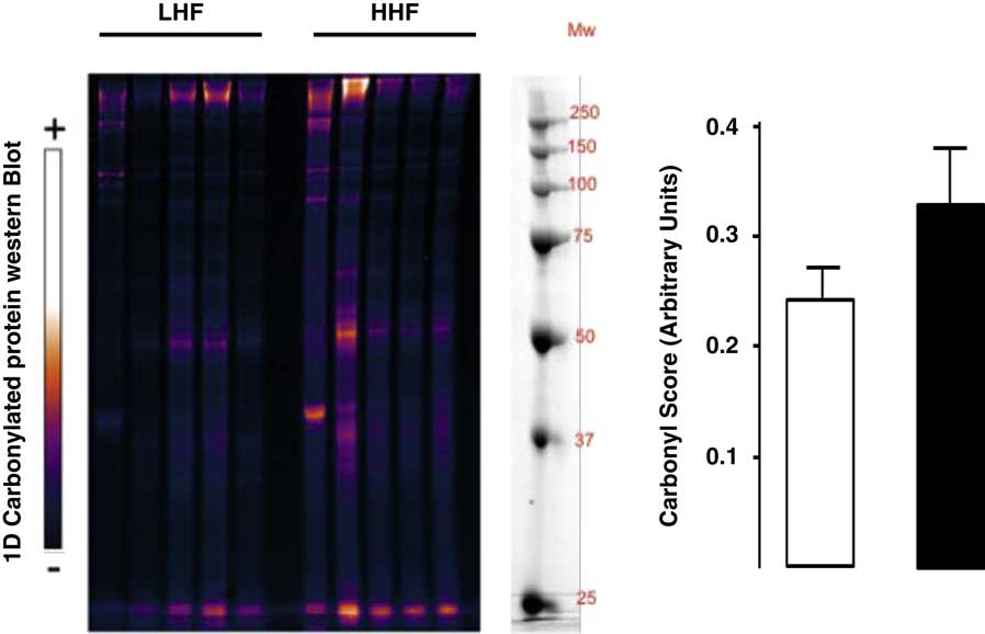
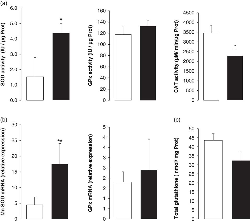
The protein carbonyl levels were slightly increased, but not significantly, in liver of the HHF group as compared to LHF group at PND54 (Fig. 1). In addition, we observed a significant increase in SOD activity (P<0.05; Fig. 2a) and Mn-SOD mRNA expression (2.9-fold and 3.8–fold, respectively) (P<0.02; Fig. 2b) in liver of HHF as compared with LHF group. HHF group also exhibited a lower CAT activity as compared with LHF group (−33%, P<0.05; Fig. 2a). The consumption of HHF did not affect the hepatic GSH levels (Fig. 2c) nor GPx (enzyme involved in GSH redox cycle) expression or activity (Fig. 2a and 2b).
Total protein carbonyl levels in liver tissue of adult intrauterine growth-restricted offspring fed LHF (□) or HHF (■) diet. Data are expressed as mean±s.d.; HHF (n=5) and LHF (n=5).

Superoxide dismutase (SOD), glutathione peroxidase (GPx), catalase (CAT) activity (a), relative expression of antioxidant enzyme Mn-SOD and GPx messenger RNA (mRNA) (b), and total glutathione content (c) in liver tissue of adult intrauterine growth-restricted offspring fed LHF (□) or HHF (■) diet. Data are expressed as mean±s.e.m. Significant difference between HHF (n=8) and LHF (n=8) groups are: *P<0.05, **P<0.02.

MicroRNAs level
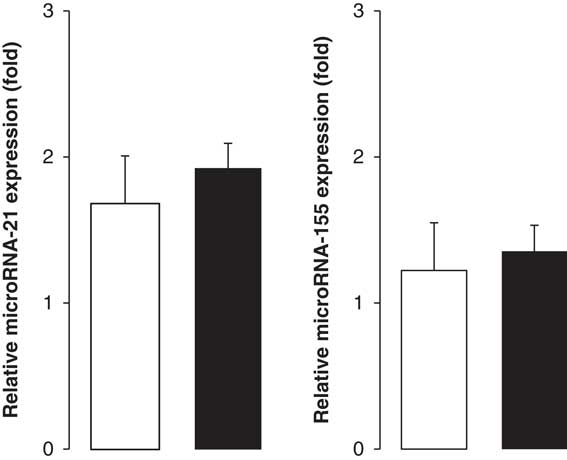
Recent studies have documented aberrant expression of several microRNAs under cellular oxidative stress conditions. In this study, we investigated the effect of HHF diet on the expression level of the ROS-sensitive microRNA-21 and microRNA-155.Reference Alinovi, Goldoni and Pinelli 31 – Reference Song, Ji, Zou and Wan 33 Thus, we measured the expression levels of these microRNAs in both HHF and LHF groups at PND54 using RT-qPCR normalized to U6. Our results indicate that both groups exhibited similar levels of miR-21 and miR-155 expression (Fig. 3).
Relative expression of microRNA-21 and microRNA-155 in liver tissue of adult intrauterine growth-restricted offspring fed LHF (□) or HHF (■) diet. Data are expressed as Mean±s.e.m.

Discussion
MRPs have been described as a source of oxidative stress in many tissues. RAGE receptor is expressed in several cell types including hepatocytes and hepatic stellate cellsReference Leung, Herath and Jia 34 , Reference Kuhla, Trieglaff and Vollmar 35 and its interaction with MRPs initiates the activation of nicotinamide adenine dinucleotide phosphate oxidase and the generation of reactive oxygen intermediates.Reference Patel, Baker and Liu 36 Our findings show that the consumption of a high load of dietary MRPs present in formula milk adversely impacts the long-term oxidative status of IUGR livers. This may promote age-related liver damage and pathologies (e.g. non-alcoholic steatohepatitis, cirrhosis and hepatic cancers) seen in IUGR offspring.Reference Basta, Navarra and De Simone 37 , Reference Hyogo and Yamagishi 38
IUGR are prone to oxidative damage due to their higher ROS metabolism combined with immature antioxidant systems as compared with the normal growth infants.Reference Hracsko, Orvos, Novak, Pal and Varga 9 Liver is one of the most important site of MRPs endogenous synthesis and a specific target of AGEs which promote oxidative stress responses.Reference Santos, Valentim, de Araújo, Ataide and Goulart 39 We have previously reported the pro-oxidant property of dietary MRPs exposure in liver of IUGR piglets.Reference Elmhiri, Mahmood and Niquet-Leridon 19 Indeed, we highlighted significant augmentation of lipid peroxidation products in liver and an accumulation of carbonylated proteins in kidneys of IUGR juvenile pigs. In the present study, livers of IUGR juvenile pigs fed HHF showed only a slight increase of protein oxidation contents. We suggest that the lower vulnerability of the liver (as compared with kidney) and its higher adaptive competence to oxidative damage is related to its higher antioxidant basal levels.
Proteins oxidation induces cellular and molecular damage, and/or conformational changes leading to impaired function.Reference Baraibar, Liu, Ahmed and Friguet 40 Protein oxidative damages have been reported to target proteins involved in ROS metabolism in liver.Reference Chaudhuri, de Waal and Pierce 41 Activity of common enzymes protecting tissues from oxidative damage may be altered by these structural modifications.
In the present study, we have focused on the expression and enzymatic activity of antioxidant enzymes (SOD, GPx and CAT). We have shown that the GSH levels and the mRNA expression and activity of GPx (known to prevent cell membrane polyunsaturated fatty acids peroxidation) was not impacted by MRPs. In contrast, the expression and activity of SOD and CAT (enzymes involved in scavenging oxygen free radicals) were markedly altered in HHF group. Indeed, HHF induces reduction of CAT activity though with increased SOD expression and enzymatic activity. These findings are consistent with observations reported in rats studies on heart and liver,Reference Gemelli, Carvalho and de Andrade 42 , Reference Borges, Nogueira, Panatieri, Rocha and Zeni 43 and in human studies on lungs.Reference Ho, Zheng and Comhair 44 These changes indicate that the liver antioxidant mechanisms are not able to efficiently detoxify ROS and preserve the redox status in IUGR juveniles.
The current study shows that the presence of a high load of MRPs in the milk formula increases the risk of oxidative stress which is known to be a risk factor for several metabolic diseases such as insulin resistance and overall inflammation. Our results are consistent with previous reports that demonstrate oxidative damage and significant changes in antioxidant enzymes expression and activity as a result of MRPs exposure in adult animals and in cell culture models.Reference Guimarães, Empsen, Geerts and van Grunsven 45 – Reference Wang, Yu and Yan 48 Although MRPs-induced oxidative stress and age-related diseases is well documented,Reference Vlassara 49 , Reference Prasad, Imrhan, Marotta, Juma and Vijayagopal 50 their potential role during development is unknown. Thus, investigation of the impact of long-term MRPs exposure on hepatic antioxidant systems of juvenile animals may constitute an important step forward in understanding its role in programmed oxidative stress. The expression and activity of antioxidant enzymes such as SOD and GPx gradually increase following birth, leading to sustained hyperoxia and oxygen metabolism acceleration, growth and maturation of organs.Reference Xu, Wang, Guo, Liu and Feng 15 , Reference Bucher and Roberts 51 Thus, abnormal developmental expression of antioxidant enzymes increases the risk of ROS escape and cell injury in infants. ROS accumulation induces a cascade of reactions that include lipids, proteins and DNA oxidative damages, leading to specific activation of redox-sensitive signaling pathways that regulate cytokine expression, mitochondrial respiration and immune response that result in hepatocytes necrosis, apoptosis, hepatic stellate cell activation and fibrosis.Reference Al-Griw, Alghazeer, Al-Azreg and Bennour 52 , Reference Singal, Jampana and Weinman 53 The hepatic antioxidant systems are controlled by epigenetic mechanisms such as changes in microRNAs expression pattern.Reference Fierro-Fernández, Miguel and Lamas 54 , Reference Yin, Guo, Zhang and Zhang 55 MicroRNAs have more recently been identified to control oxidative stress in liver and related diseases.Reference Circu and Aw 14 Excessive ROS production in cancer cell exerts a control on the microRNA processing machinery and transcription factors of microRNA genes that affect microRNAs expression level, such as those involved in the control of the cellular redox homeostasis.Reference He and Jiang 56
The exposure to MRPs during early infancy may affect the expression of microRNAs regulating antioxidant enzymes SOD, CATReference Ling, Li and Xu 32 , Reference Patel, Khan and Bhardwaj 57 and molecules such as GSH.Reference Jaquet, Gaboriau, Czernichow and Levy-Marchal 17 In turn, this amplifies redox imbalance in liver and increases the risk of metabolic disease for IUGR consuming heated infant formula. However, our hypothesis that microRNAs targeting free radical scavengers or ROS mediated signaling pathways genes may play a critical role in MRPs-induced oxidative stress in liver of animals fed HHF needs further confirmation as microRNAs expression was unaltered despite changes in the antioxidant enzymes.
Postnatal maturation of the hepatic antioxidant systems makes the developmental window a critical period of susceptibility for liver to pro-oxidant compounds. Our findings of the vulnerability of IUGR piglets to dietary MRPs in causing oxidative insults that may predispose to liver associated diseases later in life are consistent with the current theories on DOHaD effects.Reference Luo, Fraser and Julien 58
In conclusion, the present study provides the first evidence of the MRPs-induced persistent oxidative damages and a loss of antioxidant capacity in the liver of juvenile IUGR piglets. In this context, high load MRPs in milk and milk products may not be suitable in the diet of IUGR infants due to their high susceptibility to oxidative stress. MRPs intake monitoring and antioxidants supplementation during early infancy may be considered for the management of IUGR population in order to prevent developmental programming of adult diseases.
Acknowledgment
The authors thank Dr Mina Desai, David Geffen School of Medicine, for improving the document by English corrections and comments.
Financial Support
The authors have no financial relationships relevant to this article to disclose.
Conflicts of Interest
None.