Introduction
In 2017, I was invited to the Museum of Evolution of Uppsala University to sort through a collection of lichenicolous fungi left by the late Rolf Santesson. This collection contained, among others, specimens on Coccocarpia and Trachyderma hosts annotated as ‘Baryonectria coccocarpiae R. Sant. ined.’. Subsequent examination of these specimens revealed that they belong to the lichenicolous genus Lichenotubeufia Etayo (Etayo Reference Etayo2017), and are hitherto unknown to science. The aims of the present paper are: 1) to describe two new species of Lichenotubeufia; 2) to present a comparative table of diagnostic characters of species of this genus and an updated key for their identification.
Materials and Methods
Microscopy was carried out, and images were captured, using a Zeiss Axio Zoom.V16 microscope and a Zeiss Axio Imager.A1 microscope equipped with Nomarski differential interference contrast optics and fitted with a Zeiss AxioCam MRc5 digital camera. Microscopic characters were studied using sections hand-cut with a razor blade and mounted in water, 10% potassium hydroxide (K), Lugol's iodine directly (I) or after a K pretreatment (K/I), phloxine directly or after K pretreatment. Measurements were taken from water mounts unless otherwise indicated. The length, width and length/width ratio (l/w) of the ascospores, as well as the size of the perithecia, excipular hairs and asci, are given as (min‒)(x̄ − SD)‒(x̄ + SD)(‒max), where ‘min’ and ‘max’ are the extreme values observed, x̄ the arithmetic mean and SD the corresponding standard deviation. Colours were named according to Kornerup & Wanscher (Reference Kornerup and Wanscher1978). Voucher specimens are held at UPS.
Results
The new species
Lichenotubeufia etayoi Zhurb. sp. nov.
MycoBank No.: MB 841975
Differs from Lichenotubeufia cryptica mainly in the larger ascomata (150–360 μm vs 130–200 μm diam.), sometimes with a distinct papilla, a wider exciple (30–50 μm vs 15–20 μm wide) and slightly wider ascospores (3–5 μm vs 2–3.5 μm wide).
Type: Chile, Valdivia Province, Lake Rinihue, Enco, edge of the rainforest on the lake shore, on the thallus of Trachyderma gayanum growing on Ugni molinae and other shrubs, 12 October 1940, R. Santesson S422 (UPS F-857602—holotype).
(Fig. 1)
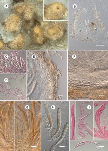
Lichenotubeufia etayoi (holotype). A, habitus of ascomata on the thallus of Trachyderma gayanum. B, ascoma in cross-section. C, excipular hairs. D, paraphysoids. E, lateral exciple in cross-section. F, basal exciple and hypothecium-like layer in cross-section. G, asci with spores. H, asci with spores. I, ascus and ascospores. B, E & F, in water; C, D & I in phloxine after K; G in I; H in K/I. Scales: A & B = 100 μm; C–I = 20 μm. In colour online.

Lichenicolous ascomycete growing on Trachyderma. Ascomata perithecia, light orange, subglobose ovoid or narrowly ampulliform, (150–)180–300(–360) μm diam. (n = 34), with a central ostiole 20–45 μm diam., sometimes with a distinct papilla 100–150 μm diam., not collapsing when dry, entirely covered with subhyaline excipular hairs; superficial, scattered or in groups, occasionally adjacent, mainly growing on the upper surface of the host lobes, often along their edge, where they also rarely occur on the lower surface, including rhizines. Excipular hairs colourless, with a thin translucent lumen, sometimes undeveloped in places, and a thick opaque wall, straight, (18–)40–84(–113) × (3–)4–5(–5.5) μm (n = 45), isodiametric or somewhat gradually tapering above, with a rounded, not enlarged apex, rarely branched below, occasionally with septa or possibly with septum-like connectors, not gathered in fascicles. Exciple continuous, same structure throughout, in surface view orange-white/very pale orange (slightly decolourizing in K), pseudoparenchymatous, composed of cells 5–14 μm diam., with walls 1.5–6 μm wide; in cross-section hyaline (inside) to orange-white (outside), 30–50 μm wide, composed of isodiametric or tangentially elongated cells 2–15 μm long, with walls 1–7.5 μm wide. Hymenium not inspersed, I−, K/I−. Hamathecium composed of abundant, persistent, branched, anastomosed paraphysoids, 1–3.5 μm wide, attached to the subhymenium and lateral exciple (along its entire length) and filling the space between the asci and the entire upper part of the perithecium. Subhymenium indistinct. Hypothecium-like layer between subhymenium and basal exciple hyaline, 35–85 μm tall, of an unclear structure partly resembling textura globulosa, composed of cells 1.5–8 μm long. Asci 8-spored, subcylindrical, gradually tapering below, short pedicellate, rounded at the apex, without any distinct apical apparatus, wall not thickened at the apex (in water), (120–)133–169(–181) × (10–)11–14(–16) μm (n = 35; in water, I or phloxine), I−, K/I− except for the orange-coloured plasma. Ascospores hyaline, acicular, gradually tapering at both ends, straight, (110–)128–157(–162) × (3–)3.5–4.5(–5) μm, l/w = (28–)32–42(–45) (n = 15, in water or I), 10–13-septate, not constricted at the septa, smooth-walled, without perispore, with many small guttules as seen in K, arranged in the ascus in one bundle.
Conidiomata not observed.
Etymology
The species is dedicated to the eminent Spanish lichenologist Javier Etayo who has made an enormous contribution to the study of the lichenicolous fungi of South America.
Distribution and host
The new species is known from the type locality in the Valdivian temperate rainforests of Chile, growing on the thallus of Trachyderma gayanum, not visibly damaging the host.
Notes
Morphologically Lichenotubeufia etayoi strongly resembles L. cryptica Etayo & Flakus growing on Lobariella; however, the latter differs in having smaller perithecia, 130–200 μm diam., a thinner exciple, 15–20 μm wide, and slightly narrower ascospores, 2–3.5 μm wide (Flakus et al. Reference Flakus, Etayo, Miadlikowska, Lutzoni, Kukwa, Matura and Rodriguez-Flakus2019). Additionally, L. cryptica is characterized by wider excipular hairs, 3–8 μm wide, a K+ slightly yellowish green exciple, the absence of a distinct hypothecium, and narrower paraphysoids, 0.5–1 μm wide (Flakus et al. op. cit.). In terms of ascospore size, it is also similar to Lichenotubeufia eriodermatis (Etayo) Etayo and L. heterodermiae (Etayo) Etayo. However, the former is well distinguished by its white perithecia, fasciculate excipular hairs and a soon disappearing hamathecium (Etayo Reference Etayo2002a), while the latter has white and smaller perithecia, 100–200 μm diam., and ascospores with more (16–22) septa (Etayo Reference Etayo2002b).
Lichenotubeufia tibellii Zhurb. sp. nov.
MycoBank No.: MB 841976
Differs from Lichenotubeufia boomiana mainly in having shorter ascospores, 50–100 μm vs 95–112 μm long, with 5–12 vs 10–15 septa.
Type: New Zealand, North Island, Taranaki, Mount Egmont National Park, at North Egmont, along Veronica Walk, 39°16ʹS, 174°05ʹE, 990 m, on the thallus of Coccocarpia palmicola growing on a decorticated stump, 26 November 1983, L. Tibell 15036 (UPS F-857606—holotype).
(Fig. 2)
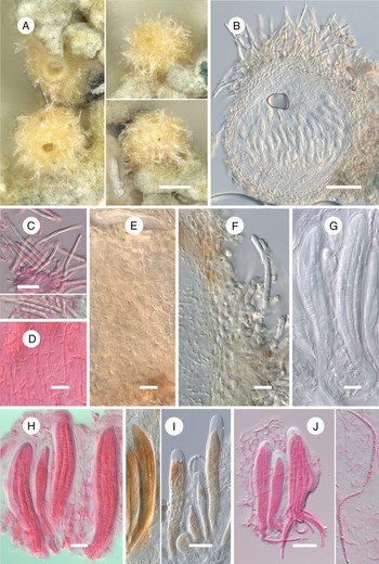
Lichenotubeufia tibellii (holotype). A, habitus of ascomata on the thallus of Cocccocarpia palmicola. B, ascoma in cross-section. C, excipular hairs; note the branched hair below. D, paraphysoids. E, exciple in surface view. F, exciple in cross-section. G, asci with spores. H, asci with spores. I, asci in I (on the left) and in K/I (on the right). J, asci, ascospores and paraphysoids. B, E & G in water; C & J in phloxine after K; D & H in phloxine; F in I. Scales: A = 100 μm; B = 50 μm; C, I & J = 20 μm; D–H = 10 μm. In colour online.

Lichenicolous ascomycete growing on Coccocarpia. Ascomata perithecia, light orange, subglobose to broadly ovoid, (95–)160–255(–275) μm diam. (n = 18), with a central ostiole 25–55 μm diam., without a papilla, not collapsing when dry, entirely covered with subhyaline excipular hairs; superficial, scattered or in groups, almost always growing on the upper surface of the host lobes, often along their edges, where they also rarely occur on the lower surface. Excipular hairs colourless, with a thin translucent lumen, sometimes underdeveloped in places, and a thick opaque wall, straight, (40–)55–95(–120) × 3–5.5 μm (n = 19), isodiametric or somewhat gradually tapering above, with a rounded, not enlarged apex, not or very rarely branched at the base, aseptate, not gathered in fascicles. Exciple continuous, same structure throughout, in surface view orange-white/very pale orange (slightly decolourizing in K), pseudoparenchymatous, composed of cells 2.5–11.5 μm diam., with walls 1–6.5 μm wide; in cross-section hyaline (inside) to orange-white (outside), 20–40 μm wide; inner cells strongly tangentially elongated, thin-walled, and outer cells isodiametric or slightly tangentially elongated, 2–10.5 μm long, with walls 1.5–6 μm wide. Hymenium not inspersed, I−, K/I−. Hamathecium composed of abundant, persistent, branched, anastomosed paraphysoids, 0.5–2.5 μm wide, attached to the subhymenium and lateral exciple (along its entire length) and filling the space between the asci and the entire upper part of the perithecium. Subhymenium indistinct. Hypothecium-like layer between subhymenium and basal exciple hyaline, 10–40 μm tall, of an unclear structure partly resembling textura globulosa, composed of cells 1–6 μm long. Asci 8-spored, subcylindrical, gradually tapering below, short pedicellate, rounded at the apex, without any distinct apical apparatus, wall not or slightly thickened at the apex (in water), (72–)86–114(–120) × 12–16(–19) μm (n = 19; in water, I or phloxine), I−, K/I− except for the orange-coloured plasma. Ascospores hyaline, acicular to long subfusiform, gradually tapering at both ends, straight, (50–)60–85(–100) × (3–)3.5–4(–4.5) μm, l/w = (11–)16–24(–30) (n = 22; in water, I or phloxine), 5–12-septate, not or only slightly constricted at the septa, smooth-walled, without perispore, with many small guttules as seen in K, arranged in the ascus in one bundle.
Conidiomata not observed.
Etymology
The species is dedicated to the eminent Swedish lichenologist Leif Tibell who collected the holotype.
Distribution and host
The new species is known from the type locality in New Zealand, growing on the thallus of Cocccocarpia palmicola, not visibly damaging the host.
Notes
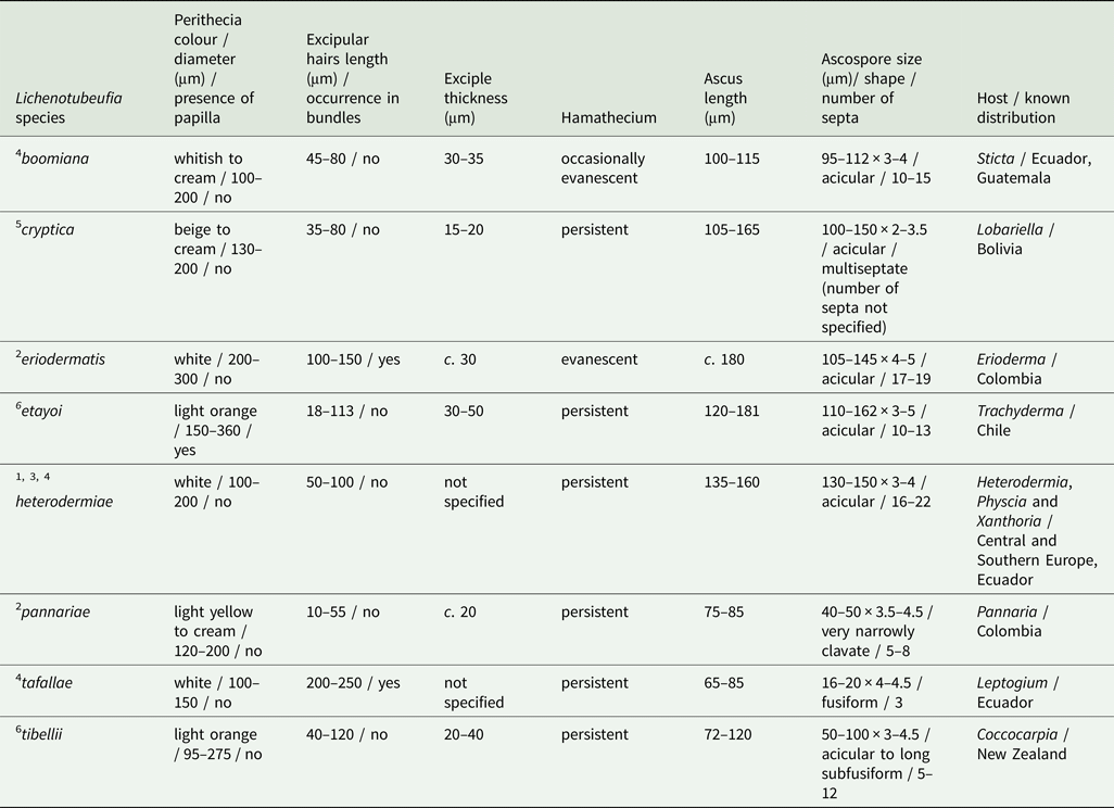
In terms of the length of the ascospores, the new species differs greatly from all other known species of Lichenotubeufia Etayo (Table 1). Morphologically it possibly most resembles L. boomiana Etayo, although this differs in having shorter excipular hairs, 45–80 μm long, an occasionally evanescent hamathecium, and on average longer ascospores, 95–112 μm long, with more (10–15) septa (Etayo Reference Etayo2017). Remarkably, this is the first species of Lichenotubeufia reported from the Australasian biogeographical realm.
Distinguishing characters of Lichenotubeufia species.

1 Brackel (Reference Brackel2014);
2 Etayo (Reference Etayo2002a);
3 Etayo (Reference Etayo2002b);
4 Etayo (Reference Etayo2017);
5 Flakus et al. (Reference Flakus, Etayo, Miadlikowska, Lutzoni, Kukwa, Matura and Rodriguez-Flakus2019);
6 present paper
Discussion
The genus Tubeufia Penz. & Sacc. (Dothideomycetes, Tubeufiales, Tubeufiaceae) was introduced to accommodate saprobic pyrenomycetes growing on decorticated or decaying woody substrata, characterized by fleshy, tender, solitary, superficial, mostly white, basally dark, glabrous, vertically oblong, non-papillate perithecia that are indefinitely split at the apex; clavate, 4–8-spored asci, extending from the base of the perithecium and usually surrounded by paraphysoids; and hyaline, cylindrical-bacilliform, pluriseptate ascospores, usually extending the full length of the ascus (Penzig & Saccardo Reference Penzig and Saccardo1898). Tubeufia was typified on T. javanica Penz. & Sacc., a detailed modern description of which is presented in Boonmee et al. (Reference Boonmee, Rossman, Liu, Li, Dai, Bhat, Jones, McKenzie, Xu and Hyde2014).
Etayo (Reference Etayo2002a, b) described three lichenicolous species of Tubeufia that he later transferred to the new genus Lichenotubeufia typified on L. eriodermatis (Etayo Reference Etayo2017). According to the diagnosis, Lichenotubeufia differs from Tubeufia in its lichenicolous lifestyle and perithecia covered by hyaline to subhyaline thick-walled hairs (Etayo Reference Etayo2017). Comparing the types of these genera, Tubeufia is additionally distinct in its oblong, subclavate to obclavate ascomata that collapse when dry and are seated on a subiculum, an exciple that is much darker at the base, asci with a distinct ocular chamber, and hyaline to pale yellow or brown ascospores (Boonmee et al. Reference Boonmee, Rossman, Liu, Li, Dai, Bhat, Jones, McKenzie, Xu and Hyde2014).
So far, six species of Lichenotubeufia have been described (Table 1). The two new species presented here match the generic concept well, although the type species differs from them in having fasciculate excipular hairs and an evanescent hamathecium (Etayo Reference Etayo2017). It is also noteworthy that papillate perithecia, as occur in Lichenotubeufia etayoi, have previously not been reported in this genus.
Six of the eight species of Lichenotubeufia are so far known from single or few finds in South and Central America, one species is known from a single find in New Zealand, and one species (L. heterodermiae) is known from many records in Europe and also from South America (Table 1). The latter has also been observed on three host genera (two of which belong to the same family), while the other species are confined to a single host genus (Table 1).
Key to the species of Lichenotubeufia
This key is based on data from Etayo (Reference Etayo2002a, b, Reference Etayo2017), Flakus et al. (Reference Flakus, Etayo, Miadlikowska, Lutzoni, Kukwa, Matura and Rodriguez-Flakus2019) and the present paper.
1 Ascospores up to 100 μm long………2
At least some ascospores more than 100 μm long………4
2(1) Ascospores 50–100 μm long, acicular to long subfusiform, 5–12-septate; on Coccocarpia …………L. tibellii
Ascospores up to 50 μm long, not acicular, 3–8-septate………3
3(2) Ascospores 16–20 μm long, fusiform, 3-septate; on Leptogium………L. tafallae
Ascospores 40–50 μm long, very narrowly clavate, 5–8-septate; on Pannaria………L. pannariae
4(1) Perithecia light orange, up to 360 μm diam., some with a distinct papilla; on Trachyderma………L. etayoi
Perithecia white, beige or cream, smaller, without papilla………5
5(4) Perithecia white, up to 300 μm diam.; excipular hairs 100–150 μm long, fasciculate; hamathecium evanescent; on Erioderma……… ………L. eriodermatis
Perithecia white, beige or cream, up to 200 μm diam.; excipular hairs up to 100 μm long, not fasciculate; hamathecium usually persistent………6
6(5) Ascospores 95–112 μm long; on Sticta………L. boomiana
Ascospores longer………7
7(6) Perithecia beige to cream, growing on the underside of the host thallus; ascospores 100–150 × 2–3.5 μm; on Lobariella……… ………L. cryptica
Perithecia white, growing on the upper side of the host thallus; ascospores 130–150 × 3–4 μm; on Heterodermia, Physcia and Xanthoria………L. heterodermiae
Acknowledgements
This study was carried out within the framework of the research project of the V. L. Komarov Botanical Institute of the Russian Academy of Sciences ‘Biodiversity, ecology, structural and functional features of fungi and fungus-like protists’ (АААА-А19-119020890079-6) using the equipment of its Core Facility Center ‘Cell and Molecular Technologies in Plant Science’. The author's visit to the UPS herbarium was financially supported by the Rolf Santesson foundation. Roland Moberg, Stefan Ekman and Martin Westberg are thanked for various kinds of help and support during his stay in Uppsala. Javier Etayo, Paul Diederich and an anonymous reviewer provided valuable comments on the manuscript.
Author ORCID
Mikhail P. Zhurbenko, 0000-0002-9839-4698.