Introduction
Myxozoans are a speciose group of obligately parasitic cnidarians (Atkinson et al., Reference Atkinson, Bartholomew and Lotan2018). Like all cnidaria, myxozoan spores have nematocysts, historically called ‘polar capsules’. Nematocysts from both free-living Cnidaria and Myxozoa consist of an intra-cellular capsule with a single opening and a coiled tubule inside (Tardent, Reference Tardent1995; Ben-David et al., Reference Ben-David, Atkinson, Pollak, Yossifon, Shavit, Bartholomew and Lotan2016). Whereas free-living cnidarians use nematocysts to capture and immobilize prey and deter invaders, myxozoans use these organelles to attach to and subsequently infect hosts. Our understanding of this initial stage of infection is from observations of Myxobolus spp. When a spore detects a host, either through mechanical or chemical cues (Kallert et al., Reference Kallert, El-Matbouli and Haas2005, Reference Kallert, Bauer, Haas and El-Matbouli2011), the tubule everts explosively, penetrates the host epithelium and contracts to pull the myxozoan alongside its target (Kallert et al., Reference Kallert, Ponader, Eszterbauer, El-Matbouli and Haas2007; Ben-David et al., Reference Ben-David, Atkinson, Pollak, Yossifon, Shavit, Bartholomew and Lotan2016). Once the parasite spore is anchored, its valve cells open along their sutures and the infectious sporoplasm migrates rapidly into the host to begin replication (El-Matbouli et al., Reference El-Matbouli, Hoffmann, Schoel, McDowell and Hedrick1999; Kallert et al., Reference Kallert, Ponader, Eszterbauer, El-Matbouli and Haas2007; Sipos et al., Reference Sipos, Ursu, Dán, Herczeg and Eszterbauer2018). Understanding the forces driving nematocyst discharge in Myxozoa could offer some means of inhibiting attachment and preventing potentially lethal infections.
Except for a few studies on the myxozoan Myxobolus cerebralis (Wagner et al., Reference Wagner, Cannon, Smith, Hillyard and Arndt2002; Kallert et al., Reference Kallert, Ponader, Eszterbauer, El-Matbouli and Haas2007), our understanding of the mechanisms of nematocyst discharge has been developed from free-living cnidarians. In these species, discharge is driven by a combination of intrinsic forces built into the coiled tubule during cnidogenesis (Jones, Reference Jones1947), and an intracapsular osmotic gradient (Weber, Reference Weber1989; Szczepanek et al., Reference Szczepanek, Cikala and David2002; Ozbek et al., Reference Ozbek, Balasubramanian and Holstein2009). When the discharge is signalled, an internal matrix of cations bound to poly-gamma glutamate dissociates, resulting in a rapid increase in Donnan potential and an influx of water, which inflates the tubule (Weber, Reference Weber1989; Tardent, Reference Tardent1995). The understanding of this process draws from observations of the effects of exogenous ions on nematocyst discharge (Cannon and Wagner, Reference Cannon and Wagner2003; Americus et al., Reference Americus, Bartholomew, Lotan and Atkinson2020). Monovalent cations (K+, Na+) increase the intra-capsular pressure of the nematocyst, and induce discharge via exchange with endogenous cations (Parker, Reference Parker1905; Yanagita, Reference Yanagita1960; McKay and Anderson, Reference McKay and Anderson1988; Weber, Reference Weber1989; Kawaii et al., Reference Kawaii, Yamashita, Nakai and Fusetani1997; Szczepanek et al., Reference Szczepanek, Cikala and David2002; Ayalon et al., Reference Ayalon, Shichor, Tal and Lotan2011). Divalent cations (Ca2+, Mg2+, Ba2+) inhibit the discharge response to physical and chemical cues via equilibrating the osmotic gradient (Blanquet, Reference Blanquet1970; Salleo et al., Reference Salleo, La Spada, Falzea and Denaro1984; Tardent, Reference Tardent1995). Trivalent cations (Gd3+, La3+) also have an inhibitory effect, possibly by blocking voltage-gated ion channels (Salleo et al., Reference Salleo, La Spada and Barbera1994). The recent description of poly-gamma glutamate-related proteins and cationic contents in the nematocysts of the myxozoan Ceratonova shasta (Piriatinskiy et al., Reference Piriatinskiy, Atkinson, Park, Morgenstern, Brekhman, Yossifon, Bartholomew and Lotan2017) suggest the molecular makeup of nematocysts in Myxozoa is similar to those of free-living Cnidaria. This was affirmed by histochemical stains of myxospores from C. shasta, M. cerebralis and seven other myxozoan species, which found nematocyst contents to be highly acidic (likely due to poly-gamma glutamate) and calcium-rich (Stilwell et al., Reference Stilwell, Griffin, Rosser, Mohammed, Sidor and Camus2020).
In this work, we continue efforts to characterize nematocyst function in Myxozoa. We hypothesized that the structural similarities between nematocysts of Myxozoa and free-living cnidarians might be conveyed in function, and myxozoans would be similarly affected by exogenous metal ions. Drawing upon the aforementioned studies on free-living Cnidaria, we screened monovalent (NaCl, KCl), divalent (CaCl2, MgCl2) and trivalent (GdCl3) salts for their ability to promote or inhibit nematocyst discharge in M. cerebralis. We tested this in vitro, using fish mucus as a proxy for live fish, and in vivo with live fish to determine if disruption of the discharge process could prevent infection. Results from the paired in vitro and in vivo tests suggested that myxozoan nematocysts respond to cationic treatments similarly to the nematocysts of free-living Cnidaria, with Na+ having the greatest effect in both assays. By inhibiting nematocyst function and spore attachment, metal salts may be potential therapeutants for prevention of myxozoan infections in fish.
Materials and methods
Source of fish
Rainbow trout (Oncorhynchus mykiss) hatched at the Roaring River Fish Hatchery and reared at the Oregon State University's Aquatic Animal Health Laboratory (AAHL) in Corvallis, Oregon were used for all experiments. Fish hatched in spring 2017 (90–100 mm) were used to isolate mucus for the in vitro assay. Fish hatched in spring 2018 (60–70 mm) were used in the subsequent in vivo assay. All procedures involving fish were in accordance with OSU institutional animal welfare guidelines under permit #5040. The authors assert that all procedures contributing to this work comply with the ethical standards of the relevant national and institutional guides on the care and use of laboratory animals.
Source of fish mucus
Individual rainbow trout were killed by overdose with MS-222 (tricaine methanesulfonate, Argent, Redmond, WA). A mucus homogenate solution was prepared using modified methods of Kallert et al. (Reference Kallert, El-Matbouli and Haas2005). Mucus was collected with a spatula, put into a 1.7 mL tube with reverse-osmosis water, and vortexed for 30 s with garnet beads to promote the transfer of soluble compounds from mucus to water. The mucus homogenate was then centrifuged for 10 min at 2200 g and the supernatant held on ice and used within 2 h of isolation.
Source of M. cerebralis actinospores
Oligochaetes (Tubifex tubifex) infected with M. cerebralis were provided for this study by Colorado Parks and Wildlife, Parvin Lakes Research Laboratory. The infection was verified with polymerase chain reaction (PCR) (Kelley et al., Reference Kelley, Zagmutt-Vergara, Leutenegger, Myklebust, Adkison, McDowell, Marty, Kahler, Bush, Gardner and Hedrick2004). Oligochaete cultures were kept at 15°C in two 3 L static tanks with aerators. Water over the oligochaetes was replaced periodically with de-chlorinated tap water, and one algae pellet added monthly (Hikari Algae Wafers Fish Food). For actinospore collection, ~150 Tubifex were removed from each tank, and placed in 200 mL plastic cups with clean mineral sand and 100 mL de-chlorinated tap water, then held for 48 h to purge their gut contents. The oligochaetes were then moved to 157 mL glass dishes in clean water, and held for an additional 36 h before the water was collected, pre-filtered through a 180 μ m wire mesh to catch worms and large debris, then poured through a 20 μ m Nitex screen, which retained the actinospores. Spores were held on ice and used within 2 h of collection.
Sources and preparation of chemical compounds
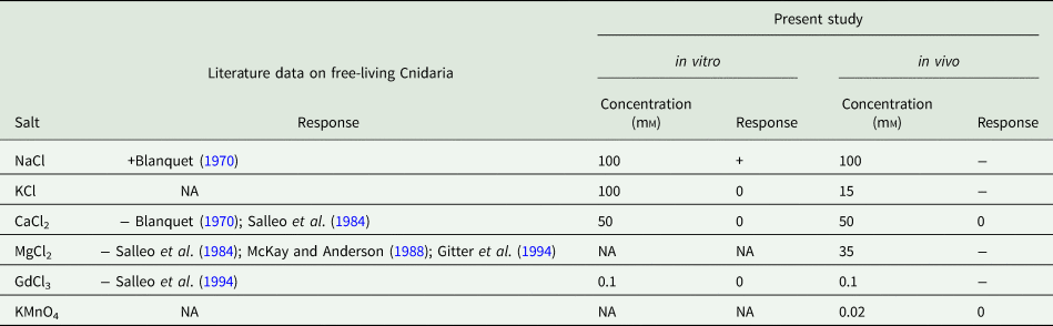
Per animal care and use guidelines, all chemicals were pharmaceutical grade and diluted with deionized water. The same salt concentrations were used in vitro and in vivo, except KCl, which was used at a lower concentration in vivo due to concerns for fish health (Trama, Reference Trama1954; Waller et al., Reference Waller, Fisher and Dabrowska1996). Concentrations were: 100 mm NaCl (Consolidated Midland Corporation); 100 mmin vitro / 15 mmin vivo KCl (Lab Alley); 50 mm CaCl2 (Research Products International); 0.1 mm GdCl3 (Alfa Aesar) (Table 1). In the in vivo assay, we also included treatments of 35 mm MgCl2 Greeway Biotech) and 0.02 mm KMnO4.
Summary of data on the effects of different salts on nematocyst discharge.

In all studies, positive control samples were induced to discharge without exposure to the treatment compounds. + indicates the treatment significantly increased the measured attribute relative to a positive control (discharge for in vitro test, infection for in vivo). − indicates treatment significantly decreased this value relative to the positive control. 0 indicates no effect. NA indicates data not available.
In vitro experiment
In addition to the salt treatments, we included a negative control with no salt or mucus addition, and positive control with no salt but receiving mucus at the same time as the other treatments. To determine actinospore concentrations in solutions from oligochaete cultures, we streaked 3 × 2 μL aliquots on an uncovered glass slide. We counted spores in each aliquot, determined an average concentration and multiplied by the total volume of solution. The spore solution was gently rocked to homogenize it, and replicate 27 μL aliquots of solution (estimated 1000 fresh M. cerebralis actinospores) were put into 1.7 mL tubes.
The in vitro experiment was performed in triplicate over 46 min at 13°C. At T = 0 min, 3 μL of one of the salts at ten times the intended concentration was added to the 1.7 mL tube containing the 27 μL of spore solution. De-chlorinated tap water was added instead of salt to control groups. At T = 15 min, actinospores were subsampled by removing 15 μL from each treatment and combining it with an equal volume of 10% formalin. At T = 16 min, 15 μL mucus homogenate or deionized tap water for the negative control, was added to each tube. At T = 46 min, the actinospore + mucus solutions were fixed by adding an equal volume of 10% formalin. Throughout the experiment, all compounds were added to the tubes in the same sequence to standardize total incubation times, while pipetting and discharging the full contents of each tube once to mix. The pH of all solutions was between 5.3 and 6.6, which was outside the reported range that can cause the pH-induced discharge of tubules in M. cerebralis (Wagner et al., Reference Wagner, Cannon, Smith, Hillyard and Arndt2002).
Counting and categorizing actinospores
From each subsample of fixed spores, triplicate 2 μL aliquots were streaked across a glass microscope slide. Slides were left uncovered to increases actinospore density for counting, and examined under a standard compound microscope at 200 × total magnification, using brightfield with the field diaphragm mostly closed to enhance contrast. Systematically in each aliquot, 50–100 actinospores were enumerated, and nematocyst conditions were assessed: actinospores were counted as ‘discharged’ if at least one of the three nematocysts had an elongated tubule, ‘undischarged’ if none of the three tubules were elongated, and were not counted if the nematocysts were obscured or if the actinospore was obviously malformed.
Statistics and data processing
For the subsamples fixed at T = 16 (after incubation with a salt solution), the proportion of actinospores with elongated tubules was used for analysis. For the subsamples fixed at T = 46 (after addition of mucus), the relative proportion of discharged spores was calculated by subtracting the number of spores discharged from the same treatment at T = 16. From these proportions, comparisons were made between all groups: those with salt and receiving mucus, the negative control without salt and not receiving mucus, and the positive control, without salt but receiving mucus at T = 16. Normality of data was checked by the Shapiro−Wilk test and normal variance was tested by Bartlett's test. Significant differences between treatments were tested by one-way ANOVA and Tukey's honestly significant difference (HSD) multiple t-test. Significance was regarded as P < 0.05.
In vivo experiments
Chemical tolerance of fish to compounds
We conducted a pre-assessment to determine what concentration of the nominated compounds could be tolerated by the fish under the proposed experimental conditions. Groups of five rainbow trout were held in 7 × 1 L containers with 250 mL aerated 16°C well water containing each salt compound. In addition to one salt-free control, we tested NaCl, KCl, CaCl2, MgCl2, GdCl3 and KMnO4 (potassium permanganate). Maximum salt concentrations were determined from relevant toxicity data in the literature (Trama, Reference Trama1954; Waller et al., Reference Waller, Fisher and Dabrowska1996; Mount et al., Reference Mount, Gulley, Russell Hockett, Garrison and Evans1997). Fish were monitored for stress (rapid movement, jumping, gasping for air and loss of equilibrium) for 30 min. Fish exhibiting signs of stress were removed and immediately euthanized by an overdose of MS-222. No stress was observed in fish treated with the initial, maximum concentration of any compound; hence these concentrations were used (Table 1).
In vivo exposures to actinospores plus compounds
After quantification using the enumeration technique described earlier, we added 50 mL aliquots of ~30 000 M. cerebralis actinospores to 1 L containers. This was followed by adding 50 mL of each salt solution at twice the concentration from the tolerance test, to make 100 mL of exposure solutions at the desired salt concentration (Table 1). The temperature of all exposure solutions was 16–20°C. To better understand the effect of divalent cations on nematocyst discharge in M. cerebralis, we included MgCl2 as a treatment in in vivo. We also included KMnO4, an oxidizing agent used to treat parasite infections in aquaculture (Straus and Griffin, Reference Straus and Griffin2002). Positive controls (fish with actinospores and no salt) and negative controls (fish with neither actinospores nor salt) were included.
The following steps were used to expose fish to M. cerebralis. First, exposure solutions were equilibrated for ~15 min prior to the addition of fish. In parallel, treatment groups of 3 fish were acclimated in 1 L aerated containers with 100 mL of each salt solution for 5 min in the absence of parasites. Groups of acclimated fish were each netted and transferred to the 1 L container with the corresponding salt and actinospore solution. Fish were exposed to spores for 15 min, netted, then dipped three times in a spore-free solution of the appropriate salt to rinse off poorly adhered actinospores. Each group of fish was then returned to the container of parasite-free solution used for the original acclimation, held for 5 min, then the fish were killed by an overdose of buffered MS-222 in the salt. Gills were removed and frozen for downstream processing. Of the known sites of attachment for M. cerebralis, gills were chosen as they accumulate more spores than skin (Eszterbauer et al., Reference Eszterbauer, Sipos, Szakály and Herczeg2019) and we suspected their rate of actinospore encounter might be less variable between fish than fins. Instruments were decontaminated with hydrogen peroxide between samples. The experiment was conducted twice, with different batches of spores from the same stock solutions and a cohort of unexposed fish.
DNA was extracted from each gill sample using a Qiagen DNeasy Blood and Tissue kit following the manufacturer's recommendations, with DNA eluted from the column with two washes of 100 μL buffer AE. Total DNA concentration was measured with a Nanodrop spectrometer (Thermo Scientific) and normalized by dilution with molecular grade water to 100–250 ng/μL, to achieve an optimal range of detection. M. cerebralis DNA was quantified using an Applied Biosystems StepOnePlus Real-Time PCR System and the amplification profile and M. cerebralis-specific TaqMan probes and primers described by Kelley et al., Reference Kelley, Zagmutt-Vergara, Leutenegger, Myklebust, Adkison, McDowell, Marty, Kahler, Bush, Gardner and Hedrick2004. Each 10 μL reaction contained 1.79 μL molecular grade water, 0.15 mm bovine serum albumin, 80 nm TaqMan probe, 400 nm each primer, 1 × TaqMan Universal PCR MasterMix, and 2 μL diluted gDNA. In a second qPCR using the same machine, we measured the concentration of a single-copy rainbow trout gene for endogenous normalization. Each 10 μL reaction contained 1 μL molecular grade water, 1 × SYBR Green Power Master Mix, 4 μ m of each ‘gDNA Salmonid EF1-α’ primers (Bland et al., Reference Bland, McIntosh, Bain and Snow2012) and 2 μL gDNA further diluted 1:10. Amplification proceeded with a 10 min incubation at 95°C and then 40 cycles of 95°C for 15 s and 60°C for 1 min. For both reactions, samples were run in triplicate wells, and included a positive control (DNA extracted from M. cerebralis actinospores and rainbow trout, respectively) and a negative control (molecular grade water).
Statistics and data processing
For each treatment, three replicate fish from the two experimental repeats (N = 6 per treatment) were included in the analysis. We used the 2−ΔΔCt method (Schmittgen and Livak, Reference Schmittgen and Livak2008) to determine relative M. cerebralis concentration, with the rainbow trout EF1-α gene used for normalization. For fold change calculations, data from each repeat were compared to the average of their respective positive control fish. These fold change values were used to compare infection between all groups. Prior to binning data from the repeats, we performed Levene's test to check for the difference in the variance of the two repeats. No significant difference in variance was observed between repeats of the in vivo experiment (Levene's test, df = 1, F = 1.034, P = 0.316) thus the two sets of three replicates were combined. Normality of data was checked by the Shapiro−Wilk test, and normal variance tested by Bartlett's test. As a non-parametric alternative to ANOVA, we used the more conservative Kruskal−Wallis test and Dunn's posthoc test.
Results
The effect of chloride salts on nematocyst discharge in vitro
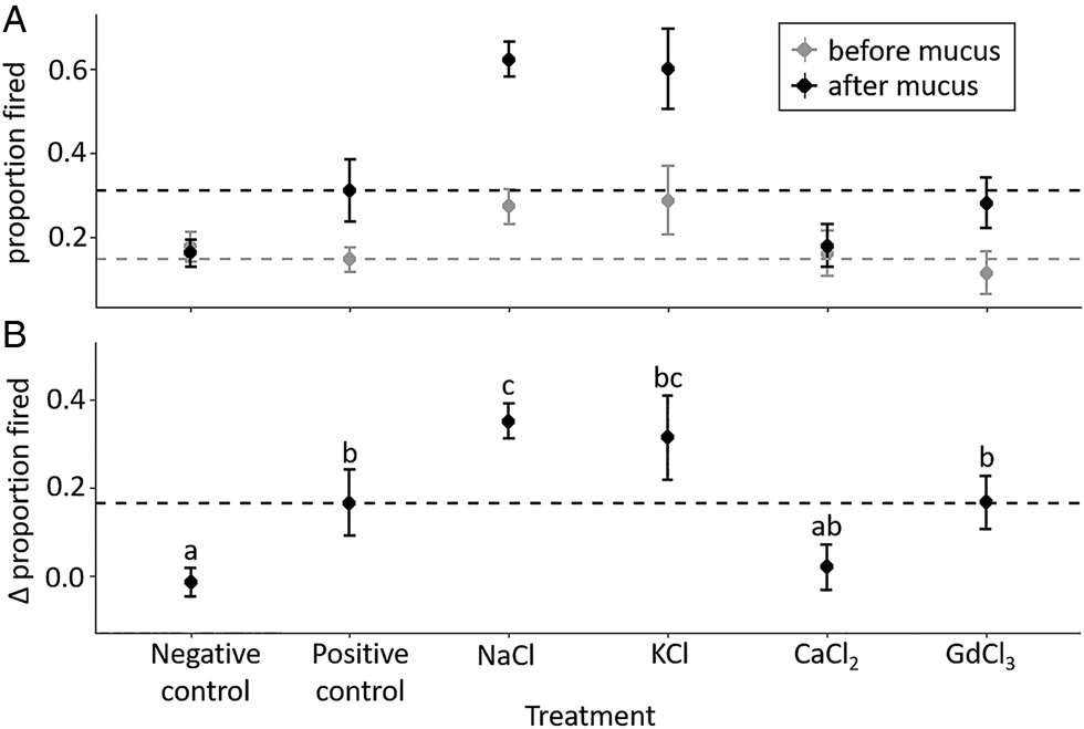
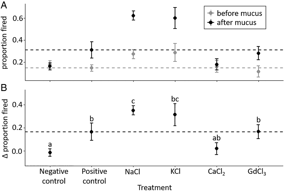
The effect of treatments on inducing nematocyst tubule discharge is displayed in Fig. 1. At T = 15 min (salt only, prior to addition of mucus), spores in Na+ and K+, had 1.5 × and 1.6 × more discharged nematocysts than the positive control (P = 0.148 and P = 0.083, respectively). When considering the change in discharge proportion from the addition of mucus, the positive control group (mucus but no salt) saw more nematocysts discharge than in the negative control group (no mucus or salt); (P = 0.047). The NaCl and KCl treatment groups had 2.1 × and 1.9 × the change of the positive control (P = 0.039; P = 0.146), respectively. Spores in CaCl2 had 0.4 × the change in discharge of the positive control (P = 0.152). GdCl3 did not have an effect on discharge before or after the addition of mucus (T = 15: P = 0.995, T = 46: P = 1.000).
In vitro effects of chemical treatments on nematocyst tubule discharge in Myxobolus cerebralis actinospores. In (A), the y-axis is the proportion of actinospores with discharged nematocysts before (T = 15 min) and after (T = 46 min) addition of mucus. In (B), proportions of discharged nematocysts at T = 15 min have been subtracted from the T = 46 min data to distinguish any additional discharge caused by the addition of mucus. Negative control was water only, no mucus. Positive control was water with mucus applied at T = 16 min. Dashed lines indicate the mean value of the positive controls. Significance from Tukey's HSD test is noted at P < 0.05.
Effect of chloride salts on M. cerebralis infection in vivo
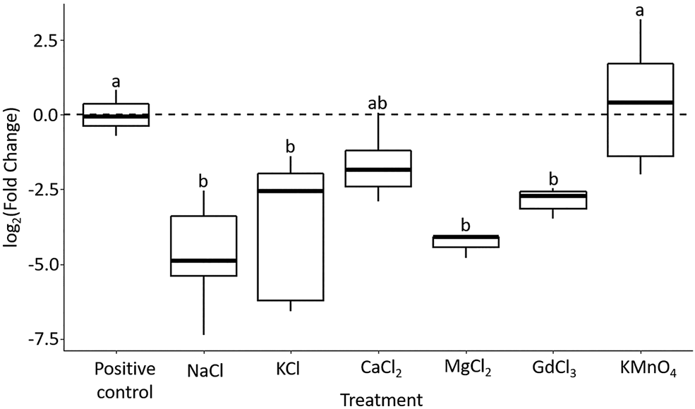
qPCR analysis was done on salt-treated fish with positive (actinospores with no addition of salts) and negative (without actinospores) controls (Fig. 2). The greatest reduction in M. cerebralis infection, relative to the positive control, was in the group exposed to NaCl (mean fold change of −50.050, P = 0.009). Likewise, significantly less parasite DNA was detected in groups exposed to KCl (mean fold change = −36.116, P = 0.030), MgCl2 (mean fold change = −20.404, P = 0.015), and GdCl3 (mean fold change = −7.551, P = 0.027). The fish in CaCl2 had slightly reduced parasite load (mean fold change = −3.566, P = 0.319), and those in KMnO4 had a similar amount of parasite DNA as the positive control (mean fold change = 1.061, P = 1.000). No M. cerebralis DNA was detected in negative control fish.
Results of in vivo exposure of rainbow trout to Myxobolus cerebralis actinospores in solutions containing different compounds. Myxobolus cerebralis DNA was detected on gills of treated fish measured by parasite-specific qPCR (Kelley et al., Reference Kelley, Zagmutt-Vergara, Leutenegger, Myklebust, Adkison, McDowell, Marty, Kahler, Bush, Gardner and Hedrick2004), normalized by Rainbow trout EF1-α gene, relativized to the average of the positive controls, and expressed as log2(Fold Change). Positive control fish were exposed to the parasite in water without the added compounds. Dashed lines indicate the mean value of the positive controls. Significance from Dunn's test is noted at P < 0.05.

Discussion
Myxozoan infections rely on the success of spores to attach to hosts, which they achieve through the timely discharge of their nematocysts. This discharge process is essential for infection, and thus could be a target for therapeutants. However, there is presently little data on the chemistry of the sensing and discharge process of myxozoan nematocysts. We designed our study to parallel what is known about the effects of exogenous cations on nematocyst discharge in free-living cnidarians (Cannon and Wagner, Reference Cannon and Wagner2003), and work that shows myxozoan nematocysts can be discharged using fish mucus preparations or inosine−arginine (Kallert et al., Reference Kallert, El-Matbouli and Haas2005, Reference Kallert, Ponader, Eszterbauer, El-Matbouli and Haas2007, Reference Kallert, Forró and Eszterbauer2014). For the in vitro assay, we used rainbow trout mucus homogenate rather than inosine−arginine (a chemical in mucus responsible for discharge; Kallert et al., Reference Kallert, Forró and Eszterbauer2014), to better align with the in vivo study. We screened a variety of salt compounds for their ability to promote or inhibit nematocyst discharge in M. cerebralis in vitro, and in vivo to determine if disruption of the discharge process could prevent infection.
Effectors of nematocyst discharge
Overall, we observed M. cerebralis nematocysts had discharge patterns analogous to those observed for nematocysts of free-living Cnidaria (Blanquet, Reference Blanquet1970; Hidaka and Afuso, Reference Hidaka and Afuso1993) (Table 1). For monovalent cations Na+ and K+, we observed induction of discharge before the addition of fish mucus. We hypothesize that this was caused by the exchange of the exogenous monovalent cations with endogenous divalent cations, which increased the nematocysts' intracapsular pressure and promoted discharge (Hidaka and Afuso, Reference Hidaka and Afuso1993). For nematocysts that did not discharge immediately in Na+ and K+, the osmotic pressure likely remained enhanced, in a hypersensitized state that made them more likely to discharge upon the addition of mucus. These in vitro observations led us to predict that Na+ and K+ enhanced tubule discharge would result in increased infectivity of the spores in the in vivo trials. Intriguingly, the in vivo results demonstrated the opposite, with exposure to Na+ and K+ significantly reducing detectable parasite DNA on fish gills. This may be due to the hypersensitized spores discharging prematurely or inaccurately when encountering fish cues, or discharging preferentially on external tissues like fins or skin. In the in vitro test, only ~10 and ~11% of the spores pre-discharged in response to Na+ and K+, respectively, therefore we suggest that spore hypersensitization rather than pre-discharge contributed more to the reductions in infection inferred from the corresponding in vivo tests.
Divalent Ca2+ was the only treatment to inhibit discharge in vitro (Fig. 1B), and likewise reduced M. cerebralis infection in vivo (Fig. 2), though its effect was not significant in either test. This was consistent with data from experiments with free-living Cnidaria (Blanquet, Reference Blanquet1970; Hidaka, Reference Hidaka1992), where Ca2+ is thought to be bound to or stabilize the anionic poly-gamma glutamate in undischarged nematocyst (Lubbock et al., Reference Lubbock, Gupta and Hall1981; Lubbock and Amos, Reference Lubbock and Amos1981; Hidaka and Afuso, Reference Hidaka and Afuso1993). Addition of exogenous divalent cations like Ca2+ and Mg2+ decreases nematocyst volume, an observable proxy for internal osmotic pressure, by reducing the Donnan potential between the interior of undischarged nematocysts and the environment (Weber, Reference Weber1989; Hidaka and Afuso, Reference Hidaka and Afuso1993). Given that we observed lower parasite DNA load in the fish treated with Ca2+ and Mg2+, and that nematocyst discharge was induced by exchange with Na+ and K+ as discussed above, we hypothesize that M. cerebralis actinospore nematocysts contain Ca2+ or another divalent cation in their native state. This aligns with staining of calcium in M. cerebralis myxospores (Stilwell et al., Reference Stilwell, Griffin, Rosser, Mohammed, Sidor and Camus2020), and suggests that nematocyst contents are conserved between myxozoan life-stages (actinospores and myxospores).
The effect of exogenous Ca2+ addition on nematocyst discharge is likewise similar between cnidarian taxa, but varies by ion concentration. In free-living species, depletion of Ca2+ inhibits nematocyst discharge (McKay and Anderson, Reference McKay and Anderson1988; Gitter et al., Reference Gitter, Oliver and Thurm1994; Kawaii et al., Reference Kawaii, Yamashita, Nakai and Fusetani1997). This was observed in the myxozoan Henneguya nuesslini, (Kallert et al., Reference Kallert, Ponader, Eszterbauer, El-Matbouli and Haas2007). Increases in Ca2+ up to 10 mm enhance discharge, however, beyond 10 mm, the additional Ca2+ inhibits discharge (Blanquet, Reference Blanquet1970; Salleo et al., Reference Salleo, La Spada, Falzea and Denaro1984). These studies and ours support the dual hypotheses that nematocysts utilize exogenous calcium for signalling discharge and endogenous calcium for driving discharge.
From similar work in free-living Cnidaria, we hypothesized that nematocysts would exhibit reduced discharge in the presence of the trivalent cation Gd3+, as this would interfere with voltage-gated calcium channels involved in the signal transduction step of nematocyst discharge (Gitter et al., Reference Gitter, Oliver and Thurm1994; Salleo et al., Reference Salleo, La Spada and Barbera1994; Weir et al., Reference Weir, Dupre, van Giesen, Lee and Bellono2020). While voltage-gated calcium channels have been identified in nematocysts of free-living Cnidaria, it is not known if they are present in myxozoan nematocysts. However, we observed that treatment with GdCl3 had no effect on tubule discharge in vitro, but reduced parasite DNA load significantly in vivo. Perhaps this was by hindering the spore's response to host cues and preventing timely attachment. Involvement of voltage-gated calcium channels in myxozoan nematocyst discharge might also explain why Mg2+ had a greater inhibitory effect on infection than Ca2+. Both are divalent cations capable of reducing Donnan potential, but Mg2+ is also a calcium channel antagonist and may interfere with signalling (Gitter et al., Reference Gitter, Oliver and Thurm1994).
Conclusions and future directions
The results of our in vitro and in vivo experiments show that like free-living cnidarians, actinospores of the parasite M. cerebralis contain nematocysts that are affected by endogenous cations. Our data support the hypothesis that M. cerebralis nematocysts contain a matrix of cations bound to poly-gamma glutamate complexes, and that these cations can be exchanged passively with ions in the environment. Our findings also suggest that the exchange of exogenous monovalent cations with the endogenous internal cations may hypersensitize spores to fish stimuli but make them less successful at infection in vivo.
Our findings support NaCl, KCl, MgCl2 and GdCl3 as potential treatment options for myxozoan parasites in freshwater aquaculture, particularly for fish that can withstand high salt concentrations. The commonalities observed across Cnidaria suggest that these treatments might similarly inhibit other pathogenic myxozoans, like Ceratonova shasta and Tetracapsuloides bryosalmonae. NaCl is already employed in salmonid aquaculture to treat infections from external parasites (Dewi, Reference Dewi2018) and minimize osmoregulatory stress (Mifsud and Rowland, Reference Mifsud and Rowland2008; Tacchi et al., Reference Tacchi, Lowrey, Musharrafieh, Crossey, Larragoite and Salinas2015). The oxidizing agent KMnO4 is also widely employed in aquaculture (Straus and Griffin, Reference Straus and Griffin2002) but had no effect on M. cerebralis infection in our tests, which highlighted the importance of targeting nematocyst function specifically to prevent infection. A salt-based therapeutant for myxozoan infections might be employed as a diffuse treatment in a contained system or as short-term dosing of a flow-through system. Salmonids are particularly vulnerable to M. cerebralis infections as juveniles (Ryce et al., Reference Ryce, Zale and McConnell2004), thus a therapeutant may be particularly useful during early hatchery rearing stages and prior to release. Before application, it will be necessary to ascertain the effect of salt treatments on infection in whole fish, not just the gills, and on myxospore development and fish morbidity.
In addition to testing treatments, the in vivo method we present here might help identify regulators and signalling pathways involved in myxozoan attachment to hosts. An important gap in our understanding of myxozoan nematocysts lies in their activation process. Nematocyst activation, specifically the role of Ca2+, has been studied in free-living cnidarians using pharmaceutical ion channel blockers like nifedipine, streptomycin and gentamicin (Watson and Hessinger, Reference Watson and Hessinger1994); any of which could be tested for efficacy in future experiments with myxozoan spores.
Further, it will be necessary to examine discharge processes in a range of myxozoan species. M. cerebralis served as an ideal model for this investigation as it is well-researched and the actinospores are relatively large (~150 μ m). However, the applicability of our findings to spores of marine myxozoans and those that infect anadromous fishes is unknown. Effect of salts on fresh-water Myxozoa is particularly relevant to understanding whether some species, like Ceratonova shasta, can infect anadromous fish in estuaries. However practical challenges exist in working with small ~10 μ mC. shasta actinospores in vitro. To overcome these challenges, myxozoan researchers could adapt tools from research on free-living cnidarians, such as microfluidics flow cells developed to study coral larvae (Van Treuren et al., Reference Van Treuren, Brower, Labanieh, Hunt, Lensch, Cruz, Cartwright, Tran and Fordyce2019) and jellyfish nematocysts (Park et al., Reference Park, Piriatinskiy, Zeevi, Ben-David, Yossifon, Shavit and Lotan2017), to study responses of individual myxozoan spores in vitro. Future work should also adapt immunohistochemistry techniques from studies of free-living Cnidaria to examine and visualize specific features (such as voltage-gated channels) in Myxozoa.
Acknowledgements
We are grateful to George Schisler, Colorado Parks and Wildlife, Parvin Lakes Research Laboratory, for providing infected Tubifex.
Financial support
This research was supported by Research grant no. IS-5001-17C from BARD, The United States-Israel Binational Agricultural Research and Development Fund.
Conflict of interest
None.
Ethical standards
All procedures involving fish were in accordance with OSU institutional animal welfare guidelines under permit #5040. The authors assert that all procedures contributing to this work comply with the ethical standards of the relevant national and institutional guides on the care and use of laboratory animals.